
Primary ciliary dyskinesia CT
Editor-In-Chief: C. Michael Gibson, M.S., M.D. [1]
Overview
Overview
CT
CT
-
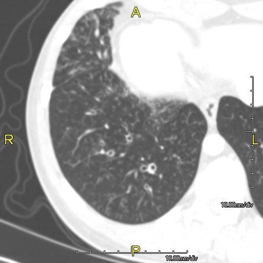 CT image showing dilated and thickened medium sized airways (bronchiectasis)in a patient with Kartagener syndrome
 -
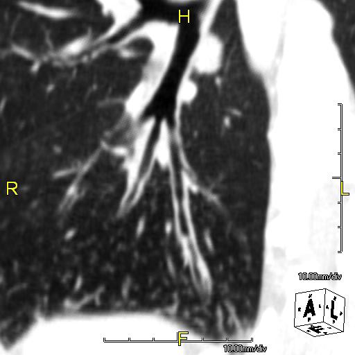 Oblique sagittal CT image showing lower lobe cylinidrical bronchiectasis in the same patient


-
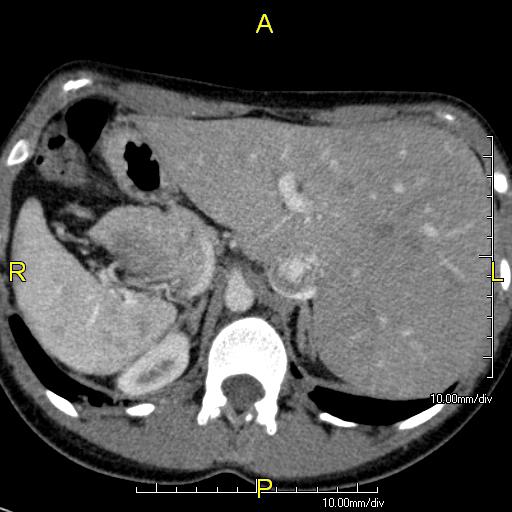 Axial CT image showing situs inversus with the liver and IVC on the left and the spleen and aorta on the right
 -
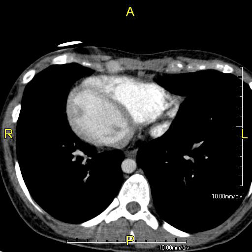 Axial CT image showing dextrocardia with the IVC and morphologic right ventricle on the left and the left ventricle on the right


-
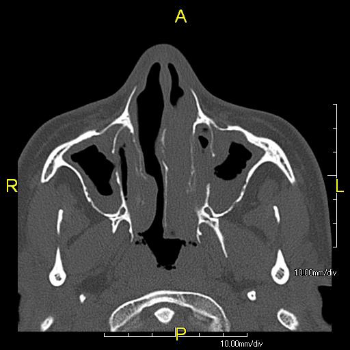 Axial CT image showing chronic sinusitis in a patient with Kartagener syndrome.
 -
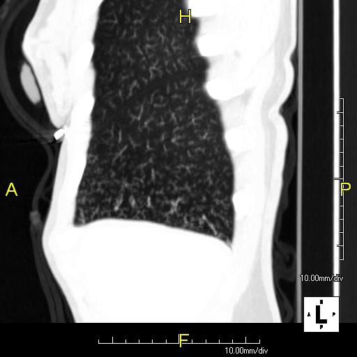 Sagittal CT image showing “tree in bud” appearance of mucous impaction in distal small airways related to primary ciliary dyskinesia
 -
 Situs inversus in a patient with Kartagener syndrome (Image courtesy of RadsWiki and copylefted)


Looking for the patient version?
© 2026 MyEClinic – IFTM Institut für Telematik in der Medizin GmbH