

Health Dictionary
Reliable, easy-to-read patient information on common medical conditions — written in plain language, free of jargon, and grounded in current evidence. Browse the most frequent diagnoses, understand what they mean for everyday life, and learn which steps and questions help you have a focused conversation with your doctor.
Sinus venarum
Introduction
- This article is on an embryological structure. For the heart defect of the same name, please see atrial septal defect.
Overview
Overview
The sinus venosus is the large quadrangular cavity located between the two venae cavae in the embryonic human heart. In the adult it is incorporated into the wall of the right atrium to form a smooth part called the sinus venarum, also know as the venarum sinus, which is separated from the rest of the atrium by a ridge of fibres called the crista terminalis.
In the embryo, the thin walls of the sinus venosus are connected below with the right ventricle, and medially with the left atrium, but are free in the rest of their extent. It receives blood from the vitelline vein, umbilical vein and common cardinal vein.
It originally starts as a paired structure but shifts towards associating only with the right atrium as the embryonic heart develops. The left portion shrinks in size and eventually forms the coronary sinus and oblique vein of the left atrium, whereas the right part becomes incorporated into the right atrium to form the sinus venarum.
Additional images
Additional images
-
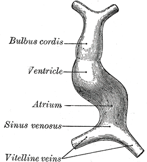 Diagram to illustrate the simple tubular condition of the heart.
 -
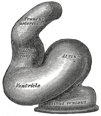 Heart of human embryo of about fourteen days.
 -
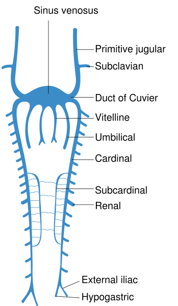 Scheme of arrangement of parietal veins.


See also
See also
External links
External links
- Template:GPnotebook
- Template:SUNYAnatomyLabs – Gross anatomy of the adult heart
- Template:UMichAtlas – “Right atrium, internal structure, anterior view”
- Template:EMedicineDictionary
Editor-In-Chief: Template:Infobox Embryology C. Michael Gibson, M.S., M.D. [1]