

Health Dictionary
Reliable, easy-to-read patient information on common medical conditions — written in plain language, free of jargon, and grounded in current evidence. Browse the most frequent diagnoses, understand what they mean for everyday life, and learn which steps and questions help you have a focused conversation with your doctor.
Submandibular ganglion
Introduction
The submandibular ganglion (or submaxillary ganglion in older texts) is part of the human autonomic nervous system. It is one of four parasympathetic ganglia of the head and neck. (The others are the otic ganglion, pterygopalatine ganglion, and ciliary ganglion).
Function
Function
The submandibular ganglion is responsible for innervation of two salivary glands: the submandibular gland and sublingual gland.
Location and relations
Location and relations
The submandibular ganglion is small and fusiform in shape. It is situated above the deep portion of the submandibular gland, on the hyoglossus muscle, near the posterior border of the mylohyoid muscle.
The ganglion ‘hangs’ by two nerve filaments from the lower border of the lingual nerve (itself a branch of the mandibular nerve, CN V3). It is suspended from the lingual nerve by two filaments, one anterior and one posterior. Through the posterior of these it receives a branch from the chorda tympani nerve which runs in the sheath of the lingual nerve.
Fibers
Fibers
Like other parasympathetic ganglia of the head and neck, the submandibular ganglion is the site of synapse for parasympathetic fibers and carries other types of nerve fiber that do not synapse in the ganglion. In summary, the fibers carried in the ganglion are:
- Sympathetic fibers from the internal carotid plexus, via the facial artery and its branches. These do not synapse in this ganglion.
- Preganglionic parasympathetic fibers from the superior salivatory nucleus of the medulla oblongata, via the chorda tympani and lingual nerve, which synapse at the origin of:
- Postganglionic parasympathetic fibers to the oral mucosa and the submandibular and sublingual salivary glands.
Additional images
Additional images
-
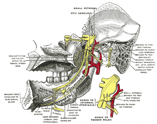 Mandibular division of trifacial nerve, seen from the middle line.
 -
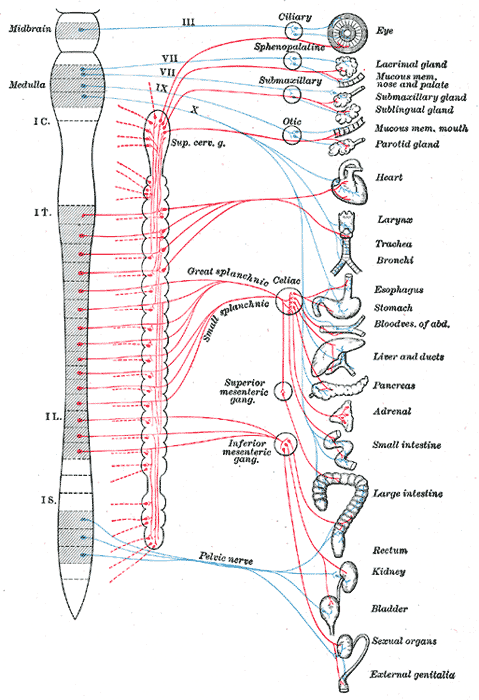 Diagram of efferent sympathetic nervous system.


Editor-In-Chief: Template:Infobox Nerve C. Michael Gibson, M.S., M.D. [1]