
The heart in oncologic disease
Editor-In-Chief: C. Michael Gibson, M.S., M.D. [1]
Associate Editor-In-Chief: Cafer Zorkun, M.D., Ph.D. [2]
Imaging
Imaging
CT
Labeled images below are courtesy of RadsWiki and copylefted.
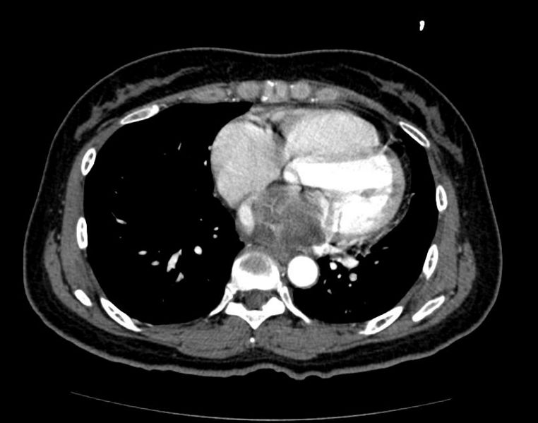
- Atrial Myxoma
-
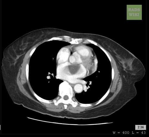 Atrial Myxoma
 -
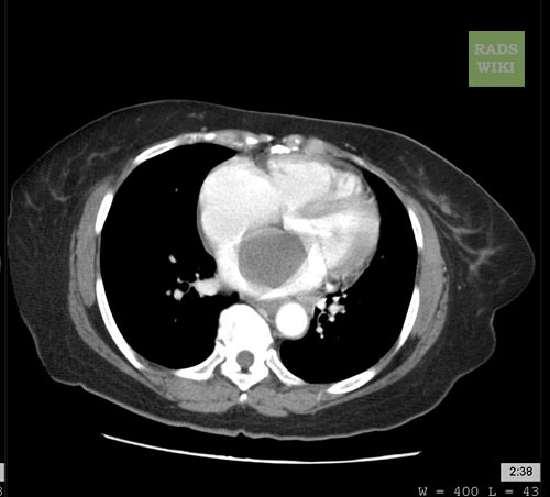 Atrial Myxoma
 -
 Atrial Myxoma


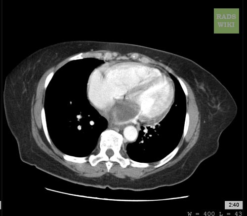
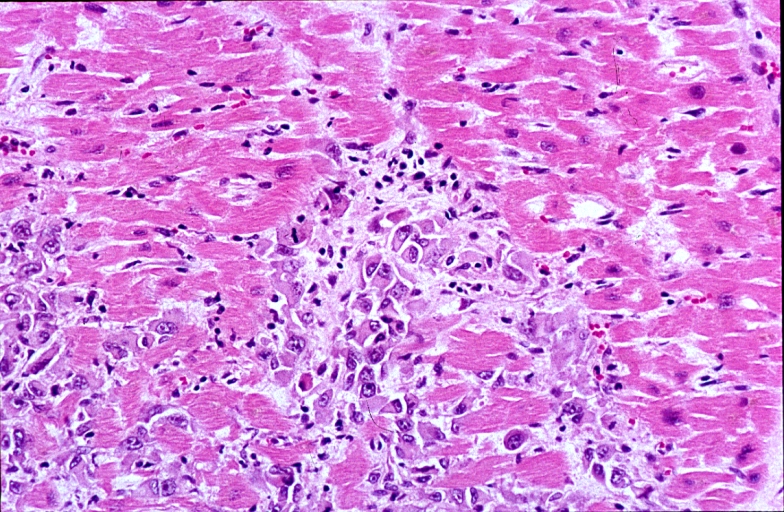
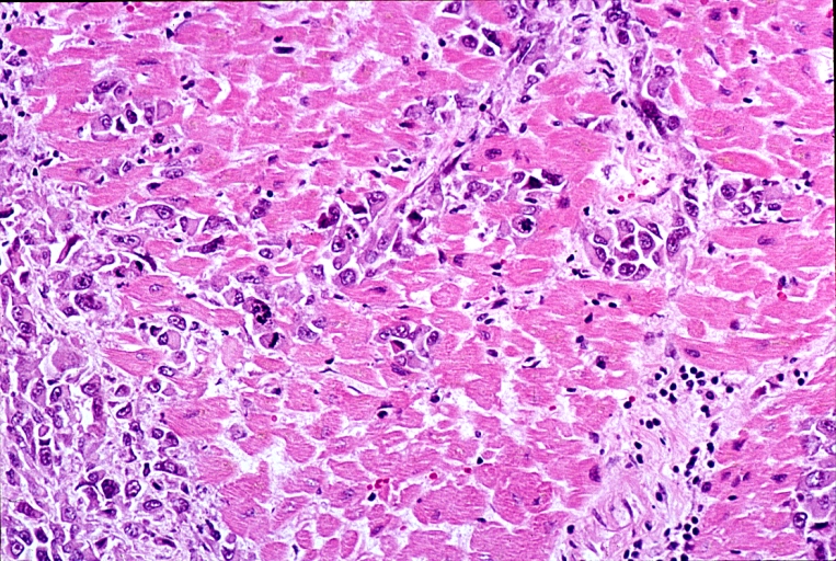
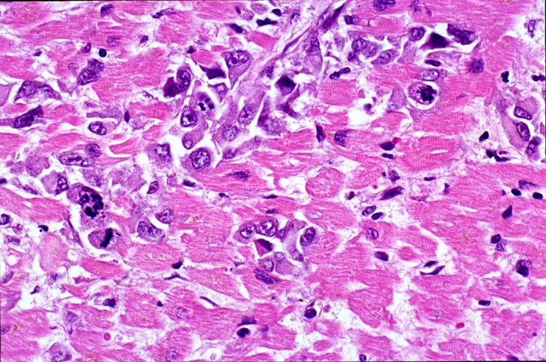
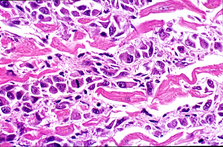
- Cardiac Rhabdomyosarcoma
-
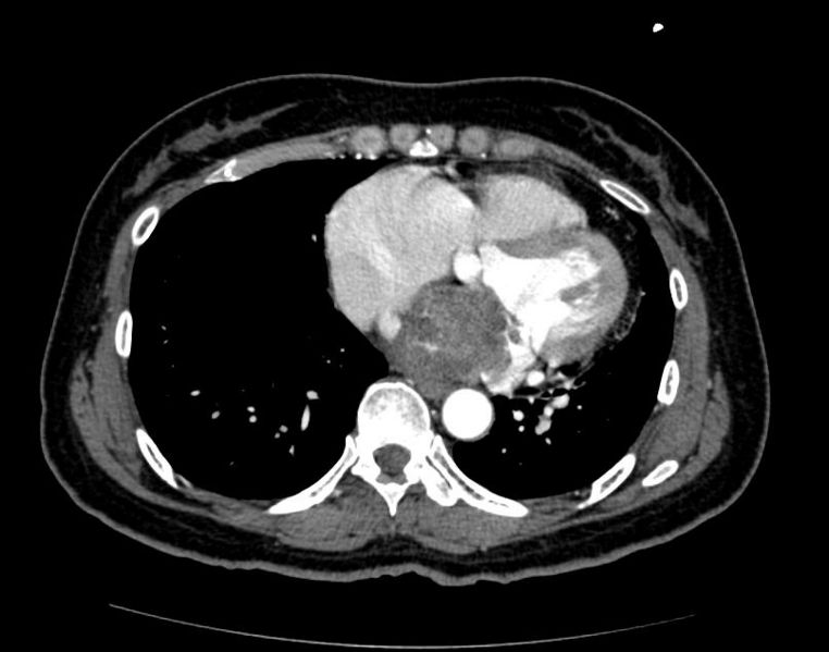 Cardiac Rhabdomyosarcoma
 -
 Cardiac Rhabdomyosarcoma
 -
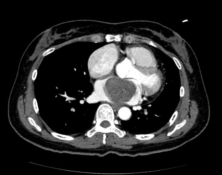 Cardiac Rhabdomyosarcoma


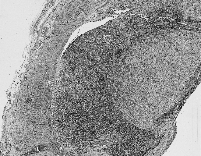
Pathological Findings
Image shown below is courtesy of Professor Peter Anderson DVM PhD and published with permission. © PEIR, University of Alabama at Birmingham, Department of Pathology
-
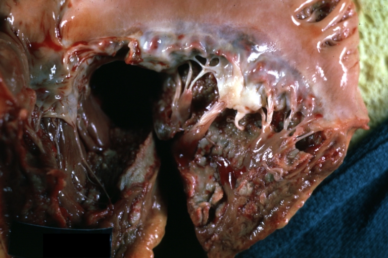
 HEART: Metastatic Tumor: Gross very unusual large metastatic carcinoid in right atrium
 -
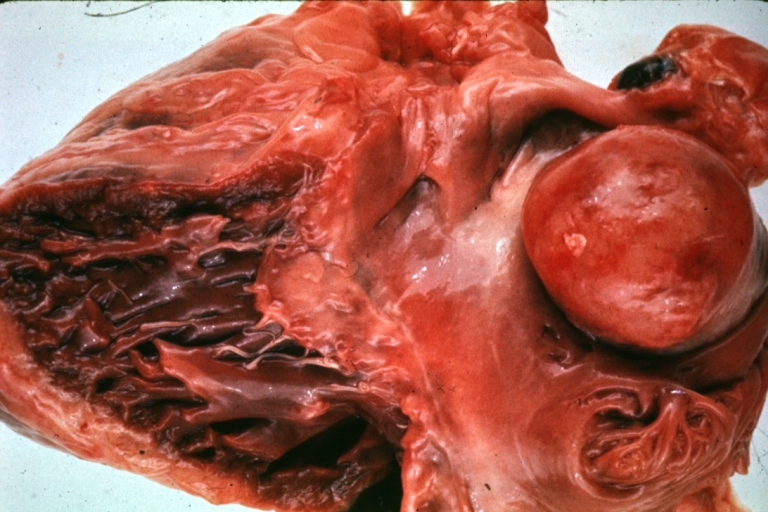
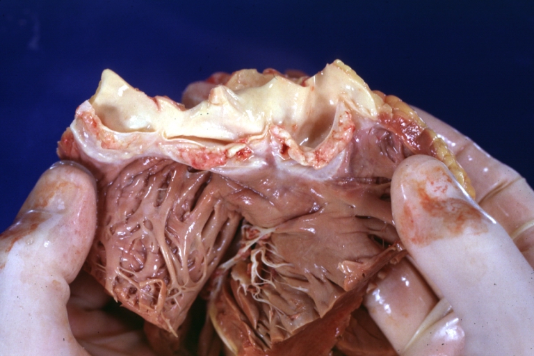
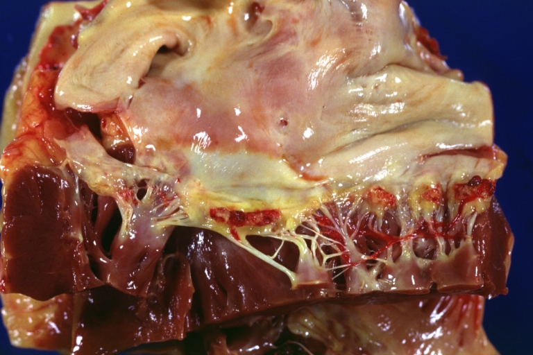
 Cardiac Myxoma A gelatinous tumor is attached by a narrow pedicle to the atrial septum. The myxoma has an irregular surface and nearly fills the left atrium.


-
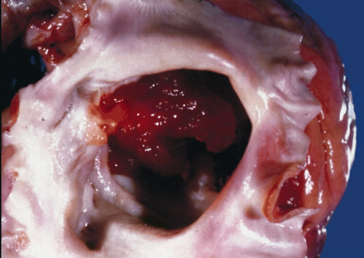
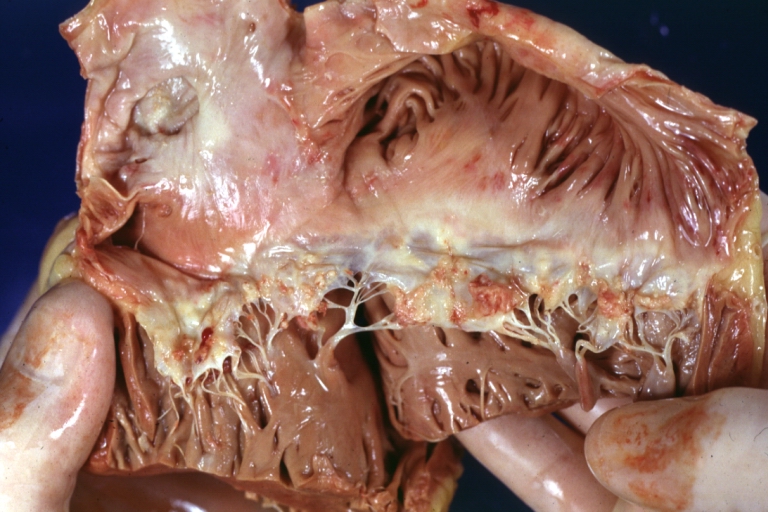
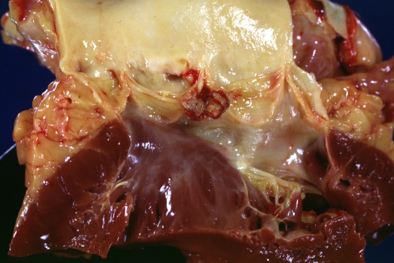
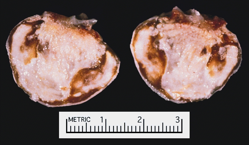 Cardiac Myxoma There was a calcified right atrial mass on the X ray of a 47-year-old man. Resection demonstrated a smooth-surfaced tumor. The gritty material seen microscopically on cut section was calcified and ossified myxoma.
 -
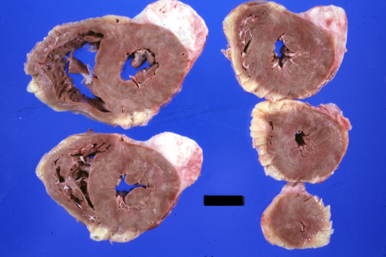
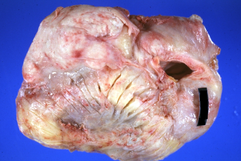
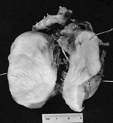 Cardiac Fibroma Cut surface of the tumor shown in figure 6-2. The left ventricular (LV) cavity is present behind the mass. The patient was a 4-month-old child who died suddenly without a previous medical history.


-
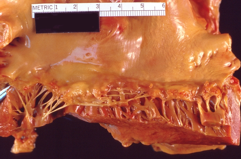
 This tumor was resected from the right atrium of a 1-year-old boy with pericardial effusions. Note areas of hemorrhage and dilated vessels. The patient was well 49 months postoperatively.
 -
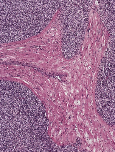
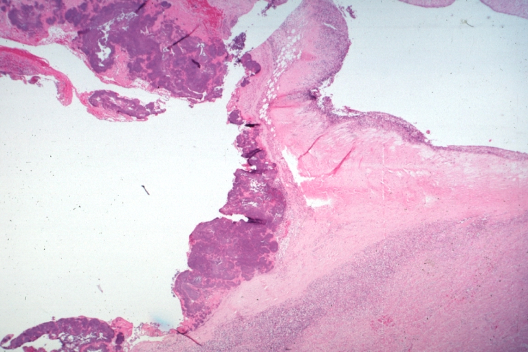
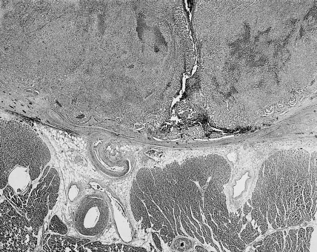
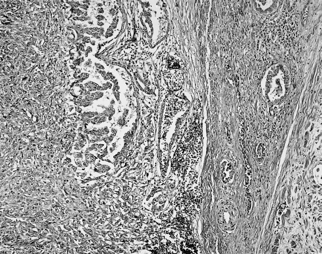
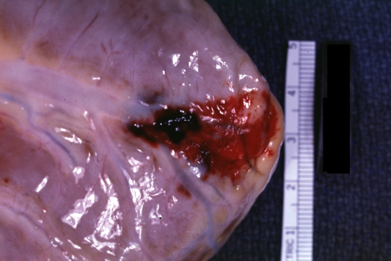
 Papillary fibroelastomas are often on the arterial surface and may project into the coronary ostium, causing ostial occlusion. This tumor is in the noncoronary sinus.


The Heart in Central and Peripheral Nervous System Cancers
Editor-In-Chief: C. Michael Gibson, M.S., M.D. [1]
Associate Editor-In-Chief: Cafer Zorkun, M.D., Ph.D. [2]
Histopathological Findings
-
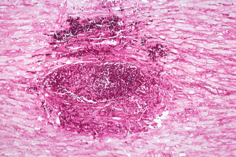
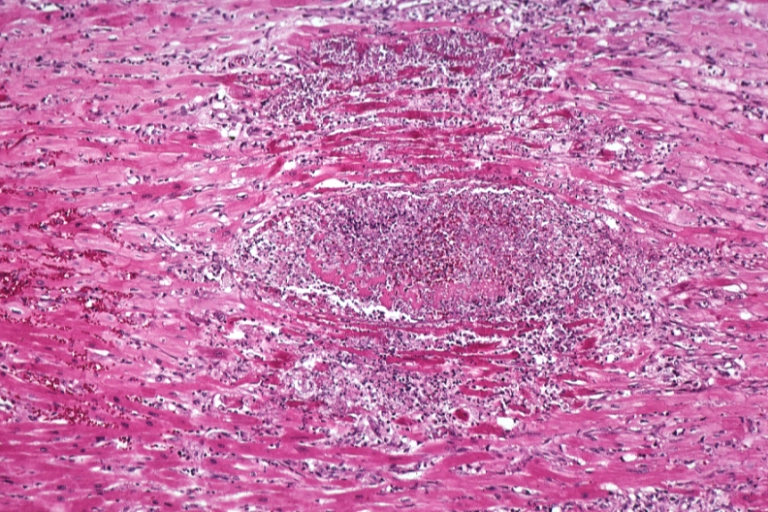
 HEART: Metastatic Tumor Malignant Schwannoma: Gross natural color good close-up view of two nodular masses of hemorrhagic tissue protruding from endocardium into right ventricle lumen (primary thought to be in esophagus)

References
Additional Resources
See Also
- The Heart in Breast Cancer
- The Heart in Gynecologic Tumors
- The Heart in Head and Neck Tumors
- The heart in leukemias
- The Heart in Lung Cancers
- The Heart in Lymphomas
- The Heart in Multiple Myeloma
- The Heart in Osteosarcomas
- The Heart in Peritoneal and Mesothelial Tumors
- The Heart in Primary Myocardial Tumors
- The Heart in Skin Cancers
- The Heart in Thyroid and Parathyroid Cancers
- The Heart in Tumors that Originated from Vascular Structure
- The Heart in Urinary System Tumors
- The Heart in Gastrointestinal, Hepatobilier & Pancreatic Tumors
The Heart in Leukemias
Editor-In-Chief: C. Michael Gibson, M.S., M.D. [1]
Associate Editor-In-Chief: Cafer Zorkun, M.D., Ph.D. [2]
Histopathological Findings
-
 HEART: Abscess: Micro PAS high mag pseudohyphae of Candida. A leukemia case
 -
 HEART: Abscess: Micro H&E high mag abscess due to Candida with marked muscle necrosis. A leukemia case


-
 HEART: Eosinophilic Endomyocarditis: Gross natural color extensive thrombi in patient with eosinophilic leukemia
 -
 HEART: Abscesses Candida: Gross natural color excellent depiction myocardial abscesses Candida tropicalis in 51yo man with acute monocytic leukemia


-
 HEART: Abscesses Candida: Gross natural color excellent example three horizontal sections myocardium with obvious abscesses Candida tropicalis in 51yo man with acute monocytic leukemia.
 -
 HEART: Candida Abscesses: Gross natural color horizontal slices of ventricle with typical bulls-eye lesions case of acute monocytic leukemia (abscess caused by Candida tropicalis)


-
 HEART: Candida Abscesses: Gross natural color close-up view of one horizontal ventricular section typical bulls-eye lesions case of acute monocytic leukemia (abscess caused by Candida tropicalis)
 -
 HEART: Note the biventricular, predominantly subepicardial diffuse infiltrates. The patient was a 45-year-old man with acute myelogenous leukemia who died with disseminated disease.


References
Additional Resources
- Extensive myocardial infiltration by hemopoietic precursors in a patient with myelodysplastic syndrome. Farrah J Mateen, Sheila R Harding, and Anurag Saxena BMC Blood Disord. 2006; 6: 4. Published online 2006 September 5. doi: 10.1186/1471-2326-6-4. PMID 1569821
See Also
- The Heart in Central and Peripheral Nervous System Cancers
- The Heart in Multiple Myeloma
- The Heart in Urinary System Tumors
- The Heart in Lung Cancers
- The Heart in Thyroid and Parathyroid Cancers
- The Heart in Osteosarcomas
- The Heart in Breast Cancer
- The Heart in Gastrointestinal, Hepatobilier & Pancreatic Tumors
- The Heart in Skin Cancers
- The Heart in Gynecologic Tumors
- The Heart in Peritoneal and Mesothelial Tumors
- The Heart in Head and Neck Tumors
- The Heart in Lymphomas
- The Heart in Tumors that Originated from Vascular Structure
- The Heart in Primary Myocardial Tumors
The Heart in Multiple Myeloma
Editor-In-Chief: C. Michael Gibson, M.S., M.D. [1]
Associate Editor-In-Chief: Cafer Zorkun, M.D., Ph.D. [2]
Histopathological Findings
-
 HEART: Thrombotic Nonbacterial Endocarditis Infected: Micro low mag H&E vegetation with no valve seen large number of superficial neutrophils is evidence of infection case of multiple myeloma
 -
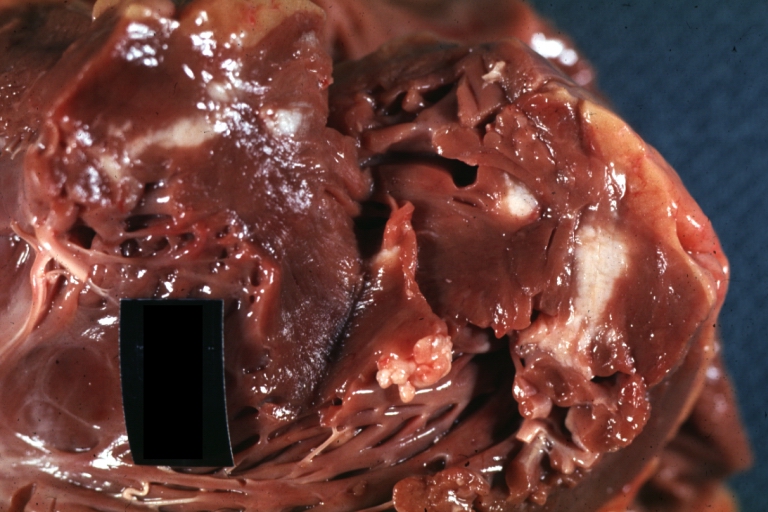
 HEART: Thrombotic Nonbacterial Endocarditis Infected: Gross natural color aortic valve vegetations excellent example case of multiple myeloma


-
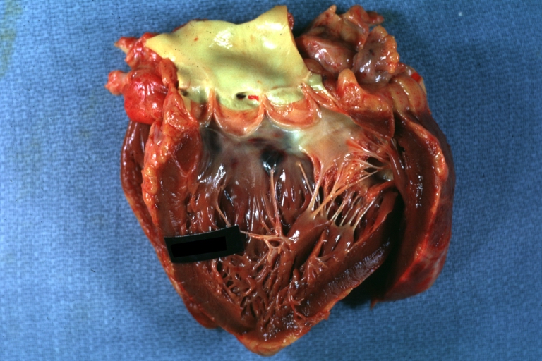
 HEART: Thrombotic Nonbacterial Endocarditis Infected: Gross natural color mitral valve lesion excellent example case of multiple myeloma
 -
 HEART: Thrombotic Nonbacterial Endocarditis Infected: Gross natural color pulmonary valve excellent example case of multiple myeloma


-
 HEART: Thrombotic Nonbacterial Endocarditis Infected: Gross natural color excellent example case of multiple myeloma

References
See Also
- The Heart in Breast Cancer
- The Heart in Gynecologic Tumors
- The Heart in Head and Neck Tumors
- The heart in Leukemias
- The Heart in Lung Cancers
- The Heart in Lymphomas
- The Heart in Osteosarcomas
- The Heart in Peritoneal and Mesothelial Tumors
- The Heart in Primary Myocardial Tumors
- The Heart in Skin Cancers
- The Heart in Thyroid and Parathyroid Cancers
- The Heart in Tumors that Originated from Vascular Structure
- The Heart in Urinary System Tumors
- The Heart in Central and Peripheral Nervous System Cancers
- The Heart in Gastrointestinal, Hepatobilier & Pancreatic Tumors
The Heart in Urinary System Tumors
Editor-In-Chief: C. Michael Gibson, M.S., M.D. [1]
Associate Editor-In-Chief: Cafer Zorkun, M.D., Ph.D. [2]
Histopathological Findings
-
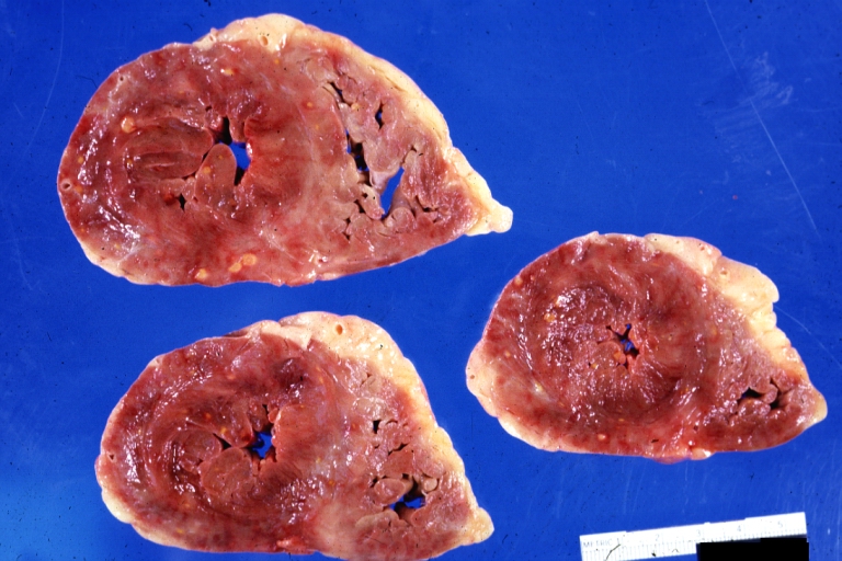
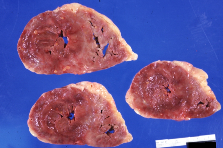
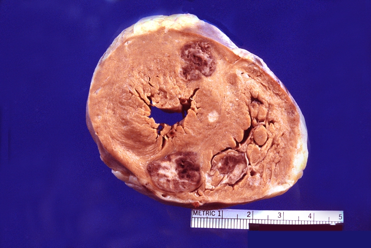
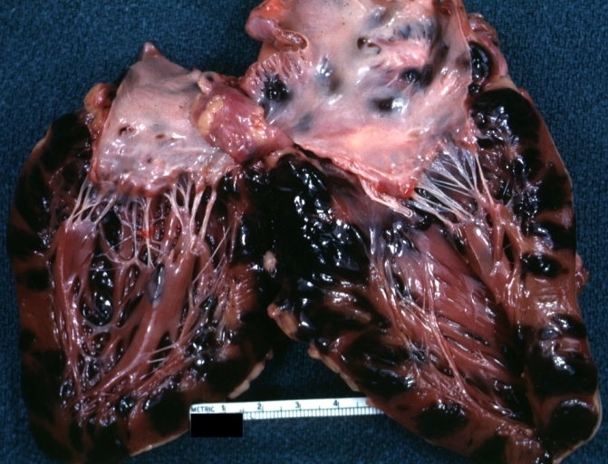
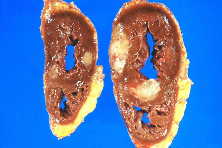
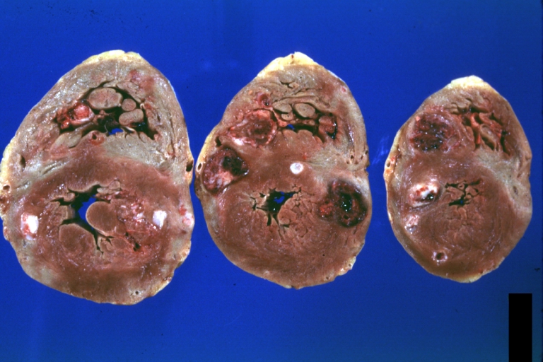 HEART: Metastatic Carcinoma: Gross natural color horizontal slices ventricles large metastatic renal cell carcinoma
 -
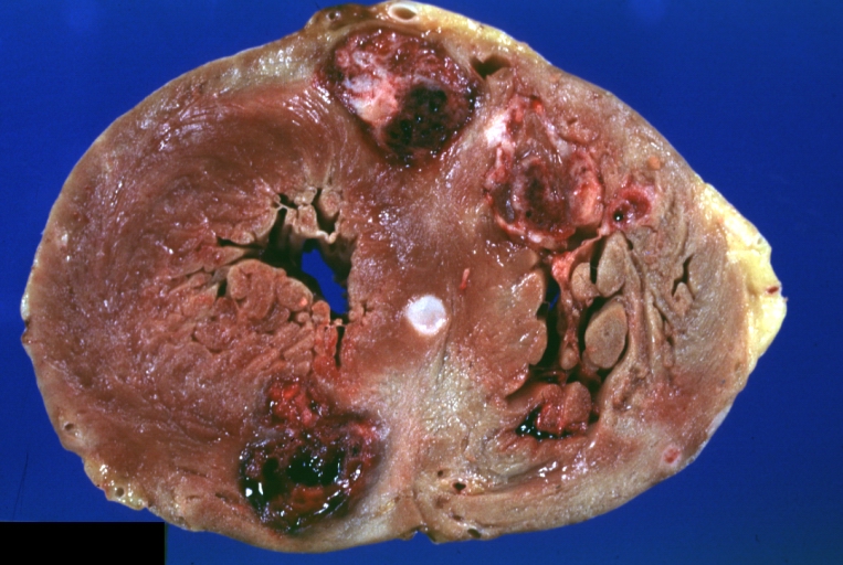 HEART: Metastatic Carcinoma: Gross natural color close-up of horizontal section ventricles with large hemorrhagic lesions (an excellent photo) in large metastatic renal cell carcinoma


-
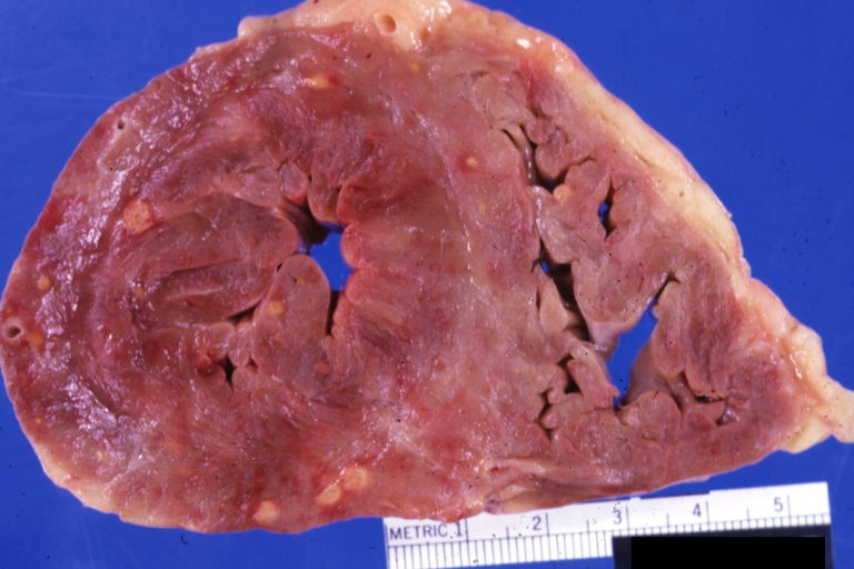
 HEART: Metastatic Carcinoma: Gross fixed tissue, color, large lesions, primer is renal cell carcinoma.
 -
 HEART: Metastatic Carcinoma: Gross fixed tissue, color, an excellent close-up of large hemorrhagic lesions primary is kidney


References
See Also
- The Heart in Breast Cancer
- The Heart in Gynecologic Tumors
- The Heart in Head and Neck Tumors
- The heart in leukemias
- The Heart in Lung Cancers
- The Heart in Lymphomas
- The Heart in Multiple Myeloma
- The Heart in Osteosarcomas
- The Heart in Peritoneal and Mesothelial Tumors
- The Heart in Primary Myocardial Tumors
- The Heart in Skin Cancers
- The Heart in Thyroid and Parathyroid Cancers
- The Heart in Tumors that Originated from Vascular Structure
- The Heart in Central and Peripheral Nervous System Cancers
- The Heart in Gastrointestinal, Hepatobilier & Pancreatic Tumors
The Heart in Lung Cancers
Editor-In-Chief: C. Michael Gibson, M.S., M.D. [1]
Associate Editor-In-Chief: Cafer Zorkun, M.D., Ph.D. [2]
Histopathological Findings
-
 Heart metastases from bronchogenic carcinoma
 -
 Heart metastases from bronchogenic carcinoma (closer look)


-
 HEART: Bronchiogenic carcinoma, smoker, right ventricular dilatation
 -
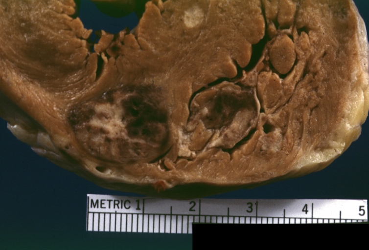
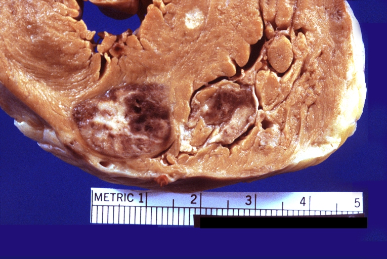
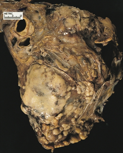
 HEART: Metastatic Carcinoma: Gross natural color horizontal sections of ventricle showing tumor in subepicardial fat which stops abruptly at myocardium which is quite typical interesting case 44yo BF with adenocarcinoma of lung occurring 25 years after successful treatment for Hodgkin’s disease


-
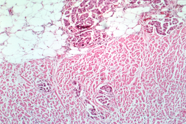 HEART: Metastatic Carcinoma: Micro low mag H&E. Lung alveolar cell carcinoma shows tumor in pericardial fat and into subepicardial myocardium
 -
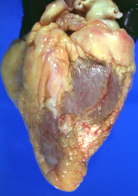
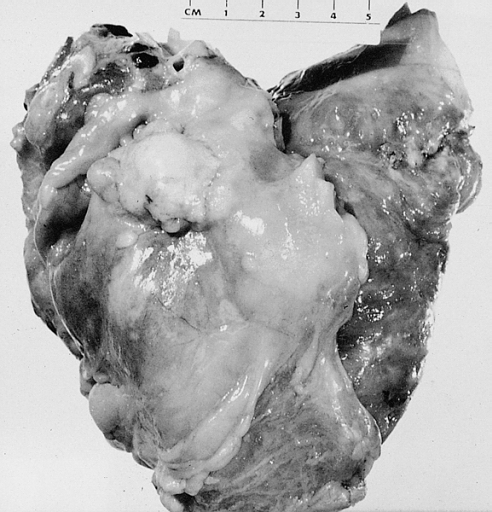
 HEART: Metastatic Carcinoma Lung: Gross natural color external view with coronaries sectioned obvious tumor all over epicardium 44yo BF adenocarcinoma giant cell type right lung occurring 25 years after treatment for Hodgkin’s disease


-
 HEART: Pericarditis, Neoplastic: Gross natural color shaggy pericarditis (primer is adenocarcinoma of lung)

References
See Also
- The Heart in Breast Cancer
- The Heart in Gynecologic Tumors
- The Heart in Head and Neck Tumors
- The heart in leukemias
- The Heart in Lymphomas
- The Heart in Multiple Myeloma
- The Heart in Osteosarcomas
- The Heart in Peritoneal and Mesothelial Tumors
- The Heart in Primary Myocardial Tumors
- The Heart in Skin Cancers
- The Heart in Thyroid and Parathyroid Cancers
- The Heart in Tumors that Originated from Vascular Structure
- The Heart in Urinary System Tumors
- The Heart in Central and Peripheral Nervous System Cancers
- The Heart in Gastrointestinal, Hepatobilier & Pancreatic Tumors
The Heart in Thyroid and Parathyroid Cancers
Editor-In-Chief: C. Michael Gibson, M.S., M.D. [1]
Associate Editor-In-Chief: Cafer Zorkun, M.D., Ph.D. [2]
Histopathological Findings

References
Additional Resource
- Giuffrida D, Gharib H. Cardiac metastasis from primary anaplastic thyroid carcinoma: report of three cases and a review of the literature. Endocrine-related Cancer (2001) 8 71–73
See Also
- The Heart in Breast Cancer
- The Heart in Gynecologic Tumors
- The Heart in Head and Neck Tumors
- The heart in leukemias
- The Heart in Lung Cancers
- The Heart in Lymphomas
- The Heart in Multiple Myeloma
- The Heart in Osteosarcomas
- The Heart in Peritoneal and Mesothelial Tumors
- The Heart in Primary Myocardial Tumors
- The Heart in Skin Cancers
- The Heart in Tumors that Originated from Vascular Structure
- The Heart in Urinary System Tumors
- The Heart in Central and Peripheral Nervous System Cancers
- The Heart in Gastrointestinal, Hepatobilier & Pancreatic Tumors
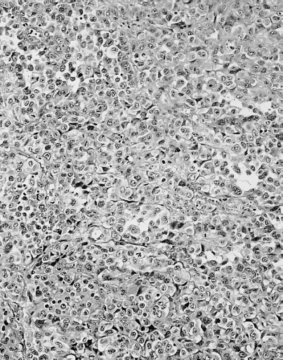
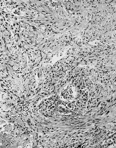
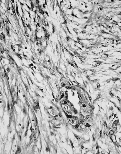
The Heart in Osteosarcomas
Editor-In-Chief: C. Michael Gibson, M.S., M.D. [1]
Associate Editor-In-Chief: Cafer Zorkun, M.D., Ph.D. [2]
Histopathological Findings
-
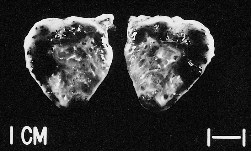
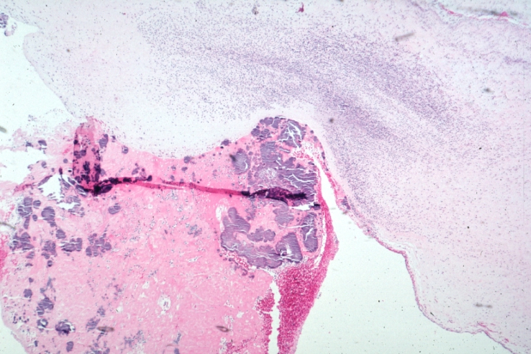
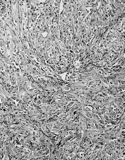
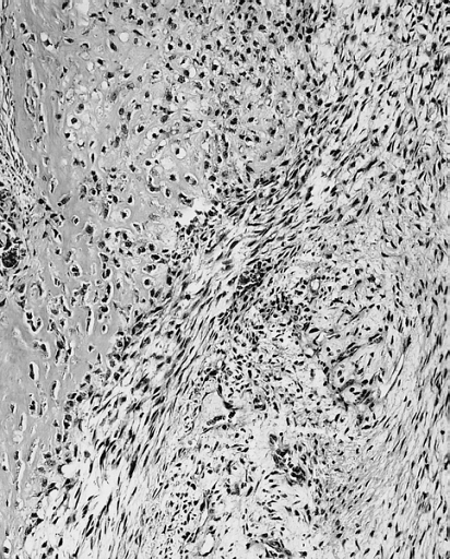 HEART; Osteosarcoma. Most cardiac osteosarcomas have spindled areas in addition to differentiated osteosarcoma (note osteoid, lower portion of field). The patient was a 16-year-old boy with a left atrial mass; the tumor recurred in 13 months at the site of the chest wall incision.
 -
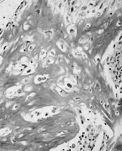 HEART; Osteosarcoma. There are atypical cells within an osteoid matrix.
 -
 HEART: Right atrium. The patient was a 21-year-old woman with a history of osteosarcoma of the humerus resected 9 years previously. Note the relatively sharp demarcation between tumor and cardiac muscle. The specimen was a surgical biopsy of a single cardiac metastasis.


References
See Also
- The Heart in Breast Cancer
- The Heart in Gynecologic Tumors
- The Heart in Head and Neck Tumors
- The heart in leukemias
- The Heart in Lung Cancers
- The Heart in Lymphomas
- The Heart in Multiple Myeloma
- The Heart in Peritoneal and Mesothelial Tumors
- The Heart in Primary Myocardial Tumors
- The Heart in Skin Cancers
- The Heart in Thyroid and Parathyroid Cancers
- The Heart in Tumors that Originated from Vascular Structure
- The Heart in Urinary System Tumors
- The Heart in Central and Peripheral Nervous System Cancers
- The Heart in Gastrointestinal, Hepatobilier & Pancreatic Tumors
The Heart in Breast Cancer
Editor-In-Chief: C. Michael Gibson, M.S., M.D. [1]; Associate Editor-In-Chief: Cafer Zorkun, M.D., Ph.D. [2]
Click here to read more about breast cancer
Histopathological Findings
-
 Heart; Breast intraductal papilloma metastasis. Thrombotic Nonbacterial Endocarditis (Infected): Gross mitral valve natural color vegetations well illustrated these were secondarily infected with staphylococcus case of 8 year survival breast intraductal papillary adenocarcinoma with extensive metastases. Aortic valve also involved
 -
 Thrombotic Nonbacterial Endocarditis Infected: Micro low mag H&E fibrin vegetation with masses of staphylococci and inflammatory cells in valve secondarily infected case 8 year survival breast papillary intraductal adenocarcinoma with extensive metastases gross is aortic valve lesions.


-
 Thrombotic Nonbacterial Endocarditis Infected: Gross close-up natural color vegetations or aortic cusps well shown secondarily infected with staphylococci micro is mitral valve also involved see and case of 8 year survival breast intraductal papillary adenocarcinoma with extensive metastases
 -
 Thrombotic Nonbacterial Endocarditis Infected: Micro low mag H&E large masses staphylococci in vegetation valvulitis is seen gross is mitral valve also involved and case of 8 year survival breast intraductal papillary adenocarcinoma with extensive metastases


-
 Heart: Metastatic Carcinoma Breast: Gross, natural color, external view with lesions over right ventricle. Typical and good example
 -
 Heart: Metastatic Carcinoma Breast: Gross, close up view, atrial epicardial lesions, breast cancer is the primary.


References
See Also
- The Heart in Gynecologic Tumors
- The Heart in Head and Neck Tumors
- The heart in leukemias
- The Heart in Lung Cancers
- The Heart in Lymphomas
- The Heart in Multiple Myeloma
- The Heart in Osteosarcomas
- The Heart in Peritoneal and Mesothelial Tumors
- The Heart in Primary Myocardial Tumors
- The Heart in Skin Cancers
- The Heart in Thyroid and Parathyroid Cancers
- The Heart in Tumors that Originated from Vascular Structure
- The Heart in Urinary System Tumors
- The Heart in Central and Peripheral Nervous System Cancers
- The Heart in Gastrointestinal, Hepatobilier & Pancreatic Tumors
The Heart in Gastrointestinal, Hepatobilier & Pancreatic Tumors
The Heart in Skin Cancers
Editor-In-Chief: C. Michael Gibson, M.S., M.D. [1]
Associate Editor-In-Chief: Cafer Zorkun, M.D., Ph.D. [2]
Histopathological Findings
-
 HEART: Metastatic Melanoma: Gross natural color right atrium and ventricle multiple tumor masses. An excellent example of metastatic carcinoma in heart.
 -
 Heart in metastatic melanoma.


References
See Also
- The Heart in Breast Cancer
- The Heart in Gynecologic Tumors
- The Heart in Head and Neck Tumors
- The heart in leukemias
- The Heart in Lung Cancers
- The Heart in Lymphomas
- The Heart in Multiple Myeloma
- The Heart in Osteosarcomas
- The Heart in Peritoneal and Mesothelial Tumors
- The Heart in Primary Myocardial Tumors
- The Heart in Thyroid and Parathyroid Cancers
- The Heart in Tumors that Originated from Vascular Structure
- The Heart in Urinary System Tumors
- The Heart in Central and Peripheral Nervous System Cancers
- The Heart in Gastrointestinal, Hepatobilier & Pancreatic Tumors
The Heart in Gynecologic Tumors
Editor-In-Chief: C. Michael Gibson, M.S., M.D. [1]
Associate Editor-In-Chief: Cafer Zorkun, M.D., Ph.D. [2]
Histopathological Findings
-
 HEART-Great Vessels: Metastatic Squamous Cell Carcinoma: Pericardium. The patient was a 68-year-old woman with a history of uterine cervical carcinoma and pericardial tamponade.

References
See Also
- The Heart in Breast Cancer
- The Heart in Head and Neck Tumors
- The heart in leukemias
- The Heart in Lung Cancers
- The Heart in Lymphomas
- The Heart in Multiple Myeloma
- The Heart in Osteosarcomas
- The Heart in Peritoneal and Mesothelial Tumors
- The Heart in Primary Myocardial Tumors
- The Heart in Skin Cancers
- The Heart in Thyroid and Parathyroid Cancers
- The Heart in Tumors that Originated from Vascular Structure
- The Heart in Urinary System Tumors
- The Heart in Central and Peripheral Nervous System Cancers
- The Heart in Gastrointestinal, Hepatobilier & Pancreatic Tumors
The Heart in Peritoneal and Mesothelial Tumors
Editor-In-Chief: C. Michael Gibson, M.S., M.D. [1]
Associate Editor-In-Chief: Cafer Zorkun, M.D., Ph.D. [2]
Histopathological Findings
-
 HEART: Mesothelioma: Gross natural color external view with white plaques over epicardium apparently spread from left side pleura
 -
 HEART: Heart, metastatic mesothelioma


-
 HEART: Heart, metastatic mesothelioma
 -
 HEART: Heart, metastatic mesothelioma


-
 HEART: Heart, metastatic mesothelioma
 -
 HEART: Heart, metastatic mesothelioma


-
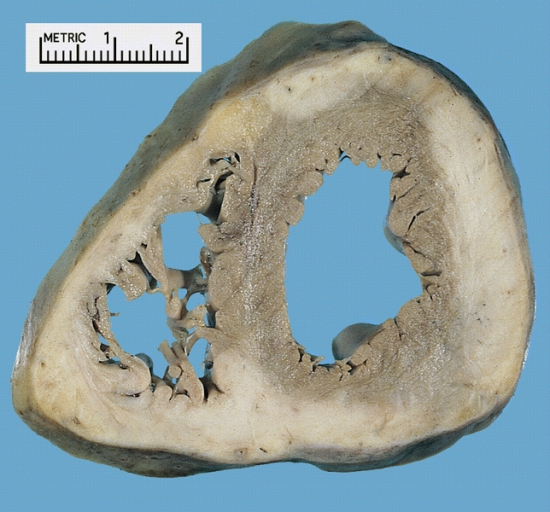
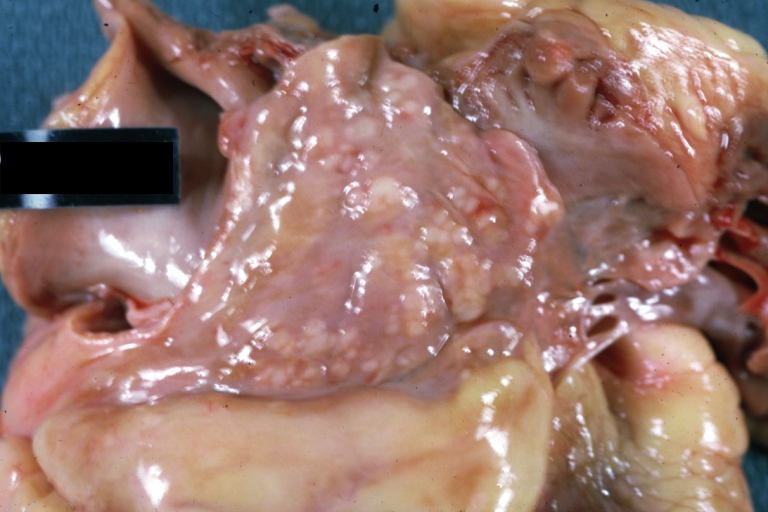
 MALIGNANT MESOTHELIOMA: PERICARDIUM Note the gross tumor studding the epicardial surface.
 -
 MALIGNANT MESOTHELIOMA: PERICARDIUM There is studding of the pericardium with tumor deposits. The patient was a 50-year-old man with a 2-year history of pericarditis (progressing) to pericardial constriction


-
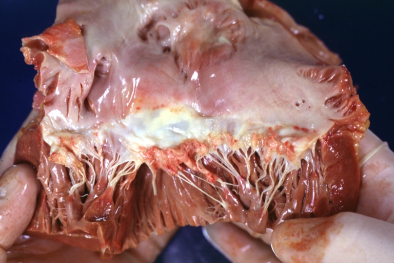
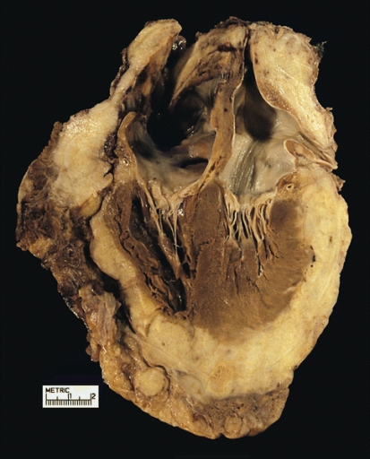 MALIGNANT MESOTHELIOMA: PERICARDIUM There is total encasement of the atria and left ventricles by firm, white tumor.
 -
 MALIGNANT MESOTHELIOMA: PERICARDIUM There is a partly necrotic mass on the surface of the epicardium.


-
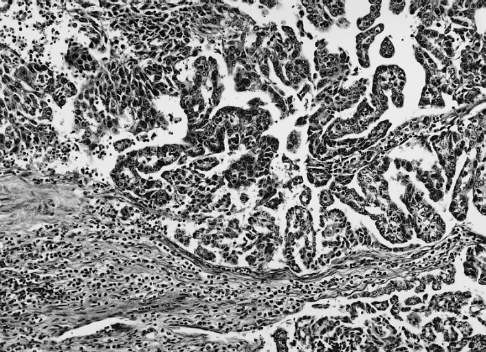
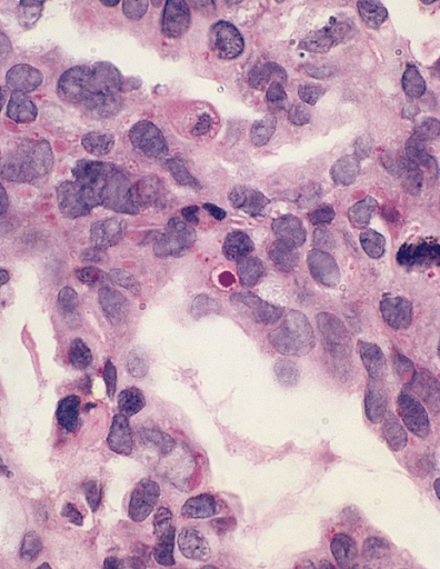
 MALIGNANT MESOTHELIOMA, EPITHELIOID TYPE: PERICARDIUM A higher magnification of figure 14-4 demonstrates a predominantly epithelioid tumor forming papillary structures on the pericardial surface (left) and tubules infiltrating stroma (right).
 -
 MALIGNANT MESOTHELIOMA, EPITHELIOID TYPE: PERICARDIUM A higher magnification of figure 14-5 demonstrates papillary areas.


-
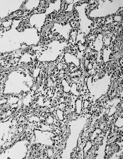
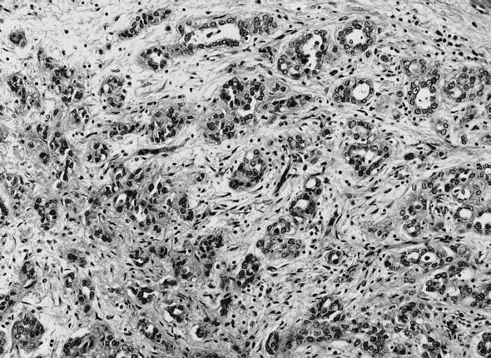 MALIGNANT MESOTHELIOMA, EPITHELIOID TYPE: PERICARDIUM The tumor infiltrates as tubules which histologically mimic adenocarcinoma.
 -
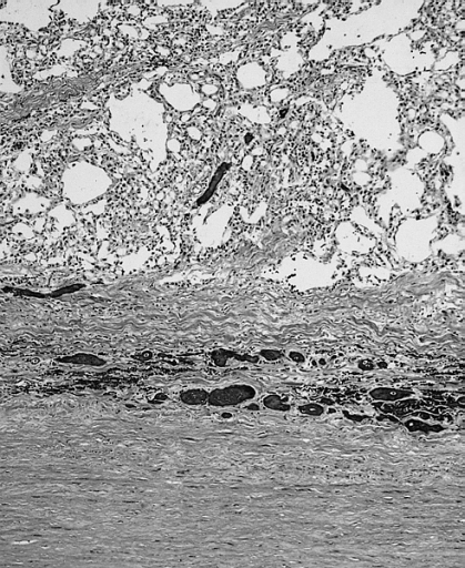
 MALIGNANT MESOTHELIOMA, EPITHELIOID TYPE: PERICARDIUM The patient was a 66-year-old male with a known history of asbestos exposure and pleural plaques who developed diffuse pericardial tumor. A low magnification view shows a section of pericardium that is markedly thickened.


-
 EPITHELIOID TYPE: PERICARDIUM: An epithelioid neoplasm mimicking carcinoma. A primary carcinoma was ruled out at autopsy.
 -
 MALIGNANT MESOTHELIOMA, BIPHASIC TYPE: PERICARDIUM: This tumor has epithelioid cells (lower half) surrounded by spindled cells. The patient was a 46-year-old woman with constrictive pericarditis; the pericardium was studded with coalescing tumor nodules.


-
 MALIGNANT MESOTHELIOMA: PERICARDIUM A higher magnification of a different area of the tumor depicted in figure 14-10 shows biphasic tumor mimicking desmoplastic adenocarcinoma.
 -
 MALIGNANT MESOTHELIOMA: PERICARDIUM The spindled and epithelioid areas may merge, imparting a sarcomatoid appearance. The patient was a 56-year-old woman with pericardial tamponade and diffuse pericardial tumor surrounding the root of the aorta.


-
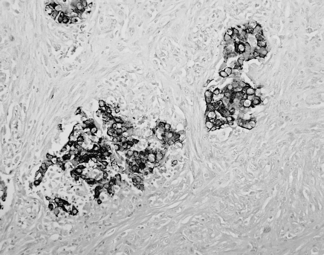 MALIGNANT MESOTHELIOMA: PERICARDIUM Immunohistochemical stain reveals epithelioid areas that strongly express cytokeratin; the spindled cells are often less strongly reactive for this antibody.
 -
 MALIGNANT MESOTHELIOMA: PERICARDIUM The patient was a 69-year-old man who died shortly after open thoracotomy to relieve pericardial constriction. There were multiple tumor nodules on the epicardial surfaces of both atria, enveloping the aortic root. This tumor has a histologic appearance reminiscent of an adenomatoid tumor.


-
 MALIGNANT MESOTHELIOMA: PERICARDIUM A higher magnification of figure 14-14 demonstrates irregular spaces suggestive of adenomatoid tumor.
 -
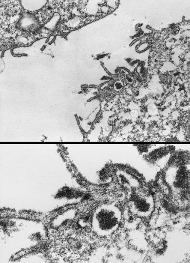 HEART-GREAT VESSELS: MALIGNANT MESOTHELIOMA: PERICARDIUM Electron micrograph demonstrates elongated microvilli characteristic of mesothelial differentiation.


-
 METASTATIC ADENOCARCINOMA: PERICARDIUM Intracytoplasmic globules containing PAS-positive material are seen in adenocarcinoma and are absent in mesothelioma and reactive mesothelial cells. (Periodic acid-Schiff after diastase pretreatment).

References
See Also
- The Heart in Breast Cancer
- The Heart in Gynecologic Tumors
- The Heart in Head and Neck Tumors
- The heart in leukemias
- The Heart in Lung Cancers
- The Heart in Lymphomas
- The Heart in Multiple Myeloma
- The Heart in Osteosarcomas
- The Heart in Primary Myocardial Tumors
- The Heart in Skin Cancers
- The Heart in Thyroid and Parathyroid Cancers
- The Heart in Tumors that Originated from Vascular Structure
- The Heart in Urinary System Tumors
- The Heart in Central and Peripheral Nervous System Cancers
- The Heart in Gastrointestinal, Hepatobilier & Pancreatic Tumors
The Heart in Head and Neck Tumors
Editor-In-Chief: C. Michael Gibson, M.S., M.D. [1]
Associate Editor-In-Chief: Cafer Zorkun, M.D., Ph.D. [2]
Histopathological Findings
-
 HEART: Metastatic Carcinoma: Gross, primary location is trachea. A good example with lesions in myocardium

References
See Also
- The Heart in Breast Cancer
- The Heart in Gynecologic Tumors
- The heart in leukemias
- The Heart in Lung Cancers
- The Heart in Lymphomas
- The Heart in Multiple Myeloma
- The Heart in Osteosarcomas
- The Heart in Peritoneal and Mesothelial Tumors
- The Heart in Primary Myocardial Tumors
- The Heart in Skin Cancers
- The Heart in Thyroid and Parathyroid Cancers
- The Heart in Tumors that Originated from Vascular Structure
- The Heart in Urinary System Tumors
- The Heart in Central and Peripheral Nervous System Cancers
- The Heart in Gastrointestinal, Hepatobilier & Pancreatic Tumors
The Heart in Lymphomas
Editor-In-Chief: C. Michael Gibson, M.S., M.D. [1]
Associate Editor-In-Chief: Cafer Zorkun, M.D., Ph.D. [2]
Histopathological Findings
-
 HEART: Hemorrhage In Region of Left Bundle Branches: Gross natural color of opened left ventricular outflow tract with subendocardial hemorrhage just below membranous septum. A case of Hodgkins disease with probable sepsis
 -
 HEART: Hemopericardium Caused By Pericardiocentesis: Gross, natural color, close-up view of apex of the heart needle apparently entered the distal posterior descending artery 26 yo men with Hodgkin’s disease or a T-cell lymphoma


-
 HEART: Metastatic Carcinoma Lung: Gross natural color external view with coronaries sectioned obvious tumor all over epicardium, a 44yo Female, an adenocarcinoma; giant cell type, right lung, occurring 25 years after treatment for Hodgkin’s disease
 -
 HEART: Hodgkin’s disease, marantic endocarditis, aortic valve


-
 HEART: Hodgkin’s disease, marantic endocarditis, aortic valve

Related Chapters
- The Heart in Breast Cancer
- The Heart in Gynecologic Tumors
- The Heart in Head and Neck Tumors
- The heart in leukemias
- The Heart in Lung Cancers
- The Heart in Multiple Myeloma
- The Heart in Osteosarcomas
- The Heart in Peritoneal and Mesothelial Tumors
- The Heart in Primary Myocardial Tumors
- The Heart in Skin Cancers
- The Heart in Thyroid and Parathyroid Cancers
- The Heart in Tumors that Originated from Vascular Structure
- The Heart in Urinary System Tumors
- The Heart in Central and Peripheral Nervous System Cancers
- The Heart in Gastrointestinal, Hepatobilier and Pancreatic Tumors
- The heart in lymphomas
The Heart in Tumors that Originated from Vascular Structure
Editor-In-Chief: C. Michael Gibson, M.S., M.D. [1]
Associate Editor-In-Chief: Cafer Zorkun, M.D., Ph.D. [2]
-
 Sarcoma of the pulmonary artery. Like aortic intimal sarcomas, there can be densely collagenized areas in pulmonary artery sarcomas

References
See Also
- The Heart in Breast Cancer
- The Heart in Gynecologic Tumors
- The Heart in Head and Neck Tumors
- The heart in leukemias
- The Heart in Lung Cancers
- The Heart in Lymphomas
- The Heart in Multiple Myeloma
- The Heart in Osteosarcomas
- The Heart in Peritoneal and Mesothelial Tumors
- The Heart in Primary Myocardial Tumors
- The Heart in Skin Cancers
- The Heart in Thyroid and Parathyroid Cancers
- The Heart in Urinary System Tumors
- The Heart in Central and Peripheral Nervous System Cancers
- The Heart in Gastrointestinal, Hepatobilier & Pancreatic Tumors
The Heart in Primary Myocardial Tumors
Editor-In-Chief: C. Michael Gibson, M.S., M.D. [1]
Associate Editor-In-Chief: Cafer Zorkun, M.D., Ph.D. [2]
CT

Histopathological Findings
-
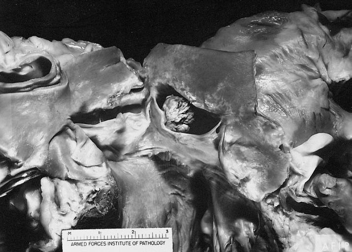
 The right side of the heart has been opened to demonstrate a large right atrial mass overlying the septal leaflet of the tricuspid valve. No metastases were demonstrated at autopsy.

-
 Myxosarcoma. This tumor was excised from a 27-year-old woman with a left atrial mass (clinically diagnosed as myxoma). She died of widespread metastases at postoperative 48th month.

-
 A 64-year-old man with back pain had surgical excision of a right atrial mass. There was a pleomorphic sarcoma with vacuolated cells. The patient died with bony metastases.

References
See Also
- The Heart in Breast Cancer
- The Heart in Gynecologic Tumors
- The Heart in Head and Neck Tumors
- The heart in leukemias
- The Heart in Lung Cancers
- The Heart in Lymphomas
- The Heart in Multiple Myeloma
- The Heart in Osteosarcomas
- The Heart in Peritoneal and Mesothelial Tumors
- The Heart in Skin Cancers
- The Heart in Thyroid and Parathyroid Cancers
- The Heart in Tumors that Originated from Vascular Structure
- The Heart in Urinary System Tumors
- The Heart in Central and Peripheral Nervous System Cancers
- The Heart in Gastrointestinal, Hepatobilier & Pancreatic Tumors
References
References
Looking for the patient version?
© 2026 MyEClinic – IFTM Institut für Telematik in der Medizin GmbH