

Health Dictionary
Reliable, easy-to-read patient information on common medical conditions — written in plain language, free of jargon, and grounded in current evidence. Browse the most frequent diagnoses, understand what they mean for everyday life, and learn which steps and questions help you have a focused conversation with your doctor.
Vitelline veins
Introduction
Path
Path
They run upward at first in front, and subsequently on either side of the intestinal canal.
They unite on the ventral aspect of the canal, and beyond this are connected to one another by two anastomotic branches, one on the dorsal, and the other on the ventral aspect of the duodenal portion of the intestine, which is thus encircled by two venous rings; into the middle or dorsal anastomosis the superior mesenteric vein opens.
The portions of the veins above the upper ring become interrupted by the developing liver and broken up by it into a plexus of small capillary-like vessels termed sinusoids.
Derivatives
Derivatives
The branches conveying the blood to this plexus are named the venae advehentes, and become the branches of the portal vein.
The vessels draining the plexus into the sinus venosus are termed the venae revehentes, and form the future hepatic veins. Ultimately the left vena revehens no longer communicates directly with the sinus venosus, but opens into the right vena revehens.
The persistent part of the upper venous ring, above the opening of the superior mesenteric vein, forms the trunk of the portal vein.
Additional images
Additional images
-
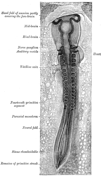 Chick embryo of thirty-three hours’ incubation, viewed from the dorsal aspect. X 30.
 -
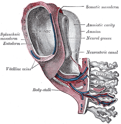 Model of human embryo 1.3 mm. long.
 -
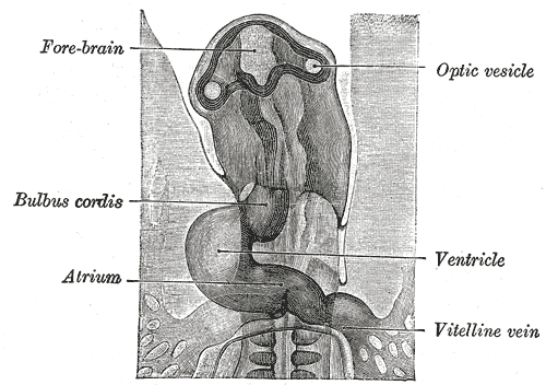 Head of chick embryo of about thirty-eight hours’ incubation, viewed from the ventral surface. X 26.
 -
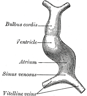 Diagram to illustrate the simple tubular condition of the heart.
 -
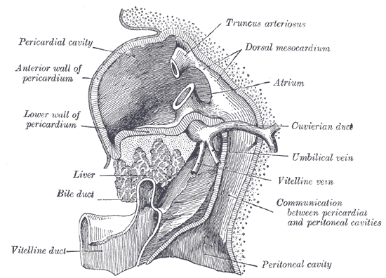 Liver with the septum transversum. Human embryo 3 mm. long.


Editor-In-Chief: Template:Infobox Embryology C. Michael Gibson, M.S., M.D. [1]