

Health Dictionary
Reliable, easy-to-read patient information on common medical conditions — written in plain language, free of jargon, and grounded in current evidence. Browse the most frequent diagnoses, understand what they mean for everyday life, and learn which steps and questions help you have a focused conversation with your doctor.
Bleb-related endophthalmitis
Introduction
For the main page on endophthalmitis, please click here
For more information on bacterial endophthalmitis, please click here
For more information on post-operative endophthalmitis, please click here
For more information on post-traumatic endophthalmitis, please click here
For more information on endogenous endophthalmitis, please click here
Synonyms and keywords: Bleb-associated endophthalmitis; BAE; BRE
Overview
Overview
Bleb-related endophthalmitis (BRE) is the second most frequent type of postoperative endophthalmitis.[1][2] Bleb-related endophthalmitis is a complication of trabeculectomy. The exact pathogenesis of bleb-related endopthalmitis following trabeculectomy is not fully understood. It is thought that bleb-related endophthalmitis is the result of bleb leakage, which allows bacterial entry from the tear film and the periocular structures into the anterior chamber or the vitreous humor.[3]
Based on latency of onset, bleb-related endophthalmitis (BRE) may be classified into early onset and late onset. Common causes of bleb-related endophthalmitis include Streptococcus spp., Staphylococcus spp., and Haemophilus influenzae. Common risk factors for the development of bleb-related endophthalmitis include bleb leakage, inappropriate use of antifibrotic agents such as 5-fluorouracil (5-FU) and mitomycin C, inferior and nasal placement of bleb, and prior history of blebitis.[1] Bleb-related endophthalmitis is a medical emergency. If left untreated, it may lead to corneal infiltration, retinal toxicity, corneal perforation, retinal detachment, and panophthalmitis that may require enucleation. Bleb-related endophthalmitis presents as an accelerated prodromal syndrome, which can progress rapidly over several hours. It is usually characterized by severe visual loss and marked pain. Endophthalmitis is a clinical diagnosis supported by culture and polymerase chain reaction (PCR) of intra-ocular fluids.[4][5]
Patients with bleb-related endophthalmitis need urgent examination by an expert ophthalmologist and/or vitreo-retinal specialist. Intravitreal injection of antibiotics, topical fortified antibiotics, and surgical repair are necessary in the management of bleb related endophthalmitis. However, the benefit of vitrectomy in the management of bleb related endophthalmitis remains unclear.[6][7] Assessment of bleb leakage following trabeculectomy surgery in every visit seems to be the most effective measure for the primary prevention of bleb-related endophthalmitis.[8]
Historical Perspective
Historical Perspective
- Since the introduction of trabeculectomy in 1968, the first phenomenon of bleb related infections was documented.[9][10]
- Since the use of anti-metabolite agents such as mitomycin C (MMC) and 5-fluorouracil (5-FU) after 1984, the second phenomenon of bleb related endophthalmitis was reported.[11]
Classification
Classification
Based on latency of onset, bleb-related endophthalmitis (BRE) can be classified into:
- Early onset (Less than 6 weeks since surgery)
- Late onset (more than 6 weeks since surgery)
Pathophysiology
Pathophysiology
Bleb-related endophthalmitis is a complication of trabeculectomy. The exact pathogenesis of bleb-related endopthalmitis following trabeculectomy is not fully understood. It is thought that bleb-related endophthalmitis is the result of bleb leakage, which allows bacteria from the tear film and the periocular structures access into the anterior chamber or the vitreous humor.
Trabeculectomy is performed to achieve low intraocular pressure (IOP) in glaucoma that has failed medical management. A filtering bleb is a surgically created defect in the sclera which allows excess aqueous humor to leak out of the anterior chamber and be absorbed into the systemic circulation. There is a serious concern for bleb-related endophthalmitis (BRE) even after successful trabeculectomy.[1][2]
Early onset bleb leakage is commonly caused by either wound dehiscence or incomplete conjunctival closure. Late onset bleb leakage is caused by the use of adjunctive anti-metabolites such as mitomycin C (MMC) and 5-fluorouracil (5-FU), which are usually used to prevent fibrosis and scarring of the scleral flap and bleb.
Anti-metabolites may result in bleb leakage by the following mechanism:[3]
- Reduced mucin production (secondary to loss of goblet cell)
- General conjunctival thinning
- Reduced cellularity
- Avascular bleb
Even in the absence of a leak, it is also thought that more virulant organisms, such as streptococcus spp, can penetrate an intact bleb into the anterior chamber and vitreous humor.[12][13]
Causes
Causes
Common causes of bleb-related endophthalmitis include:[1]
- Early onset BRE
- Late onset BRE
- Streptococcus spp. (31%)
- Staphylococcus spp. (7%-22%)
- Haemophilus influenzae( 23%)
- Enterococcus
- Pseudomonas
- Moraxella
Differentiating Bleb-related Endophthalmitis from Other Diseases
Differentiating Bleb-related Endophthalmitis from Other Diseases
Bleb-related endophthalmitis must be differentiated from:[14]
- Blebitis (bleb-related endophthalmitis is characterized by severe loss of vision, marked pain, and presence of vitritis)
- Anterior uveitis
- Uveitis-glaucoma-hyphema syndrome
Epidemiology and Demographics
Epidemiology and Demographics
Incidence
- The incidence of bleb-related endophthalmitis is approximately range from 170 to 1,300 per 100,000 individuals with trabeculectomy surgery.[15][16]
- The incidence of bleb-related endophthalmitis is approximately 3,000 per 100,000 individuals with the use of antiproliferative agent.[15][17]
- The incidence of bleb-related endophthalmitis is approximately 9,000 per 100,000 individuals with inferior placement of bleb.[15][18]
Age
The incidence of bleb-related endophthalmitis decreases with age. Many studies have shown a higher prevalence of blebitis in younger patients (younger than 40 years old).
Gender
Males are more commonly affected with bleb-related endophthalmitis than females.[19]
Race
African-Americans are more commonly affected with bleb-related endophthalmitis than other races.[19]
Developed countries
In the United States, the incidence of bleb-related endophthalmitis is approximately range from 450 to 1,300 per 100,000 individuals with trabeculectomy after up to 5 years follow up.[2]
Risk Factors
Risk Factors
Common risk factors in the development of bleb-related endophthalmitis include:[1][2][3][20][19]
- Bleb-related risk factors
- Bleb leakage (increase the risk of bleb infection 26 fold)
- Inappropriate use of of The antifibrotic agents (such as 5-fluorouracil (5-FU) and mitomycin C (MMC))
- Inferior and nasal placement of bleb
- Prior history of blebitis
- Bleb manipulation/trauma
- Bleb revision surgery
- Ocular risk factors
- Conjunctivitis
- Blepharitis
- Nasolacrimal duct obstruction
- Contact lens wear
- Abnormal lid position
- Systemic risk factors
- Diabetes
- Chronic antibiotic use
- Juvenile glaucoma
- Male
- Age under 60 years
- Black race
- Upper respiratory infection (URI)
- Malnutrition
Screening
Screening
Screening for bleb-related endophthalmitis is not recommended following trabeculactomy surgery.[21] However, screening for bleb leakage by ophthalmologist following each visit is recommended among patients with trabeculactomy surgery.
Natural History, Complications, and Prognosis
Natural History, Complications, and Prognosis
Natural History
Bleb-related endophthalmitis is a medical emergency. If left untreated, It may lead to corneal infiltration, retinal toxicity, corneal perforation, retinal detachment, and panophthalmits that may require enucleation.[22]
Complications
Complications to bleb resalted endophthalmitis include:
- Corneal perforation
- Corneal edema
- Persistence intraocular inflammation
- Cataract
- Hypotony
- Residdual vitreous opacity
Prognosis
Despite intensive topical, systemic, and intravitreal antibiotics in combination with vitrectomy. Bleb-related endophthalmitis is associated with a poor visual outcome.[6] Bleb-related endophthalmitis caused by moraxella and coagulase negative staphylococcus is associated with more favorable outcomes compared with those caused by streptococcus.[23]
Diagnosis
Diagnosis
Endophthalmitis is a clinical diagnosis, supported by culture and polymerase chain reaction (PCR) of intra-ocular fluids.[4][5]
History
Specific areas of focus when obtaining a history from the patient with bleb-related endophthalmitis include:
- Prior history of trabeculectomy
- History of blebitis
- Use of adjunctive anti-metabolites, such as mitomycin C(MMC) and 5-fluorouracil (5-FU)
Symptoms
Patients with bleb-related endophthalmitis present with accelerated prodromal syndrome, which can progress rapidly over several hours. Symptoms of bleb-related endophthalmitis may include the following:[1][2]
- Ocular pain and discomfort
- Redness
- Blurred vision
- Eye Discharge
- Loss of vision
- Eyebrow ache
- Headache
- External ocular inflammation
Physical Examination
A thorough physical and eye examination from the patient is necessary. Common ophthalmoscopic examination findings of bleb-related endophthalmitis include:[1][2]
- Ocular pain
- Redness
- Decreased vision
- Conjunctival injection
- Chemosis
-
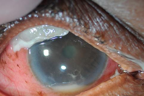 Hyperemic conjunctiva, opacified bleb with purulent adherent material, hypopyon, and fibrinous AC reaction.
 -
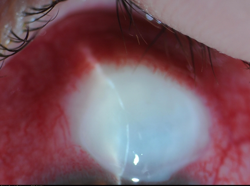 “White-on-red” appearance of acute blebitis
 -
![Thin walled, transparent, tense, avascular bleb[24]](https://www.wikidoc.org/images/8/89/Thin_bleb_for_article.jpg) Thin walled, transparent, tense, avascular bleb[24]
Thin walled, transparent, tense, avascular bleb[24]


![Thin walled, transparent, tense, avascular bleb[24]](https://www.wikidoc.org/index.php/File%3AThin_bleb_for_article.jpg)
Laboratory Findings
Laboratory studies consistent with the diagnosis of bleb-related endophthalmitis include:[25] [26]
- Culture and gram stain of aqueous humor as well as the vitreous humor
- Polymerase chain reaction (PCR) of aqueous humor as well as the vitreous humor
- Conjunctival and eyelid cultures
Imaging Findings
X Ray
There are no diagnostic x ray findings associated with bleb-related endophthalmitis.
CT
There are no diagnostic CT scan findings associated with bleb-related endophthalmitis.
MRI
There are no diagnostic MRI findings associated with bleb-related endophthalmitis.
Ultrasound
B scan ultrasonography may be helpful in diagnosis of coexisting posterior segment problem, such as vitreous changes or retinal detachment, in patient with bleb-related endophthalmitis.
Other Imaging Findings
There are no other Imaging Findings associated with bleb-related endophthalmitis.
Other Diagnostic Studies
Slit lamp examination
- A mucopurulent infiltrate
- Anterior chamber and vitreous inflammation
- Hypopyon
- Inflamed bleb
- Bleb leak
Treatment
Treatment
The patient needs urgent examination by an expert ophthalmologist and/or vitreo-retina specialist who will usually decide for urgent intervention to provide intravitreal injection of potent antibiotics. Enucleation may be required to remove a blind and painful eye.
- Bacterial and fungal cultures from vitreous samples are necessary in the management of bleb related endophthalmitis
- Surgical repair may be considered in the presence of poor bleb morphology
- Topical fortified antibiotics should be considered
- The benefit of vitrectomy in the management of bleb related endophthalmitis remains unclear
Medical Therapy
Antimicrobial Regimens
- Infectious endophthalmitis[4]
- 1. Causative pathogens
- 2.Empiric antimicrobial therapy
- Preferred regimen: Vancomycin 1 mg per 0.1 mL normal saline intravitreal injection, single dose AND Vancomycin 1 g IV bid for 2 weeks AND Ceftazidime 2.25 mg per 0.1 mL normal saline intravitreal injection, single dose AND Ceftazidime 1 g IV bid for 2 weeks AND Clindamycin 600-1200 mg IV bid to qid for 2 weeks
- Note (1): Re-injection should be considered if the infection does not improve beyond 48 hours of the first injection. Re-injection significantly increases the risk of retinal toxicity.
- Note (2): In addition to intravitreal and systemic antibiotic therapy, vitrectomy is usually necessary
- 3. Pathogen-directed antimicrobial therapy
- 3.1 Bacillus spp.
- Preferred regimen: Vancomycin 1 mg per 0.1 mL normal saline intravitreal injection, single dose AND Vancomycin 1 g IV bid for 2 weeks AND Clindamycin 600-1200 mg IV bid to qid for 2 weeks
- Note: In addition to antimicrobial therapy, vitrectomy is usually necessary
- 3.2 Non-Bacillus gram-positive bacteria
- Preferred regimen: Vancomycin 1 mg per 0.1 mL normal saline intravitreal injection, single dose AND Vancomycin 1 g IV bid for 2 weeks
- Note: In addition to antimicrobial therapy, vitrectomy is usually necessary
- 3.3 Gram-negative bacteria
- Preferred regimen: Ceftazidime 2.25 mg per 0.1 mL normal saline intravitreal injection, single dose AND Ceftazidime 1 g IV bid for 2 weeks
- Note: In addition to antimicrobial therapy, vitrectomy is usually necessary
- 3.4 Candida spp.
- Preferred regimen: (Fluconazole 400-800 mg IV/PO qd for 6-12 weeks OR Voriconazole 400 mg IV/PO bid for 2 doses followed by 200-300 mg IV/PO bid for 6-12 weeks OR Amphotericin B 0.7-1.0 mg/kg IV qd for 6-12 weeks) AND Amphotericin B 5-10 microgram in 0.1 mL in normal saline intravitreal injection, single dose
- Note (1): In addition to antimicrobial therapy, vitrectomy is usually necessary
- 3.5 Aspergillus spp.
- Preferred regimen: Amphotericin B 5-10 microgram in 0.1 mL normal saline intravitreal injection, single dose AND Dexamethasone 400 microgram intravitreal injection, single dose
- Note (1): In addition to antimicrobial therapy, vitrectomy is usually necessary
- Note (2): Repeat antimicrobial regimen in 2 days post-vitrectomy
Surgery
Vitrectomy
- The benefit of vitrectomy in the management of bleb related endophthalmitis remains unclear.[27][28]
Prevention
Primary Prevention
Effective measures for the primary prevention of bleb-related endophthalmitis include:[29]
- Assessment of bleb leakage following tabeculectomy surgery in every visit
- Aggressive treatment of blebitis
- Bleb revision with conjunctival advancement to manage avascular leaking blebs (100% success rate)
- Amiotic membrane grafting as a possible alternative to conjunctival advancement (45% success rate)
- Surgical repair in the presence of poor bleb morphology without an apparent leak
Secondary prevention
There are no secondary preventive measures available for bleb-related endophthalmitis.
References
References
- ↑ 1.0 1.1 1.2 1.3 1.4 1.5 1.6 Ohtomo K, Mayama C, Ueta T, Nagahara M (2015). “Outcomes of Late-Onset Bleb-Related Endophthalmitis Treated with Pars Plana Vitrectomy”. J Ophthalmol. 2015: 923857. doi:10.1155/2015/923857. PMC 4606135. PMID 26495137.
- ↑ 2.0 2.1 2.2 2.3 2.4 2.5 Vaziri K, Kishor K, Schwartz SG, Maharaj AS, Moshfeghi DM, Moshfeghi AA; et al. (2015). “Incidence of bleb-associated endophthalmitis in the United States”. Clin Ophthalmol. 9: 317–22. doi:10.2147/OPTH.S75286. PMC 4334336. PMID 25709395.
- ↑ 3.0 3.1 3.2 Matsuo H, Tomidokoro A, Suzuki Y, Shirato S, Araie M (2002). “Late-onset transconjunctival oozing and point leak of aqueous humor from filtering bleb after trabeculectomy”. Am J Ophthalmol. 133 (4): 456–62. PMID 11931778.
- ↑ 4.0 4.1 4.2 Durand ML (2013). “Endophthalmitis”. Clin Microbiol Infect. 19 (3): 227–34. doi:10.1111/1469-0691.12118. PMC 3638360. PMID 23438028.
- ↑ 5.0 5.1 Kernt M, Kampik A (2010). “Endophthalmitis: Pathogenesis, clinical presentation, management, and perspectives”. Clin Ophthalmol. 4: 121–35. PMC 2850824. PMID 20390032.
- ↑ 6.0 6.1 Song A, Scott IU, Flynn HW Jr, et al. Delayed-onset bleb-associated endophthalmitis: clinical features and visual acuity outcomes. Oph- thalmology 2002;109(5):985–91.
- ↑ Sharma T, Chen SD, Salmon JF. Bleb-associated endophthalmitis. Oph- thalmology 2005;112(6):1168; author reply 1169.
- ↑ Burstein AL, WuDunn D, Knotts SL, et al. Conjunctival advancement versus nonincisional treatment for late-onset glaucoma filtering bleb leaks. Ophthalmology 2002;109:71–5.
- ↑ Hattenhauer JM, Lipsich MP. Late endophthalmitis after filtering surgery. Am J Ophthalmol 1971;72(6):1097–101.
- ↑ Tabbara KF. Late infections following filtering procedures. Ann Oph- thalmol 1976;8(10):1228–31.
- ↑ ShieldsMB,ScroggsMW,SloopCM,etal.Clinicalandhistopathologic observations concerning hypotony after trabeculectomy with adjunc- tive mitomycin C. Am J Ophthalmol 1993;116(6):673–83
- ↑ Ayyala RS, Bellows AR, Thomas JV, et al. Bleb infections: clinically different courses of ‘blebitis’ and endophthalmitis. J Ophthal Nurs Technol 1997;16(6):292–300.
- ↑ Waheed S, Ritterband DC, Greenfield DS, et al. New patterns of infecting organisms in late bleb-related endophthalmitis: a ten year review. Eye (London, England) 1998;12(Pt 6):910–15.
- ↑ Mac, Ivan, and Joern B. Soltau. “Glaucoma-filtering bleb infections.” Current opinion in ophthalmology 14.2 (2003): 91-94.
- ↑ 15.0 15.1 15.2 Ba’arah BT, Smiddy WE (2009). “Bleb-related Endophthalmitis: Clinical Presentation, Isolates, Treatment and Visual Outcome of Culture-proven Cases”. Middle East Afr J Ophthalmol. 16 (1): 20–4. doi:10.4103/0974-9233.48862. PMC 2813581. PMID 20142955.
- ↑ Collignon-Brach J (1996). “[Surgery for glaucoma and endophthalmitis]”. Bull Soc Belge Ophtalmol. 260: 73–7. PMID 9026310.
- ↑ Wolner B, Liebmann JM, Sassani JW, Ritch R, Speaker M, Marmor M (1991). “Late bleb-related endophthalmitis after trabeculectomy with adjunctive 5-fluorouracil”. Ophthalmology. 98 (7): 1053–60. PMID 1891213.
- ↑ Higginbotham EJ, Stevens RK, Musch DC, Karp KO, Lichter PR, Bergstrom TJ; et al. (1996). “Bleb-related endophthalmitis after trabeculectomy with mitomycin C.” Ophthalmology. 103 (4): 650–6. PMID 8618766.
- ↑ 19.0 19.1 19.2 Soltau JB, Rothman RF, Budenz DL, Greenfield DS, Feuer W, Liebmann JM; et al. (2000). “Risk factors for glaucoma filtering bleb infections”. Arch Ophthalmol. 118 (3): 338–42. PMID 10721955.
- ↑ Yamamoto T, Sawada A, Mayama C, Araie M, Ohkubo S, Sugiyama K; et al. (2014). “The 5-year incidence of bleb-related infection and its risk factors after filtering surgeries with adjunctive mitomycin C: collaborative bleb-related infection incidence and treatment study 2”. Ophthalmology. 121 (5): 1001–6. doi:10.1016/j.ophtha.2013.11.025. PMID 24424248.
- ↑ US Preventivre Services Task Force http://www.uspreventiveservicestaskforce.org/BrowseRec/Search?s=endophthalmitis Accessed on August 5, 2016
- ↑ Song A, Scott IU, Flynn HW Jr, et al. Delayed-onset bleb-associated endophthalmitis: clinical features and visual acuity outcomes. Oph- thalmology 2002;109(5):985–91.
- ↑ Berrocal AM, Scott IU, Miller D, et al. Endophthalmitis caused by Moraxella species. Am J Ophthalmol 2001;132(5):788–90.
- ↑ American Academy of Ophthalmology/EyeWiki (2016) http://eyewiki.aao.org/Bleb_Associated_Infections Accessed on August 12, 2016
- ↑ Mac, Ivan, and Joern B. Soltau. “Glaucoma-filtering bleb infections.” Current opinion in ophthalmology 14.2 (2003): 91-94.
- ↑ Kresloff MS, Castellarin AA, Zarbin MA: Endophthalmitis. Surv Ophthal- mol 43:193–224, 1998.
- ↑ Song A, Scott IU, Flynn HW Jr, et al. Delayed-onset bleb-associated endophthalmitis: clinical features and visual acuity outcomes. Oph- thalmology 2002;109(5):985–91.
- ↑ Sharma T, Chen SD, Salmon JF. Bleb-associated endophthalmitis. Oph- thalmology 2005;112(6):1168; author reply 1169.
- ↑ Burstein AL, WuDunn D, Knotts SL, et al. Conjunctival advancement versus nonincisional treatment for late-onset glaucoma filtering bleb leaks. Ophthalmology 2002;109:71–5.
[Category:Disease]]
Editor-In-Chief: C. Michael Gibson, M.S., M.D. [1]; Associate Editor(s)-in-Chief: Sara Mehrsefat, M.D. [2]