
Blepharitis
For patient information, click here
Editor-In-Chief: C. Michael Gibson, M.S., M.D. [1]; Associate Editor(s)-in-Chief: Sara Mehrsefat, M.D. [2]
Synonyms and keywords: Eyelid inflammation; Inflammation of the eyelids; Redness of the eyelids; Tarsitis; Staphylococcal blepharitis; Seborrheic blepharitis; Anterior blepharitis; Posterior blepharitis; Acute blepharitis; Chronic blepharitis; Demodex blepharitis
Overview
Editor-In-Chief: C. Michael Gibson, M.S., M.D. [1]; Associate Editor(s)-in-Chief: Sara Mehrsefat, M.D. [2]
Overview
Blepharitis is characterized by inflammation of the eyelid margins. Blepharitis may be classified according to the anatomic location of the disease into 2 subtypes: anterior and posterior. Additionally, blepharitis may be classified based on the duration of symptoms into either acute or chronic.[1] The exact pathogenesis of blepharitis is not fully understood. It is thought that blepharitis is caused by either bacterial colonization (Staphylococcus aureus or Staphylococcus epidermidis) in the eyelids or meibomian gland dysfunction. Blepharitis may also caused by allergens and mites that affect the eyelashes.[2] Blepharitis must be differentiated from conjunctivitis, trichiasis, dry eye syndrome, keratitis, hordeolum, and chalazion.[3] Blepharitis is usually asymptomatic until the disease progresses. As it progresses, the patient begins to notice a foreign body sensation, itching, irritation, and crusting of the eyelids. Blepharitis can frequently be improved but is rarely eliminated. If left untreated, severe blepharitis may cause loss of eyelashes, scarring of the eyelids, corneal involvement, and ultimately blindness. Common complications of blepharitis include loss of eyelashes, hordeolum, chalazion, corneal ulcer, and conjunctivitis. Blepharitis is associated with a favorable long-term prognosis.[4] Physical examination of patients with blepharitis is usually remarkable for irritated eyelid edges, crusting of the lashes, entropion, ectropion, poliosis, and diffuse conjunctival injection.[5] In general, blepharitis is diagnosed based on clinical features alone. There are no diagnostic lab findings associated with blepharitis. Blepharitis is diagnosed based on clinical features alone.[5][6] Eyelid hygiene and regular cleaning are the mainstay of therapy for blepharitis. Antimicrobial topical therapy may be indicated in some cases depending on the causative pathogen and the underlying cause.[7][8]
Historical Perspective
Blepharitis was first described in the late 19th century as “conjunctivitis meibomianae” in a patient with accumulated sebaceous-like material in the meibomian glands.[3]
Classification
Blepharitis may be classified according to the anatomic location of the disease into 2 subtypes: anterior and posterior. Additionally, blepharitis may be classified based on the duration of symptoms into either acute or chronic.[1]
Pathophysiology
The exact pathogenesis of blepharitis is not fully understood. It is thought that blepharitis is caused by either bacterial colonization (Staphylococcus aureus or Staphylococcus epidermidis) in the eyelids or meibomian gland dysfunction. Blepharitis may also caused by allergens and lice that affect the eyelashes.[2][9]
Causes
Common causes of blepharitis include bacterial infections, dysfunctional meibomian glands, allergies (less common), and Demodex folliculorum (small parasitic mites).[3]
Differentiating Blepharitis from other Diseases
Blepharitis must be differentiated from allergic contact dermatitis, conjunctivitis, trichiasis, dry eye syndrome, keratitis, hordeolum, chalazion, rosacea, and sebaceous carcinoma.[10]
Epidemiology and Demographics
Blepharitis is one of the most common ocular disorders encountered in clinical practice. Up to 20,000 per 100,000 adults over the age of 45 report some discomfort from blepharitis and meibomian gland dysfunction (MGD).[11]
Risk Factors
Common risk factors in the development of blepharitis are dry eye syndrome, dermatologic conditions (acne rosacea, seborrheic dermatitis), demodicosis (chronic blepharitis), lice, and allergies.[12][13][14]
Screening
Screening for blepharitis is not recommended.[15]
Natural History, Complications and Prognosis
Blepharitis is usually asymptomatic until the disease progresses. As it progresses, the patient begins to notice a foreign body sensation, itching, irritation, and crusting of the eyelids. Blepharitis can frequently be improved but are rarely eliminated. If left untreated, severe blepharitis may cause alterations in the eyelid margin, loss of eyelashes, scarring of the eyelids, conjunctivitis, corneal involvement, superficial keratopathy, and ultimately blindness. Common complications of blepharitis include loss of eyelashes, hordeolum, chalazion, corneal ulcer, and conjunctivitis. In general, blepharitis is associated with a favorable long-term prognosis. Severe blepharitis is rarely associated with permanent alterations in the eyelid margin or vision loss from superficial keratopathy. Therefore, severe blepharitis is associated with a poor prognosis.[4]
Diagnosis
History and Symptoms
A detailed and thorough history from the patient is necessary. Specific areas of focus when obtaining a history from the patient include history of smoking, use of retinoids, prior history of acne rosacea, and history of seborrheic dermatitis. Common symptoms of blepharitis include red, swollen,and itchy eyelids, burning sensation, crusting or matting of eyelashes in the morning, light sensitivity, blurred vision, and oily skin.[16][8]
Physical Examination
Physical examination of patients with blepharitis is usually remarkable for irritated eyelid edges, hard crusting of the lashes, entropion, ectropion, poliosis, and diffuse conjunctival injection.[5]
Laboratory Findings
There are no diagnostic lab findings associated with blepharitis. Blepharitis is diagnosed based on clinical features alone. Culture of eyelid margins may be indicated for some patients with blepharitis who have recurrent anterior blepharitis with severe inflammation, as well as patients who are not responding to therapy.[5][6]
Other imaging findings
Posterior blepharitis overlaps with meibomian gland dysfunction (MGD). Dynamic meibomian imaging (DMI) can be used to obtain a distinct picture of the entire everted inferior tarsal plate in a patient with blepharitis.[17][18] There are no electrocardiogram, X ray, CT scan, MRI, and ultrasound findings associated with blepharitis.
Other diagnostic studies
Other diagnostic studies for blepharitis include Slit lamp examination, tear break up time (TBUT), and measurement of tear osmolarity.[18][19][20]
Treatment
Medical Therapy
Eyelid hygiene and regular cleaning are the mainstay of therapy for blepharitis. Antimicrobial topical therapy may be indicated in some cases depending on the causative pathogen and the underlying cause.[7]
Surgery
Surgery is not the firstline treatment option for patients with blepharitis. Surgery is usually reserved for patients with complications, such as chalazion, entropion, ectropion, or horizontal eyelid laxity.[7][21]
Primary Prevention
Effective measures for the primary prevention of blepharitis include good self-hygiene and anti-dandruff shampoo.[22] [23]
Secondary Prevention
Secondary prevention strategies following blepharitis include good self-hygiene, anti-dandruff shampoo, removing eye makeup before bedtime, and avoiding eyeliner.[24]
References
- ↑ 1.0 1.1 Cheung J, Sharma S (2000). “Ophthaproblem. Blepharitis”. Can Fam Physician. 46: 2393, 2400. PMC 2145002. PMID 11153404.
- ↑ 2.0 2.1 Bunya VY, Brainard DH, Daniel E, Massaro-Giordano M, Nyberg W, Windsor EA; et al. (2013). “Assessment of signs of anterior blepharitis using standardized color photographs”. Cornea. 32 (11): 1475–82. doi:10.1097/ICO.0b013e3182a02e0e. PMC 3947496. PMID 24055901.
- ↑ 3.0 3.1 3.2 Lemp MA, Nichols KK (2009). “Blepharitis in the United States 2009: a survey-based perspective on prevalence and treatment”. Ocul Surf. 7 (2 Suppl): S1–S14. PMID 19383269.
- ↑ 4.0 4.1 Nemet AY, Vinker S, Kaiserman I (2011). “Associated morbidity of blepharitis”. Ophthalmology. 118 (6): 1062–8. doi:10.1016/j.ophtha.2010.10.015. PMID 21276617.
- ↑ 5.0 5.1 5.2 5.3 Jackson WB (2008). “Blepharitis: current strategies for diagnosis and management”. Can J Ophthalmol. 43 (2): 170–9. doi:10.1139/i08-016. PMID 18347619.
- ↑ 6.0 6.1 McCulley JP, Shine WE (2000). “Changing concepts in the diagnosis and management of blepharitis”. Cornea. 19 (5): 650–8. PMID 11009317.
- ↑ 7.0 7.1 7.2 Geerling G, Tauber J, Baudouin C, Goto E, Matsumoto Y, O’Brien T; et al. (2011). “The international workshop on meibomian gland dysfunction: report of the subcommittee on management and treatment of meibomian gland dysfunction”. Invest Ophthalmol Vis Sci. 52 (4): 2050–64. doi:10.1167/iovs.10-6997g. PMC 3072163. PMID 21450919.
- ↑ 8.0 8.1 Blepharitis. American Academy of Ophthalmology. (2013). http://www.aao.org/preferred-practice-pattern/blepharitis-ppp–2013 Invalid parameter “ppp” in
<ref>tag. The supported parameters are: dir, follow, group, name. - ↑ Dougherty JM, McCulley JP (1986). “Bacterial lipases and chronic blepharitis”. Invest Ophthalmol Vis Sci. 27 (4): 486–91. PMID 3957566.
- ↑ Mathers WD, Choi D (2004). “Cluster analysis of patients with ocular surface disease, blepharitis, and dry eye”. Arch Ophthalmol. 122 (11): 1700–4. doi:10.1001/archopht.122.11.1700. PMID 15534133.
- ↑ Macsai MS (2008). “The role of omega-3 dietary supplementation in blepharitis and meibomian gland dysfunction (an AOS thesis)”. Trans Am Ophthalmol Soc. 106: 336–56. PMC 2646454. PMID 19277245.
- ↑ McCulley JP, Dougherty JM (1985). “Blepharitis associated with acne rosacea and seborrheic dermatitis”. Int Ophthalmol Clin. 25 (1): 159–72. PMID 3156100.
- ↑ Bhandari V, Reddy JK (2014). “Blepharitis: always remember demodex”. Middle East Afr J Ophthalmol. 21 (4): 317–20. doi:10.4103/0974-9233.142268. PMC 4219223. PMID 25371637.
- ↑ Bowman RW, Dougherty JM, McCulley JP (1987). “Chronic blepharitis and dry eyes”. Int Ophthalmol Clin. 27 (1): 27–35. PMID 3818198.
- ↑ American Academy of Ophthalmology/eyewiki (2014) http://eyewiki.org/Blepharitis Accessed on July 14, 2016
- ↑ Pelletier JS, Stewart KP, Capriotti K, Capriotti JA (2015). “Rosacea Blepharoconjunctivitis Treated with a Novel Preparation of Dilute Povidone Iodine and Dimethylsulfoxide: a Case Report and Review of the Literature”. Ophthalmol Ther. 4 (2): 143–50. doi:10.1007/s40123-015-0040-4. PMC 4675729. PMID 26525679.
- ↑ Schaumberg DA, Nichols JJ, Papas EB, Tong L, Uchino M, Nichols KK (2011). “The international workshop on meibomian gland dysfunction: report of the subcommittee on the epidemiology of, and associated risk factors for, MGD”. Invest Ophthalmol Vis Sci. 52 (4): 1994–2005. doi:10.1167/iovs.10-6997e. PMC 3072161. PMID 21450917.
- ↑ 18.0 18.1 Driver PJ, Lemp MA (1996). “Meibomian gland dysfunction”. Surv Ophthalmol. 40 (5): 343–67. PMID 8779082.
- ↑ Tomlinson A, Bron AJ, Korb DR, Amano S, Paugh JR, Pearce EI; et al. (2011). “The international workshop on meibomian gland dysfunction: report of the diagnosis subcommittee”. Invest Ophthalmol Vis Sci. 52 (4): 2006–49. doi:10.1167/iovs.10-6997f. PMC 3072162. PMID 21450918.
- ↑ Bachmeyer C, Bégon E (2013). “Chronic blepharitis”. Neth J Med. 71 (5): 259–63. PMID 23799315.
- ↑ Qiao J, Yan X (2013). “Emerging treatment options for meibomian gland dysfunction”. Clin Ophthalmol. 7: 1797–803. doi:10.2147/OPTH.S33182. PMC 3772773. PMID 24043929.
- ↑ Benitez-Del-Castillo JM (2012). “How to promote and preserve eyelid health”. Clin Ophthalmol. 6: 1689–98. doi:10.2147/OPTH.S33133. PMC 3484726. PMID 23118519.
- ↑ Guillon M, Maissa C, Wong S (2012). “Eyelid margin modification associated with eyelid hygiene in anterior blepharitis and meibomian gland dysfunction”. Eye Contact Lens. 38 (5): 319–25. doi:10.1097/ICL.0b013e318268305a. PMID 22890229.
- ↑ Beare JM (1969). “Blepharitis and related conditions”. Proc R Soc Med. 62 (1): 5–7. PMC 2279072. PMID 4236660.
Historical Perspective
Editor-In-Chief: C. Michael Gibson, M.S., M.D. [1]; Associate Editor(s)-in-Chief: Sara Mehrsefat, M.D. [2]
Overview
Blepharitis was first described in the late 19th century as “conjunctivitis meibomianae” in a patient with accumulated sebaceous-like material in the meibomian glands.
Historical Perspective
Terminology
The terminology for blepharitis has evolved alongside the advance in its pathophysiology and treatment. Early terms used to describe related meibomian gland conditions include ophthalmia tarsi, puriform palpebral flux, polyadenitis meibomiana chronica suppurativa, conjunctivitis meibomianae, meibomian seborrhea, keratitis meibomiana, seborrheic blepharoconjunctivitis, meibomian keratoconjunctivitis, meibomianitis, and meibomitis.
The earliest description of blepharitis dates back to 1894, when Lydston reported the clinical entity “conjunctivitis meibomianae” in a patient with accumulated sebaceous-like material in the meibomian glands.[1]
In 1901, Maklahoff reported another case characterized by dilated meibomian gland openings with pus formation in the glands.[2]
In 1908, Elschnig described the symptom of meibomian gland hypersecretion which could be relieved by emptying of the glands and the use of astringents.[3]
In 1921, Gifford isolated Staphylococcus aureus and Bacillus xerosis from meibomian gland cultures of young and elderly individuals, respectively.[4]
In 1922, Cowper reported a case of “meibomian seborrhea” treated by astringents, yellow oxide, and radiation with only temporary relief.[5]
In 1942, Scobee noted frequent isolation of staphylococci from meibomian gland cultures in both conjunctivitis patients and normal controls.[6] This finding suggested that colonization of microorganisms may play a role in the pathogenesis. Scobee also recommended the use of lid massage and adrenalin in conjunction with antiseptic eyedrops to promote drainage of the meibomian glands.
Several terms indicating the site of involvement have been used extensively in the later studies. The term “meibomian gland dysfunction”, suggested by Gutgesell et al. in 1982, gained general acceptance and has been used to describe the spectrum of meibomian gland abnormalities associated with blepharitis.[7]
Classification
Several classifications for blepharitis have been developed on the basis of etiology, anatomy, clinical manifestations, meibography findings, tear osmolarity, and Schirmer testing.
In 1921, Gifford proposed the earliest classification of blepharitis, which divided chronic meibomian gland disease into six categories: 1) hypersecretion, 2) chronic meibomitis, 3) chronic meibomitis with hypertrophy, 4) chronic meibomitis with chalazia, 5) chronic meibomitis secondary to chronic conjunctivitis, and 6) chronic meibomitis with concretions.[8]
In 1946, Thygeson developed a classification system according to the etiology: 1) seborrheic, 2) staphylococcal, 3) mixed seborrheic and staphylococcal, and 4) blepharitis due to Hemophilus duplex.[9]
In 1982, the classification was superseded by a more precise scheme developed by McCulley et al. on the basis of clinical criteria: 1) staphylococcal, 2) seborrheic, 3) mixed seborrheic/staphylococcal, 4) seborrheic with meibomian seborrhea, 5) seborrheic with secondary meibomitis, 6) primary meibomitis (also known as meibomian keratoconjunctivitis), and 7) other including atopic, psoriatic, and fungal.[10]
In 1991, Mathers et al. classified meibomian gland dysfunction based on meibomian gland morphology, tear osmolarity, and Schirmer test: 1) seborrheic, 2) obstructive, 3) obstructive with sicca, and 4) sicca.[11] Bron et al. utilized morphologic features on slit-lamp biomicroscopy to categorize meibomian gland diseases.[12]
In 1992, Wilhelmus described a clinically useful approach to classify blepharitis based on the affected location of the lid margin delineated by the gray line (the muscle of Riolan): anterior blepharitis vs. posterior blepharitis.[13]
In 2003, Foulks and Bron devised a classification for meibomian glad dysfunction that integrated anatomic changes, gland expressibility, biochemical alterations of meibomian gland lipids, and the underlying etiology.[14]
In 2011, the International Workshop on Meibomian Gland Dysfunction presented a detailed classification system that distinguishes among the subgroups by the level of secretions and further subdivides those categories by potential consequences and manifestations.[15]
References
- ↑ Lydston, James A. “CONJUNCTIVITIS MEIBOMIANÆ.” Journal of the American Medical Association 23.6 (1894): 241-242.
- ↑ Maklahoff, AA. “Zur Bactderchron eitrigen Entzund der Gland Meib des Lides.” Arch fur Augenheilkd. 13.10 (1901).
- ↑ Elschnig, A. “Beitrag zur Aethiologie und Therapie der cronischen Konjunctivitis.” Deuts Med Wochenschr 26 (1908): 1133-1135.
- ↑ Gifford, Sanford R. “The etiology of chronic meibomitis.” American Journal of Ophthalmology 4.8 (1921): 566-570.
- ↑ Cowper, H. W. “Meibomian seborrhea.” American Journal of Ophthalmology 5.1 (1922): 25-30.
- ↑ Scobee, Richard G. “The Role of the Meibomian Glands in Recurrent Conjunctivitis: A Review with Experimental Observations.” American Journal of Ophthalmology 25.2 (1942): 184-192.
- ↑ Gutgesell, Vicki J., George A. Stern, and C. Ian Hood. “Histopathology of meibomian gland dysfunction.” American journal of ophthalmology 94.3 (1982): 383-387.
- ↑ Gifford, Sanford R. “Meibomian glands in chronic blepharo-conjunctivitis.” American Journal of Ophthalmology 4.7 (1921): 489-494.
- ↑ Thygeson, Phillips. “Etiology and treatment of blepharitis: a study in military personnel.” Archives of Ophthalmology 36.4 (1946): 445-477.
- ↑ McCulley, James P., Joel M. Dougherty, and David G. Deneau. “Classification of chronic blepharitis.” Ophthalmology 89.10 (1982): 1173-1180.
- ↑ Mathers, William D., et al. “Meibomian gland dysfunction in chronic blepharitis.” Cornea 10.4 (1991): 277-285.
- ↑ Bron, A. J., L. Benjamin, and G. R. Snibson. “Meibomian gland disease. Classification and grading of lid changes.” Eye 5.Pt 4 (1991): 395-411.
- ↑ Wilhelmus, K. R. “Inflammatory disorders of the eyelid margins and eyelashes.” Ophthalmol Clin North Am 5.2 (1992): 187.
- ↑ Foulks, Gary N., and Anthony J. Bron. “Meibomian gland dysfunction: a clinical scheme for description, diagnosis, classification, and grading.” The ocular surface 1.3 (2003): 107-126.
- ↑ Nelson, J. Daniel, et al. “The international workshop on meibomian gland dysfunction: report of the definition and classification subcommittee.” Investigative ophthalmology & visual science 52.4 (2011): 1930-1937.
Classification
Editor-In-Chief: C. Michael Gibson, M.S., M.D. [1]; Associate Editor(s)-in-Chief: Sara Mehrsefat, M.D. [2]
Overview
Blepharitis may be classified according to the affected lid structure delineated by the gray line (the muscle of Riolan) into anterior and posterior blepharitis. Blepharitis may be acute or chronic, depending on the acuity of onset and time course of clinical presentation. Alternatively, blepharitis can be classified based on meibomian gland morphology, tear osmolarity, and Schirmer test result into (1) seborrheic, (2) obstructive, (3) obstructive with sicca, and (4) sicca.
Classification
Classification by anatomic location
A clinically useful approach is to classify blepharitis based on the affected location of the lid margin delineated by the gray line (the muscle of Riolan), which divides the area into the anterior lamella (skin, muscle, eyelash follicles, and glands of Zeis) and posterior lamella (tarsus, conjunctiva, and meibomian glands). According to the affected lid structure and the location of the predominant inflammation, marginal blepharitis can be divided into:[1][2][3][4]
- Anterior blepharitis
- Anterior blepharitis describes an inflammation of the lid margin anterior to the gray line and concentrated around the lashes. It may be accompanied by squamous debris or collarettes around the lashes, and inflammation may spill onto the posterior lid margin.
- Posterior blepharitis
- Posterior blepharitis describes an inflammation of the posterior lid margin, which may have different causes, including meibomian gland dysfunction, conjunctival inflammation (allergic or infective), and/or other conditions, such as acne rosacea.
Classification by time course
Blepharitis can also be classified as acute or chronic, depending on the acuity of onset and time course of clinical presentation:[5]
- Acute blepharitis
- Acute ulcerative blepharitis
- Acute ulcerative blepharitis is typically caused by staphylococcal infection. It may also be caused by herpes simplex virus or varicella zoster virus.
- Acute nonulcerative blepharitis is usually caused by an allergic reaction (e.g., atopic blepharodermatitis and seasonal allergic blepharoconjunctivitis) or contact hypersensitivity (e.g., dermatoblepharoconjunctivitis).
- Chronic blepharitis
- Chronic blepharitis refers to non-infectious inflammation of unknown cause.
Classification by meibomian gland morphology, tear osmolarity, and Schirmer test result
Alternatively, blepharitis may be classified based on three objective criteria—meibomian gland morphology, tear osmolarity, and Schirmer test result:[6]
- Seborrheic
- Characterized by hypersecretion, normal gland morphology, and low or normal tear osmolarity
- Obstructive
- Characterized by low excretion or high gland dropout on meibography, increased tear osmolarity, and normal Schirmer test result
- Obstructive with sicca
- Characterized by low excretion or high gland dropout on meibography, increased tear osmolarity, and low Schirmer test result
- Sicca
- Characterized by normal gland morphology, increased tear osmolarity, and low Schirmer test result
References
- ↑ Wilhelmus, K. R. “Inflammatory disorders of the eyelid margins and eyelashes.” Ophthalmol Clin North Am 5.2 (1992): 187.
- ↑ Keith, C. G. “Seborrhoeic blepharo-kerato-conjunctivitis.” Transactions of the ophthalmological societies of the United Kingdom 87 (1966): 85-103.
- ↑ Gutgesell, Vicki J., George A. Stern, and C. Ian Hood. “Histopathology of meibomian gland dysfunction.” American journal of ophthalmology 94.3 (1982): 383-387.
- ↑ Foulks, Gary N., and Anthony J. Bron. “Meibomian gland dysfunction: a clinical scheme for description, diagnosis, classification, and grading.” The ocular surface 1.3 (2003): 107-126.
- ↑ Porter, Robert (2011). The Merck manual of diagnosis and therapy. Whitehouse Station, N.J: Merck Sharp & Dohme Corp. ISBN 978-0911910193.
- ↑ Mathers, William D., et al. “Meibomian gland dysfunction in chronic blepharitis.” Cornea 10.4 (1991): 277-285.
Pathophysiology
Editor-In-Chief: C. Michael Gibson, M.S., M.D. [1]; Associate Editor(s)-in-Chief: Sara Mehrsefat, M.D. [2]
Overview
The exact pathogenesis of blepharitis is not fully understood. It is thought that blepharitis is caused by either bacterial colonization in the eyelids or meibomian gland dysfunction. Blepharitis may also be caused by allergens and mites that affect the eyelashes.[1][2]
Pathophysiology
Pathogenesis
The exact pathogenesis of blepharitis is not fully understood. It is thought that blepharitis is caused by either bacterial colonization in the eyelids or meibomian gland dysfunction. However, in most cases of blepharitis, there is a considerable pathogenesis overlap between anterior and posterior blepharitis.
Anterior blepharitis
Anterior blepharitis is often associated with staphylococcal infection or seborrhoeic dermatitis. The exact pathogenesis of anterior blepharitis is not fully understood. It is thought that blepharitis is caused by bacterial colonization (Staphylococcus aureus or Staphylococcus epidermidis) in the eyelids. Following bacterial colonization, bacterial lipase changes meibomian gland secretions and increases cholesterol concentration. These changes may result in an environment that affects the ocular surface and tear evaporation. It is thought that anterior blepharitis may also be caused by allergic response to bacterial antigens (mostly staphylococcal antigens).[1][2]
Posterior blepharitis
The exact pathogenesis of posterior blepharitis is not fully understood. It is thought that the abnormal meibomian gland secretions may result in posterior blepharitis. Posterior blepharitis is often associated with skin conditions, such as rosacea or seborrhoeic dermatitis. These conditions are associated with sebaceous glands and gland orifices abnormalities. It is thought that the abnormal meibomian gland secretions may cause a direct toxic effect on the ocular surface. Sebaceous glands abnormalities may also result in an environment that affect bacterial growth.[3]
Acute blepharitis
Acute blepharitis may be caused by irritant or allergen exposure.[4]
Chronic blepharitis
Demodex folliculorum is an external parasite in hair follicles, sebaceous glands, and meibomian glands. It may cause either anterior or posterior blepharitis. The exact pathogenesis of chronic blepharitis caused by the Demodex mites is not fully understood. It is thought that over-proliferation of Demodex folliculorum in meibomian glands may result in lid-margin infection and ocular discomfort. It is also thought demodex blepharitis may be caused by immune system reactions to the mites leading to inflammation. [5][6]
Associated Conditions
Many diseases are associated with blepharitis such as skin, systemic, and ocular disease. These include:
- Systemic or skin disease:[7][8]
- Ocular disease:[7][8][9]
- Conjunctivitis
- Meibomianitis (inflammation of the meibomian glands)
- Keratitis
- Dry eye syndrome
- Chalazion
- Trichiasis
Gross Pathology
On gross pathology, lid margin swelling, misdirection of lashes, loss of lashes, oily or greasy deposits and crusting of anterior lid margin, , and conjunctival hyperaemia are characteristic findings of blepharitis.[10]
Microscopic Pathology
On microscopic histopathological analysis, non granulomatous inflammation with neutrophils, acanthosis, or parakeratosis are characteristic findings of staphylococcal blepharitis.[11]
On microscopic histopathological analysis, hyperkeratinization of the meibomian gland ductal epithelium, mononuclear cellular infiltrates, and spongiosis in eyelids are characteristic findings of seborrheic blepharitis.[12]
On microscopic histopathological analysis, chronic inflammatory changes, epithelial hyperplasia, and follicular plugging are characteristic findings of chronic blepharitis.[13]
Images
The following are gross images associated with blepharitis:
-
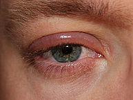 Blepharitis of the right eye – By clubtable – Own work (Original text: eigenes Foto), Public Domain, https://commons.wikimedia.org/w/index.php?curid=20138102
 -
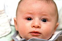 Infant with blepharitis – By Sage Ross – Own work, CC BY-SA 3.0, https://commons.wikimedia.org/w/index.php?curid=8978907


References
- ↑ 1.0 1.1 Bunya VY, Brainard DH, Daniel E, Massaro-Giordano M, Nyberg W, Windsor EA; et al. (2013). “Assessment of signs of anterior blepharitis using standardized color photographs”. Cornea. 32 (11): 1475–82. doi:10.1097/ICO.0b013e3182a02e0e. PMC 3947496. PMID 24055901.
- ↑ 2.0 2.1 Dougherty JM, McCulley JP (1986). “Bacterial lipases and chronic blepharitis”. Invest Ophthalmol Vis Sci. 27 (4): 486–91. PMID 3957566.
- ↑ Blepharitis. American Academy of Ophthalmology/eyewiki (2014). http://eyewiki.aao.org/Blepharitis Accessed on July 14, 2016
- ↑ Lemp MA, Nichols KK (2009). “Blepharitis in the United States 2009: a survey-based perspective on prevalence and treatment”. Ocul Surf. 7 (2 Suppl): S1–S14. PMID 19383269.
- ↑ Bhandari V, Reddy JK (2014). “Blepharitis: always remember demodex”. Middle East Afr J Ophthalmol. 21 (4): 317–20. doi:10.4103/0974-9233.142268. PMC 4219223. PMID 25371637.
- ↑ Viswalingam M, Rauz S, Morlet N, Dart JK (2005). “Blepharokeratoconjunctivitis in children: diagnosis and treatment”. Br J Ophthalmol. 89 (4): 400–3. doi:10.1136/bjo.2004.052134. PMC 1772603. PMID 15774912.
- ↑ 7.0 7.1 McCulley JP, Dougherty JM (1985). “Blepharitis associated with acne rosacea and seborrheic dermatitis”. Int Ophthalmol Clin. 25 (1): 159–72. PMID 3156100.
- ↑ 8.0 8.1 Nemet AY, Vinker S, Kaiserman I (2011). “Associated morbidity of blepharitis”. Ophthalmology. 118 (6): 1062–8. doi:10.1016/j.ophtha.2010.10.015. PMID 21276617.
- ↑ Blepharitis. Diseases Database (2016). http://www.diseasesdatabase.com/relationship.asp?glngUserChoice=1455&bytRel=2&blnBW=0&strBB=RL&blnClassSort=255 Accessed on July 15, 2016
- ↑ Benitez-Del-Castillo JM (2012). “How to promote and preserve eyelid health”. Clin Ophthalmol. 6: 1689–98. doi:10.2147/OPTH.S33133. PMC 3484726. PMID 23118519.
- ↑ Seal D, Ficker L, Ramakrishnan M, Wright P (1990). “Role of staphylococcal toxin production in blepharitis”. Ophthalmology. 97 (12): 1684–8. PMID 2087299.
- ↑ THYGESON P, VAUGHAN DG (1954). “Seborrheic blepharitis”. Trans Am Ophthalmol Soc. 52: 173–88. PMC 1312591. PMID 13274422.
- ↑ Wesolowska M, Knysz B, Reich A, Blazejewska D, Czarnecki M, Gladysz A; et al. (2014). “Prevalence of Demodex spp. in eyelash follicles in different populations”. Arch Med Sci. 10 (2): 319–24. doi:10.5114/aoms.2014.42585. PMC 4042053. PMID 24904668.
Causes
Editor-In-Chief: C. Michael Gibson, M.S., M.D. [1]; Associate Editor(s)-in-Chief: Ogheneochuko Ajari, MB.BS, MS [2], Sara Mehrsefat, M.D. [3]
Overview
Common causes of blepharitis include bacterial infections, dysfunctional meibomian glands, allergies, and Demodex folliculorum.[1][2]
Causes
Common causes of blepharitis include:[1][2][3][4][5]
- Bacterial colonization
- Staphylococcus aureus (most common)
- Corynebacterium
- Staphylococcus epidermidis
- Dysfunctional meibomian glands
- Allergies
- Demodex folliculorum
Blepharitis may be also caused by:[6]
- Lipoid proteinosis (autosomal recessive condition)
- Periorbital cellulitis
- Dipivefrin
Causes by Organ System
Causes in Alphabetical Order
- Acitretin
- Allergies
- Apraclonidine
- Atopic dermatitis [7]
- Atopic keratoconjunctivitis
- Atropine (systemic)
- Betaxolol (systemic)
- Bexarotene (systemic)
- Brimonidine (ophthalmic)
- Brinzolamide
- Cetuximab [8]
- Chalazion
- Chemical irritants
- Chronic graft-versus-host disease
- Clomipramine
- Congenital erythropoietic porphyria
- Contact dermatitis
- Contact lens
- Crohn’s disease [9]
- Demodex
- Diabetes
- Difluprednate
- Dipivefrin
- Dry eyes
- Dysfunctional meibomian glands [5]
- Dyskeratosis congenita
- Epidermolysis bullosa
- Erythromycin and benzoyl peroxide topical (patient information)
- Fluocinolone (ophthalmic)
- Gatifloxacin
- Gefitinib
- Glucagonoma syndrome
- Herpes simplex virus infection
- HIV [10]
- Iloperidone
- Ipilimumab
- Isotretinoin
- Lansoprazole
- Lice
- Lipoid proteinosis
- Lodoxamide tromethamine
- Loteprednol
- Methotrexate
- Metipranolol
- Mirtazapine
- Molluscum contagiosum
- Mupirocin
- Mustard gas [11]
- Myotonic dystrophy
- Nisoldipine
- Nitisinone
- Pediculosis
- Pegaptanib
- Pentamidine (oral inhalation)
- Periorbital cellulitis
- Phthiriasis
- Poor hygiene
- Pramipexole
- Pregabalin
- Propionibacterium acnes [4]
- Pseudomonas aeruginosa
- Psoriasis [12]
- Quetiapine
- Ranibizumab
- Rasagiline
- Rosacea [7]
- Saquinavir
- Seborrheic dermatitis [7]
- Sjögren’s syndrome
- Skin cancer
- Staphylococcus aureus [4]
- Syphilis
- Tacrolimus (topical)
- Tiagabine
- Timolol (ophthalmic)
- Travoprost
- Unoprostone
- Varicella zoster
- Venlafaxine
- Vernal keratoconjunctivitis
- Verteporfin
- Xeroderma pigmentosum [13]
- Zaleplon
- Ziprasidone
References
- ↑ 1.0 1.1 Lemp MA, Nichols KK (2009). “Blepharitis in the United States 2009: a survey-based perspective on prevalence and treatment”. Ocul Surf. 7 (2 Suppl): S1–S14. PMID 19383269.
- ↑ 2.0 2.1 Blepharitis. American Academy of Ophthalmology/eyewiki (2014). http://eyewiki.org/Blepharitis Accessed on July 14, 2016
- ↑ Bernardes TF, Bonfioli AA (2010). “Blepharitis”. Semin Ophthalmol. 25 (3): 79–83. doi:10.3109/08820538.2010.488562. PMID 20590417.
- ↑ 4.0 4.1 4.2 McCulley JP, Dougherty JM (1986). “Bacterial aspects of chronic blepharitis”. Trans Ophthalmol Soc U K. 105 ( Pt 3): 314–8. PMID 3466455.
- ↑ 5.0 5.1 Driver PJ, Lemp MA (1996). “Meibomian gland dysfunction”. Surv Ophthalmol. 40 (5): 343–67. PMID 8779082.
- ↑ Blepharitis. Diseases Database. (2016). http://www.diseasesdatabase.com/relationship.asp?glngUserChoice=1455&bytRel=2&blnBW=0&strBB=RL&blnClassSort=255 Accessed on July 15, 2016
- ↑ 7.0 7.1 7.2 Auw-Haedrich C, Reinhard T (2007). “[Chronic blepharitis. Pathogenesis, clinical features, and therapy]”. Ophthalmologe. 104 (9): 817–26, quiz 827-8. doi:10.1007/s00347-007-1608-8. PMID 17762935.
- ↑ Dranko S, Kinney C, Ramanathan RK (2006). “Ocular toxicity related to cetuximab monotherapy in patients with colorectal cancer”. Clin Colorectal Cancer. 6 (3): 224–5. doi:10.3816/CCC.2006.n.040. PMID 17026793.
- ↑ Yilmaz S, Aydemir E, Maden A, Unsal B (2007). “The prevalence of ocular involvement in patients with inflammatory bowel disease”. Int J Colorectal Dis. 22 (9): 1027–30. doi:10.1007/s00384-007-0275-1. PMID 17262200.
- ↑ Biswas J, Sudharshan S (2008). “Anterior segment manifestations of human immunodeficiency virus/acquired immune deficiency syndrome”. Indian J Ophthalmol. 56 (5): 363–75. PMC 2636142. PMID 18711264.
- ↑ Karimian F, Zarei-Ghanavati S, A BR, Jadidi K, Lotfi-Kian A (2011). “Microbiological evaluation of chronic blepharitis among Iranian veterans exposed to mustard gas: a case-controlled study”. Cornea. 30 (6): 620–3. doi:10.1097/ICO.0b013e3181e16f7c. PMID 21282998.
- ↑ Rehal B, Modjtahedi BS, Morse LS, Schwab IR, Maibach HI (2011). “Ocular psoriasis”. J Am Acad Dermatol. 65 (6): 1202–12. doi:10.1016/j.jaad.2010.10.032. PMID 21550135.
- ↑ Brooks BP, Thompson AH, Bishop RJ, Clayton JA, Chan CC, Tsilou ET; et al. (2013). “Ocular manifestations of xeroderma pigmentosum: long-term follow-up highlights the role of DNA repair in protection from sun damage”. Ophthalmology. 120 (7): 1324–36. doi:10.1016/j.ophtha.2012.12.044. PMC 3702678. PMID 23601806.
Differentiating Blepharitis from other Diseases
Editor-In-Chief: C. Michael Gibson, M.S., M.D. [1]; Associate Editor(s)-in-Chief: Sara Mehrsefat, M.D. [2]
Overview
Blepharitis must be differentiated from other diseases that cause eye itching, irritation, burning, and foreign body sensation such as allergic contact dermatitis, conjunctivitis, trichiasis, dry eye syndrome, keratitis, hordeolum, chalazion, rosacea, and sebaceous carcinoma.
Differentiating Blepharitis from Other Diseases
Blepharitis must be differentiated from other conditions associated with eyelid inflammation, including:[1]
| Differential diagnosis of blepharitis | |
|---|---|
| Condition | Entity |
| Bacterial infections |
|
| Viral infections | |
| Parasitic infections |
|
| Immunologic conditions |
|
| Dermatoses | |
| Benign eyelid tumors |
|
| Malignant eyelid tumors |
|
| Trauma |
|
| Toxic conditions |
|
Blepharitis must be differentiated from other diseases that cause red, swollen, and itchy eyes, including:[2][3][4][5][6][7]
- Allergic contact dermatitis
- Allergic conjunctivitis (atopic keratoconjunctivitis)
- Bacterial conjunctivitis
- Viral conjunctivitis
- Trichiasis
- Dry eye syndrome
- Keratitis
- Hordeolum
- Chalazion
- Rosacea
- Superior limbic keratoconjunctivitis
- Contact lens complications
- Sebaceous carcinoma
References
- ↑ “Blepharitis – Preferred Practice Pattern Guideline – 2013 – American Academy of Ophthalmology.” http://www.aao.org/preferred-practice-pattern/blepharitis-ppp–2013.
- ↑ Mathers WD, Choi D (2004). “Cluster analysis of patients with ocular surface disease, blepharitis, and dry eye”. Arch Ophthalmol. 122 (11): 1700–4. doi:10.1001/archopht.122.11.1700. PMID 15534133.
- ↑ Lemp MA, Nichols KK (2009). “Blepharitis in the United States 2009: a survey-based perspective on prevalence and treatment”. Ocul Surf. 7 (2 Suppl): S1–S14. PMID 19383269.
- ↑ Beare JM (1969). “Blepharitis and related conditions”. Proc R Soc Med. 62 (1): 5–7. PMC 2279072. PMID 4236660.
- ↑ Bowman RW, Dougherty JM, McCulley JP (1987). “Chronic blepharitis and dry eyes”. Int Ophthalmol Clin. 27 (1): 27–35. PMID 3818198.
- ↑ Blepharitis. American Academy of Ophthalmology/eyewiki (2014) http://eyewiki.org/Blepharitis Accessed on July 14, 2016
- ↑ Leibowitz HM (2000). “The red eye”. N Engl J Med. 343 (5): 345–51. doi:10.1056/NEJM200008033430507. PMID 10922425.
Epidemiology and Demographics
Editor-In-Chief: C. Michael Gibson, M.S., M.D. [1]; Associate Editor(s)-in-Chief: Sara Mehrsefat, M.D. [2]
Overview
Blepharitis is one of the most common ocular disorders encountered in clinical practice and affects more than 180 million people in the United States.
Epidemiology and Demographics
Prevalence
Blepharitis is one of the most common ocular disorders encountered by eye care practitioners.[1] In the United States, blepharitis is estimated to affect more than 180 million people.[2] In a United States survey of ophthalmologists and optometrists, it was estimated that 37% and 47% of the patients presented with certain form of blepharitis in clinical practice, respectively.[3]
incidence
The true incidence of blepharitis remains undetermined.
Age
Blepharitis can affect all age groups.[4][5][6] A single-center study of patients with chronic blepharitis reported a mean age of 50 years.[7] Compared with other forms of blepharitis, patients affected by staphylococcal blepharitis are relatively young, with a mean age of 42 years.[8]
Gender
Compared with other forms of blepharitis, patients affected by staphylococcal blepharitis are mostly female.[9]
Race
Blepharitis can affect all ethnic groups.[10][11][12] There is no racial predilection for blepharitis.
References
- ↑ Macsai MS (2008). “The role of omega-3 dietary supplementation in blepharitis and meibomian gland dysfunction (an AOS thesis)”. Trans Am Ophthalmol Soc. 106: 336–56. PMC 2646454. PMID 19277245.
- ↑ Guttman, C. (2009). Recent surveys draw attention to blepharitis. Ophthalmology Times, 34(15), 30-31.
- ↑ Lemp MA, Nichols KK (2009). “Blepharitis in the United States 2009: a survey-based perspective on prevalence and treatment”. Ocul Surf. 7 (2 Suppl): S1–S14. PMID 19383269.
- ↑ Lindsley K, Matsumura S, Hatef E, Akpek EK. Interventions for chronic blepharitis. Cochrane Database Syst Rev. 2012;5:CD005556.
- ↑ Pflugfelder SC, Karpecki PM, Perez VL. Treatment of blepharitis: recent clinical trials. Ocul Surf. 2014;12(4):273-84.
- ↑ Lemp MA, Nichols KK. Blepharitis in the United States 2009: a survey-based perspective on prevalence and treatment. Ocular Surface 2009;7(Suppl 2):S1–14.
- ↑ McCulley JP, Dougherty JM, Deneau DG. Classification of chronic blepharitis. Ophthalmology 1982;89:1173-80.
- ↑ McCulley JP, Dougherty JM. Blepharitis associated with acne rosacea and seborrheic dermatitis. International Ophthalmology Clinics 1985;25(1):159–72.
- ↑ McCulley JP, Dougherty JM. Blepharitis associated with acne rosacea and seborrheic dermatitis. International Ophthalmology Clinics 1985;25(1):159–72.
- ↑ Lindsley K, Matsumura S, Hatef E, Akpek EK. Interventions for chronic blepharitis. Cochrane Database Syst Rev. 2012;5:CD005556.
- ↑ Pflugfelder SC, Karpecki PM, Perez VL. Treatment of blepharitis: recent clinical trials. Ocul Surf. 2014;12(4):273-84.
- ↑ Lemp MA, Nichols KK. Blepharitis in the United States 2009: a survey-based perspective on prevalence and treatment. Ocular Surface 2009;7(Suppl 2):S1–14.
Risk Factors
Editor-In-Chief: C. Michael Gibson, M.S., M.D. [1]; Associate Editor(s)-in-Chief: Sara Mehrsefat, M.D. [2]
Overview
Common risk factors for the development of blepharitis include dry eye, acne rosacea, seborrheic dermatitis, and demodicosis.
Risk factors
Risk factors for the development of blepharitis include:
- Dry eye[1]
- Dermatologic conditions (acne rosacea, seborrheic dermatitis)[2]
- Demodicosis[3]
- Isotretinoin[4][5]
- Giant papillary conjunctivitis[6]
References
- ↑ Bowman RW, Dougherty JM, McCulley JP (1987). “Chronic blepharitis and dry eyes”. Int Ophthalmol Clin. 27 (1): 27–35. PMID 3818198.
- ↑ McCulley JP, Dougherty JM (1985). “Blepharitis associated with acne rosacea and seborrheic dermatitis”. Int Ophthalmol Clin. 25 (1): 159–72. PMID 3156100.
- ↑ Kemal M, Sumer Z, Toker MI, et al. The prevalence of Demodex folliculorum in blepharitis patients and the normal population. Ophthalmic Epidemiol 2005;12:287-90.
- ↑ McCulley JP, Dougherty JM, Deneau DG. Classification of chronic blepharitis. Ophthalmology 1982;89:1173-80.
- ↑ Bozkurt B, Irkec MT, Atakan N, et al. Lacrimal function and ocular complications in patients treated with systemic isotretinoin. Eur J Ophthalmol 2002;12:173-6.
- ↑ Martin NF, Rubinfeld RS, Malley JD, Manzitti V. Giant papillary conjunctivitis and meibomian gland dysfunction blepharitis. CLAO J 1992;18:165-9.
Screening
Editor-In-Chief: C. Michael Gibson, M.S., M.D. [1]; Associate Editor(s)-in-Chief: Sara Mehrsefat, M.D. [2]
Overview
Screening for blepharitis is not recommended.[1]
Screening
Screening for blepharitis is not recommended.[1]
References
- ↑ 1.0 1.1 U.S. Preventive Services task force. Blepharitis. https://www.uspreventiveservicestaskforce.org/BrowseRec/Search?s=blepharitis
Natural History, Complications and Prognosis
Editor-In-Chief: C. Michael Gibson, M.S., M.D. [1]; Associate Editor(s)-in-Chief: Sara Mehrsefat, M.D. [2]
Overview
Blepharitis is usually asymptomatic until the disease progresses. As it progresses, the patient begins to notice a foreign body sensation, eyelid crusting, itching and irritation of the eyelids. If left untreated, severe blepharitis may cause alterations in the eyelid margin, loss of eyelashes, scarring of the eyelids, conjunctivitis, corneal involvement, superficial keratopathy, and ultimately blindness. Common complications of blepharitis include loss of eyelashes, hordeolum or stye, chalazion, corneal ulcer, and conjunctivitis. Blepharitis is generally associated with a favorable long-term prognosis.
Natural History
Earlier in the course of blepharitis, patients may be asymptomatic and present with findings of eyelid margin telangiectasia and meibomian gland orifice narrowing.[1] As the disease progresses, patients usually develop symptoms of foreign body sensation, eyelid crusting, matting of the lashes, tearing, and burning. If left untreated, blepharitis may lead to alterations in the eyelid margin, loss of eyelashes, scarring of the eyelids, corneal involvement (corneal neovascularization and scarring), superficial keratopathy, and eventually blindness.[2][3]
Complications
Complications to blepharitis include:[4][5][6]
Prognosis
In general, blepharitis is associated with a favorable long-term prognosis. Severe blepharitis is rarely associated with permanent alterations in the eyelid margin or vision loss from superficial keratopathy. However, severe blepharitis cases with these complications are generally associated with a poor prognosis.[7][8]
References
- ↑ Hykin, P. G., and A. J. Bron. “Age-related morphological changes in lid margin and meibomian gland anatomy.” Cornea 11.4 (1992): 334-342.
- ↑ Nemet AY, Vinker S, Kaiserman I (2011). “Associated morbidity of blepharitis”. Ophthalmology. 118 (6): 1062–8. doi:10.1016/j.ophtha.2010.10.015. PMID 21276617.
- ↑ Ficker L, Ramakrishnan M, Seal D, Wright P. Role of cell-mediated immunity to staphylococci in blepharitis. Am J Ophthalmol 1991;111:473-9.
- ↑ Dougherty JM, McCulley JP (1984). “Comparative bacteriology of chronic blepharitis”. Br J Ophthalmol. 68 (8): 524–8. PMC 1040405. PMID 6743618.
- ↑ PubMed Health (2009). http://www.ncbi.nlm.nih.gov/pubmedhealth/PMHT0023008/ Accessed on July, 13 2016
- ↑ Sharma S (1998). “Ophthaproblem. Chalazion”. Can Fam Physician. 44: 1249, 1254, 1257. PMC 2278269. PMID 9640516.
- ↑ Lindsley K, Matsumura S, Hatef E, Akpek EK (2012). “Interventions for chronic blepharitis”. Cochrane Database Syst Rev (5): CD005556. doi:10.1002/14651858.CD005556.pub2. PMC 4270370. PMID 22592706.
- ↑ Raskin EM, Speaker MG, Laibson PR (1992). “Blepharitis”. Infect Dis Clin North Am. 6 (4): 777–87. PMID 1460262.
Diagnosis
Diagnosis
History and Symptoms | Physical Examination | Laboratory Findings | Electrocardiogram | X Ray | CT | MRI | Ultrasound | Other Imaging Findings | Other Diagnostic Studies
Treatment
Treatment
Medical Therapy | Surgery | Primary Prevention |Secondary Prevention | Cost-Effectiveness of Therapy |Future or Investigational Therapies
Looking for the patient version?
© 2026 MyEClinic – IFTM Institut für Telematik in der Medizin GmbH