
Bursitis
For patient information page click here
Editor-In-Chief: C. Michael Gibson, M.S., M.D. [1]; Associate Editor-In-Chief: Sara Mehrsefat, M.D. [2]; Cafer Zorkun, M.D., Ph.D. [3]
Synonyms and keywords: Inflammation of bursa
Overview
Editor-In-Chief: C. Michael Gibson, M.S., M.D. [1]; Associate Editor(s)-in-Chief: Sara Mehrsefat, M.D. [2]
Overview
Bursitis is characterized by the inflammation of a bursa and the buildup of the fluid in the bursal sac. A bursa is a small, fluid-filled sac that acts as a cushion between a bone and other moving parts: muscles, tendons, or skin. Over 160 bursa are found throughout the body but relatively few of them can cause bursitis. Based on the nature of the inflammation, bursitis may be classified into 2 subtypes: septic and aseptic. The most common bursitis subtypes include subacromial, olecranon, trochanteric, prepatellar, and retrocalcaneal. Aseptic bursitis can be caused by overuse or repetitive injury to the joint, abnormal and bony structure, and/or crystal deposit in the bursa. Additionally, septic bursitis can be caused by bacterial infection of the bursa through a skin injury following repetitive trauma. Common causes of septic bursitis include Staphylococcus aureus, Staphylococcus epidermidis, and Streptococcus spp. Bursitis must be differentiated from tendonitis, cellulitis, osteoarthritis, ligamentous injuries, and septic arthritis. The symptoms of bursitis differ based on the location of the inflammation. Focal swelling, pain, and redness are symptoms common to all forms of bursitis. The diagnosis of bursitis is usually made clinically. There are no diagnostic lab findings associated with bursitis. However, patients with septic bursitis may have elevated ESR, CRP, and white blood cell count. Ultrasonography may be a useful tool for confirming the diagnosis of bursitis, while the aspiration of bursal fluids is usually reserved for the diagnosis of septic bursitis.[1][2] Medical therapy for non-septic bursitis depends on the involved bursa and includes the RICE regimen (rest, ice, compression, elevation), NSAIDs, and/or corticosteroid injections. Restriction of activity is encouraged to prevent further injury and promote healing. Antimicrobials are the mainstay of therapy for septic bursitis. Surgical management is often reserved for non-responders.[3][4][5]
Classification
Based on the nature of the inflammation, bursitis may classified into 2 subtypes: septic and aseptic. Common anatomic locations of bursitis include the shoulder, elbow, hip, knee, and ankle. The most common bursitis subtypes include:[6][7][8][9]
- Subacromial bursitis
- Olecranon bursitis
- Trochanteric bursitis
- Prepatellar bursitis
- Retrocalcaneal bursitis
Pathophysiology
Bursitis is characterized by acute or chronic inflammation of a bursa and buildup of fluid in the bursa sac. A bursa is a small, fluid-filled sac that acts as a cushion between a bone and other moving parts: muscles, tendons, or skin. Over 160 bursae are found throughout the body, though relatively few of them can cause bursitis. Aseptic bursitis can be caused by overuse and repetitive injuries to the joint, abnormal bony structure and crystal deposit in the bursa. It commonly affects the knee or elbow as a result of kneeling or leaning on the elbows longer than usual. Moreover, septic bursitis can be caused by bacterial infection of the bursa through the skin injury following repetitive trauma.[7][10][11]
Images
The following images depict different cases of bursitis:[12]
-
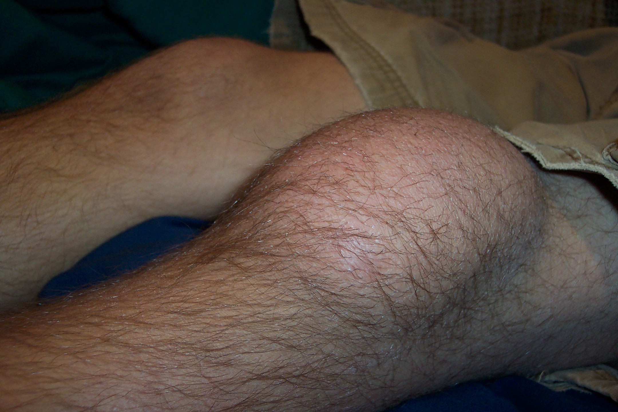 Prepatellar bursitis – By Thomas Kees – Own work (Original text: eigenes Archiv (selbst photographiert)), CC BY-SA 3.0 de, https://commons.wikimedia.org/w/index.php?curid=25418133
 -
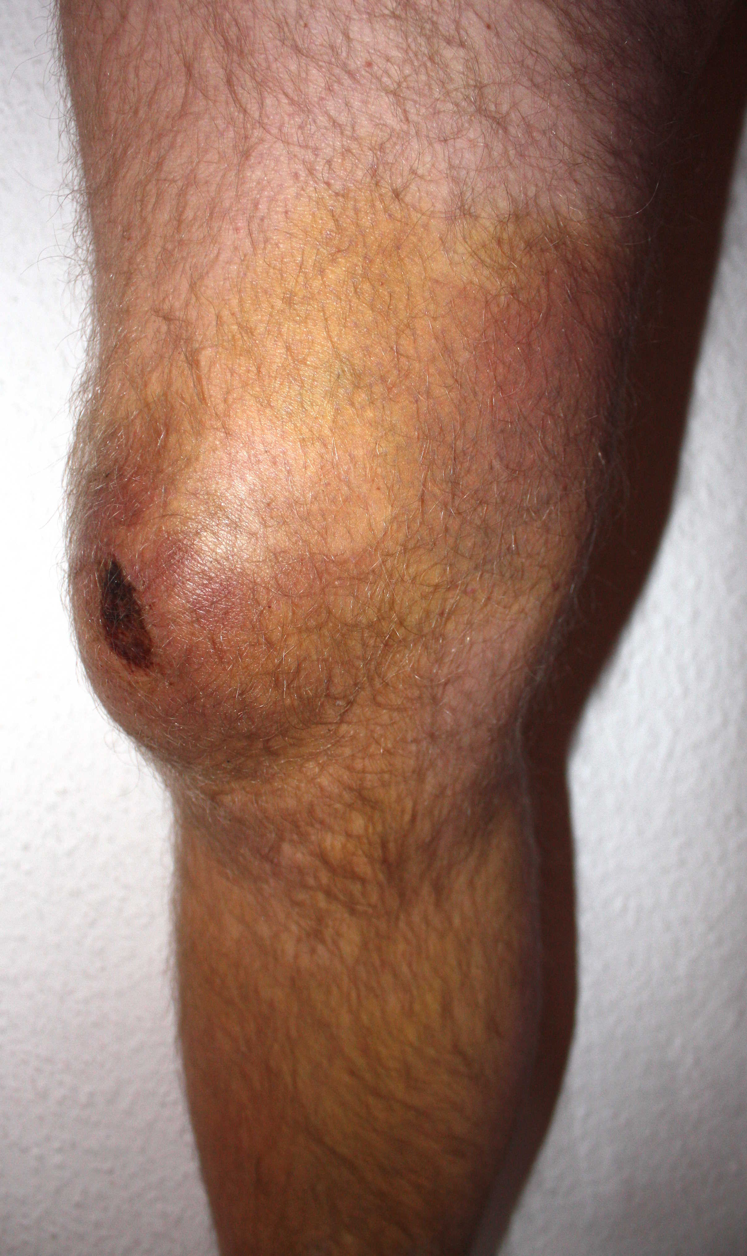 Prepatellar bursitis – By Atropos235 – Own work, CC BY-SA 3.0, https://commons.wikimedia.org/w/index.php?curid=4079323
 -
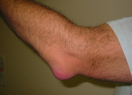 Olecranon bursitis – By en:User:NJC123 – en:Image:Bursitis_Elbow_WC.JPG, Public Domain, https://commons.wikimedia.org/w/index.php?curid=2814450


Causes
Common causes of bursitis include:[1][8][13][14][15]
Aseptic bursitis:
- Prolonged pressure, overuse, or strenuous activity
- Other inflammatory conditions (e.g., rheumatoid arthritis and spondyloarthritis)
- Gout and pseudogout
Septic bursitis:
Differential Diagnosis
Bursitis symptoms and signs are relatively non-specific. Even after detailed history and physical examination, imaging studies are often necessary to rule out other musculoskeletal conditions. Bursitis must be differentiated from tendonitis, cellulitis, osteoarthritis, ligamentous injuries, and septic arthritis.[7][8][9]
Epidemiology and Demographics
Bursitis accounts for 400 visits per 100,000 visits to primary care clinic. The exact prevalence and incidence of bursitis are unknown.[15]
Risk Factors
Common risk factors in the development of bursitis include:[15]
- rheumatoid arthritis
- osteoarthritis
- gout or pseudogout
- cellulitis
- diabetes mellitus
- use of systemic glucocorticoids
- alcoholism
- malignancy
- leukopenia
- having a hobby or job that involves repetitive motions (e.g., bicycling, playing baseball, gardening, setting tiles)
Screening
Screening for bursitis is not recommended.[16]
Natural History, Complications, and Prognosis
Bursitis is often caused by overuse and repetitive injuries to the joint. Symptoms of bursitis may develop rapidly within 2 to 3 days in an acute form. Patients with bursitis usually present with edema, erythema, and tenderness over the involved joint. In most cases, after an appropriate lifestyle adjustment, bursitis will gradually clear within a few days to weeks without any long-term consequences. If left untreated, acute bursitis may lead to chronic bursitis, which can result in cicatricial adhesions, reduced mobility, and progressive pain. With proper treatment and an activities adjustment, septic and aseptic bursitis are associated with an excellent prognosis.
Diagnosis
History
Obtaining a complete history will be helpful in determining whether the bursitis is associated with any specific activities.[7][8][9]
Symptoms and Physical Examination
| Type of Bursitis | Symptoms | Physical examination |
|---|---|---|
| Subacromial bursitis[8] |
|
|
| Olecranon bursitis[17][18] |
|
|
| Trochanteric bursitis[19][20] |
|
|
| Prepatellar bursitis[9][21] |
|
|
| Retrocalcaneal bursitis[7][22] |
|
Laboratory Findings
The diagnosis of bursitis is usually made clinically. There are no diagnostic lab findings associated with bursitis. However, patients with septic bursitis may have elevated ESR, CRP, and white blood cells.[23][8][9]
X ray
X ray is rarely required in patients with bursitis. X ray may be used as a diagnostic measure to support a clinical diagnosis of bursitis. Joint x ray is generally reserved for patients with histories of significant trauma. A standard x ray may be helpful in diagnosing a fracture or dislocation.[24][21][25]
CT
CT scans are rarely required in patients with bursitis. CT scans are usually reserved for patients who do not respond to initial treatment. On a CT scan, superficial bursitis may be characterized by fluid density at the subcutaneous tissue.[24][21]
MRI
MRI is rarely required in patients with bursitis. Due to the associated cost and time requirements, the utility of MRI is limited compere to ultrasound. MRI is often reserved for patients who are likely to have other medical conditions such as tumors, ligamentous injures, or tendon injuries. On MRI, bursitis is characterized by bursal fluid collection, subcutaneous edema, and joint effusion.[24][21]
Ultrasound
Ultrasonography may be helpful in confirming a diagnosis of bursitis. On an ultrasound, bursitis may be characterized by bursal wall distention with presence of local hypoechoic or anechoic intra-bursal material, synovial proliferation, calcifications, and rheumatoid nodules.[26][27]
Other Diagnostic Studies
Other diagnostic studies for bursitis include aspiration of the bursal fluid. Aspiration of bursal fluids is not recommended for the diagnosis of all types of bursitis. It is usually reserved for confirming a diagnosis of septic bursitis.[1][2]
Treatment
Medical Therapy
Medical therapy for non-septic bursitis depends on the involved bursa and can include the RICE regimen (rest, ice, compression, elevation), NSAIDs, and/or corticosteroid injections. Restriction of activity is encouraged to prevent further injury and promote healing. Antimicrobials are the mainstay of therapy for septic bursitis. Surgical intervention is generally reserved for non-responders.[3][4][5]
| Septic[3][4] | Aseptic |
|---|---|
|
|
Surgery
Surgical intervention is not recommended for the management of bursitis. However, surgical techniques including bursectomy or longitudinal band release are usually reserved for patients with chronic, recurrent, or septic bursitis.[28][29]
Primary Prevention
Effective measures for the primary prevention of bursitis include maintaining a healthy weight, taking regular breaks from repetitive tasks, using foam for knee- and elbow-pads, and practicing good posture.[30]
Secondary Prevention
There are no established methods for secondary prevention of bursitis. However, a fast recovery may be facilitated by an adjustment in activities, the consistent use of foam for knee- or elbow-pads, and regular breaks during repetitive tasks.[30]
References
- ↑ 1.0 1.1 1.2 Stell IM, Gransden WR (1998). “Simple tests for septic bursitis: comparative study”. BMJ. 316 (7148): 1877. PMC 28586. PMID 9632407.
- ↑ 2.0 2.1 Shell, Donald, Rob Perkins, and Andrew Cosgarea. “Septic olecranon bursitis: recognition and treatment.” The Journal of the American Board of Family Practice 8.3 (1995): 217-220.
- ↑ 3.0 3.1 3.2 Reilly D, Kamineni S (2016). “Olecranon bursitis”. J Shoulder Elbow Surg. 25 (1): 158–67. doi:10.1016/j.jse.2015.08.032. PMID 26577126.
- ↑ 4.0 4.1 4.2 Zimmermann B, Mikolich DJ, Ho G (1995). “Septic bursitis”. Semin Arthritis Rheum. 24 (6): 391–410. PMID 7667644.
- ↑ 5.0 5.1 Aaron DL, Patel A, Kayiaros S, Calfee R (2011). “Four common types of bursitis: diagnosis and management”. J Am Acad Orthop Surg. 19 (6): 359–67. PMID 21628647.
- ↑ Chatra PS (2012). “Bursae around the knee joints”. Indian J Radiol Imaging. 22 (1): 27–30. doi:10.4103/0971-3026.95400. PMC 3354353. PMID 22623812.
- ↑ 7.0 7.1 7.2 7.3 7.4 Fauci, Anthony S., and Carol Langford. Harrison’s rheumatology. McGraw Hill Professional, 2010.
- ↑ 8.0 8.1 8.2 8.3 8.4 8.5 Walker‐Bone, Karen, et al. “Prevalence and impact of musculoskeletal disorders of the upper limb in the general population.
- ↑ 9.0 9.1 9.2 9.3 9.4 Aaron, Daniel L., et al. “Four common types of bursitis: diagnosis and management.” Journal of the American Academy of Orthopaedic Surgeons 19.6 (2011): 359-367.
- ↑ Hellmann DB, Imboden JB., Jr. Musculoskeletal and immunologic disorders. In: McPhee SJ, Papadakis MA, editors. Current Medical Diagnosis & Treatment. McGraw-Hill Lange; 2010. pp. 2056–2061.
- ↑ García-Porrúa C, González-Gay MA, Ibañez D, García-País MJ (1999). “The clinical spectrum of severe septic bursitis in northwestern Spain: a 10 year study”. J Rheumatol. 26 (3): 663–7. PMID 10090179.
- ↑ Wikimedia Commons. Bursitis. (2012) https://commons.wikimedia.org/wiki/Category:Bursitis Accessed on August 31, 2016
- ↑ Wang JP, Granlund KF, Bozzette SA, Botte MJ, Fierer J (2000). “Bursal sporotrichosis: case report and review”. Clin Infect Dis. 31 (2): 615–6. doi:10.1086/313983. PMID 10987734.
- ↑ National Institute of Arthritis and Musculoskeletal and Skin disease, Bursitis. http://www.niams.nih.gov/Health_Info/Bursitis/default.asp Accessed August 25, 2016
- ↑ 15.0 15.1 15.2 McAfee JH, Smith DL (1988). “Olecranon and prepatellar bursitis. Diagnosis and treatment”. West J Med. 149 (5): 607–10. PMC 1026560. PMID 3074561.
- ↑ U.S. Preventive Services Task Force http://www.uspreventiveservicestaskforce.org/BrowseRec/Search?s=bursitis Accessed on August 29, 2016
- ↑ Stell IM (1996). “Septic and non-septic olecranon bursitis in the accident and emergency department–an approach to management”. J Accid Emerg Med. 13 (5): 351–3. PMC 1342774. PMID 8894865.
- ↑ Lockman L (2010). “Treating nonseptic olecranon bursitis: a 3-step technique”. Can Fam Physician. 56 (11): 1157. PMC 2980436. PMID 21075998.
- ↑ Brinks A, van Rijn RM, Bohnen AM, Slee GL, Verhaar JA, Koes BW; et al. (2007). “Effect of corticosteroid injection for trochanter pain syndrome: design of a randomised clinical trial in general practice”. BMC Musculoskelet Disord. 8: 95. doi:10.1186/1471-2474-8-95. PMC 2045096. PMID 17880718.
- ↑ Karpinski MR, Piggott H (1985). “Greater trochanteric pain syndrome. A report of 15 cases”. J Bone Joint Surg Br. 67 (5): 762–3. PMID 4055877.
- ↑ 21.0 21.1 21.2 21.3 Huang, Yu-Chih, and Wen-Lin Yeh. “Endoscopic treatment of prepatellar bursitis.” International orthopaedics 35.3 (2011): 355-358.
- ↑ Lyman, Jeffrey, Paul S. Weinhold, and Louis C. Almekinders. “Strain behavior of the distal Achilles tendon implications for insertional Achilles tendinopathy.” The American Journal of Sports Medicine 32.2 (2004): 457-461.
- ↑ Approach to Articular and Musculoskeletal Disorders, Harrison’s Internal Medicine, 2011
- ↑ 24.0 24.1 24.2 Radiopedia. Olecranon Bursitis. http://radiopaedia.org/articles/olecranon-bursitis Accessed on August 23, 2016
- ↑ Blankstein A, Cohen I, Diamant L, Heim M, Dudkiewicz I, Israeli A; et al. (2001). “Achilles tendon pain and related pathologies: diagnosis by ultrasonography”. Isr Med Assoc J. 3 (8): 575–8. PMID 11519381.
- ↑ Blankstein A, Ganel A, Givon U, Mirovski Y, Chechick A. Ultrasonographic findings in patients with olecranon bursitis. Ultraschall Med 2006; 27: 568-571.
- ↑ Martinoli C, Bianchi S, Giovagnorio F, Pugliese F. Ultrasound of the elbow. Skeletal Radiol 2001; 30: 605-614
- ↑ Huang YC, Yeh WL (2011). “Endoscopic treatment of prepatellar bursitis”. Int Orthop. 35 (3): 355–8. doi:10.1007/s00264-010-1033-5. PMC 3047636. PMID 20521045.
- ↑ Lustenberger DP, Ng VY, Best TM, Ellis TJ (2011). “Efficacy of treatment of trochanteric bursitis: a systematic review”. Clin J Sport Med. 21 (5): 447–53. doi:10.1097/JSM.0b013e318221299c. PMC 3689218. PMID 21814140.
- ↑ 30.0 30.1 National Institute of Arthritis and Musculoskeletal and Skin disease. Bursitis. http://www.niams.nih.gov/Health_Info/Bursitis/default.asp Accessed August 25, 2016
Historical Perspective
Please help WikiDoc by adding content here. It’s easy! Click here to learn about editing.
References
Classification
Editor-In-Chief: C. Michael Gibson, M.S., M.D. [1]; Associate Editor(s)-in-Chief: Sara Mehrsefat, M.D. [2]
Overview
Based on the nature of the inflammation, bursitis may classified into two subtypes: septic and aseptic. Common anatomic location include the shoulder, elbow, hip, knee, and ankle. The most common bursitis subtypes include subacromial, olecranon, trochanteric, prepatellar, and retrocalcaneal. Moreover, based on the location of the affected bursa in relation to the skin, bursitis may be further classified into two additional subtypes: superficial and deep. Superficial bursa are more prone to get infected with bacteria and develop septic bursitis. Common locations of septic bursitis include the knee (prepatellar bursitis) and elbow (olecranon bursitis).[1][2][3][4]
Classification
Based on the nature of inflammation bursitis may classified as:[1][2][3][4]
Common anatomic locations include:
- Elbow bursae
- Shoulder bursae
- Subacromial/subdeltoid bursa
- Subscapularis recess
- Subcoracoid bursa
- Coracoclavicular bursa
- Supra-acromial bursa
- Hip bursae
- Trochanteric bursa
- Iliopsoas bursa
- Subgluteus medius bursa
- Subgluteus minimus bursa
- Knee bursae
- Prepatellar bursa
- Infrapatellar bursa
- Suprapatellar bursa
- Medial collateral ligament bursa
- Baker’s cyst
- pes anserine bursa
- Ankle bursae
- Retrocalcaneal bursa
- Achilles bursea
Based on the location of the affected bursa in relation to the skin, bursitis may be further classified into two subtypes: superficial and deep.
- Common superficial forms of bursitis include:
- Olecranon bursitis
- Prepatellar bursitis
- Infrapatellar bursitis
- Retrocalcanea bursitis
- Common deep forms of bursitis include:
- Trochanteric bursitis
- Anserine bursitis
- Subacromial bursitis
Additionally, based on duration of symptoms and presentation bursitis may classified as acute, subacute or chronic.
Images
The following are images associated with different type of bursitis.[1]
-
 Subacromial-subdeltoid bursa – By Zameer Hirji – Zameer Hirji, Jaspal S Hunjun, Hema N Choudur (2011). “Imaging of the Bursae”. Journal of Clinical Imaging Science 1 (1): 22. DOI:10.4103/2156-7514.80374. ISSN 2156-7514.Figure 1, CC BY 3.0, https://commons.wikimedia.org/w/index.php?curid=25309551

References
- ↑ 1.0 1.1 1.2 Chatra PS (2012). “Bursae around the knee joints”. Indian J Radiol Imaging. 22 (1): 27–30. doi:10.4103/0971-3026.95400. PMC 3354353. PMID 22623812.
- ↑ 2.0 2.1 Fauci, Anthony S., and Carol Langford. Harrison’s rheumatology. McGraw Hill Professional, 2010.
- ↑ 3.0 3.1 Walker‐Bone, Karen, et al. “Prevalence and impact of musculoskeletal disorders of the upper limb in the general population.
- ↑ 4.0 4.1 Aaron, Daniel L., et al. “Four common types of bursitis: diagnosis and management.” Journal of the American Academy of Orthopaedic Surgeons 19.6 (2011): 359-367.
Pathophysiology
Editor-In-Chief: C. Michael Gibson, M.S., M.D. [1]; Associate Editor(s)-in-Chief: Sara Mehrsefat, M.D. [2]
Overview
Bursitis is characterized by acute or chronic inflammation of a bursa and buildup of fluid in the bursal sac. A bursa is a small, fluid-filled sac that acts as a cushion between a bone and other moving parts: muscles, tendons, or skin. Over 160 bursae are found throughout the body, though relatively few of them can cause bursitis. Aseptic bursitis can be caused by overuse and repetitive injuries to the joint, abnormal bony structure, and crystal deposit in the bursa. It commonly affects the knee or the elbow as a result of kneeling or leaning on the elbows for a longer period of time than usual. Moreover, septic bursitis can be caused by bacterial infection of the bursa through skin injury following repetitive trauma.[1][2][3]
Pathophysiology
Bursitis is characterized by acute or chronic inflammation of a bursa and buildup of fluid in the bursal sac. A bursa is a small, fluid-filled sac that acts as a cushion between a bone and other moving parts: muscles, tendons, or skin. Over 160 bursae are found throughout the body, though relatively few of them can cause bursitis.
Aseptic
- Bursitis commonly affects a knee or elbow, from kneeling or leaning on the elbows longer than usual on a hard surface.
- Aseptic bursitis can be caused by the following factors:[3]
- Injuries, overuse, and repetitive stress to the joint
- Abnormal bony structures or soft-tissue changes that affect the movement of the joint
- Crystal deposit in the bursa in patients with gout and pseudogout
Septic
- Septic bursitis can be caused by bacterial infection of the bursa via the following routes:[1][2][3]
- Through a skin injury following repetitive trauma
- Via fistula (vascular access) in chronic hemodialysis patients
- Bursa close to the surface of the skin are the most likely to become infected with bacteria. Common locations of septic bursitis include:
- Olecranon bursitis (in carpenters, athletes, or hemodialysis patients)
- Prepatellar or infrapatellar septic bursitis (in athletes and those whose occupations involve regular kneeling)
- Ischiogluteal bursitis (in weavers and patients with spinal cord injuries)
- A bursa on medial aspect of the first metatarsophalangeal joint (due to skin breakdown in patients with hallux valgus and inappropriate shoes)
Gross Pathology
On gross pathology, characteristic findings of bursitis include a thickened, erythematous, and shaggy bursal wall with fibrinous exudates.[4]

Microscopic histopathological analysis
On microscopic histopathological analysis, chronic inflammation and scarring are characteristic findings of bursitis.
References
- ↑ 1.0 1.1 Fauci, Anthony S., and Carol Langford. Harrison’s rheumatology. McGraw Hill Professional, 2010.
- ↑ 2.0 2.1 Hellmann DB, Imboden JB., Jr. Musculoskeletal and immunologic disorders. In: McPhee SJ, Papadakis MA, editors. Current Medical Diagnosis & Treatment. McGraw-Hill Lange; 2010. pp. 2056–2061.
- ↑ 3.0 3.1 3.2 García-Porrúa C, González-Gay MA, Ibañez D, García-País MJ (1999). “The clinical spectrum of severe septic bursitis in northwestern Spain: a 10 year study”. J Rheumatol. 26 (3): 663–7. PMID 10090179.
- ↑ Wikimedia Commons. Bursitis. (2012) https://commons.wikimedia.org/wiki/Category:Bursitis Accessed on August 31, 2016
Causes
Editor-In-Chief: C. Michael Gibson, M.S., M.D. [1]; Associate Editor(s)-in-Chief: Sara Mehrsefat, M.D. [2] Luke Rusowicz-Orazem, B.S.
Overview
Aseptic bursitis is commonly caused by prolonged pressure, overuse, or strenuous activity. Elbows and knees are the most commonly affected because they are rested upon more than many other parts of the body with bursae and they also get the most repetitive use. Shoulder bursitis is more commonly due to overuse of the shoulder joint and muscles. Inflammation of bursae can also be caused by other inflammatory conditions such as rheumatoid arthritis and Spondyloarthritis. Gout and pseudogout can also be a cause of bursitis. Common causes of septic bursitis include Staphylococcus aureus, Staphylococcus epidermidis, and Streptococcus spp.[1][2][3][4][5]
Common Causes
Aseptic
Common causes of aseptic bursitis include:[1][2][3]
- Direct injury or trauma
- Prolonged pressure (can occur after prolonged kneeling or leaning on an elbow)
- Overuse or excessive strenuous activity
- Systemic inflammatory disease
- A crystal-depositing condition
Septic
Common causes of septic bursitis include:[2][4][5]
Less commonly, septic bursitis is caused by:
Causes by Organ System
| Cardiovascular | No underlying causes |
| Chemical/Poisoning | No underlying causes |
| Dental | No underlying causes |
| Dermatologic | No underlying causes |
| Drug Side Effect | Celexa, Lexapro, Prozac, Sarafem |
| Ear Nose Throat | No underlying causes |
| Endocrine | No underlying causes |
| Environmental | No underlying causes |
| Gastroenterologic | No underlying causes |
| Genetic | No underlying causes |
| Hematologic | Blood disease |
| Iatrogenic | No underlying causes |
| Infectious Disease | Haemophilus influenzae, Joint infection , Mcobacteria, Sporothrix schenckii, Staphylococcus aureus, Staphylococcus epidermidis, Streptococcus spp |
| Musculoskeletal/Orthopedic | Scoliosis |
| Neurologic | No underlying causes |
| Nutritional/Metabolic | Obesity, Overweight |
| Obstetric/Gynecologic | No underlying causes |
| Oncologic | No underlying causes |
| Ophthalmologic | No underlying causes |
| Overdose/Toxicity | No underlying causes |
| Psychiatric | No underlying causes |
| Pulmonary | No underlying causes |
| Renal/Electrolyte | Renal disease |
| Rheumatology/Immunology/Allergy | Arthritis, Crystal-induced arthropathy, Gout, Joint infection , Joint injury, Joint overuse, Pseudogout, Pyrophosphate arthropathy, Rheumatoid arthritis, Rheumatoid disease, Scleroderma , Spondyloarthritis, Systemic lupus erythematosus, Tophaceous gout |
| Sexual | No underlying causes |
| Trauma | Joint injury, Joint overuse, Overuse or excessive strenuous activity, Prolonged pressure, Trauma |
| Urologic | No underlying causes |
| Miscellaneous | No underlying causes |
Causes in Alphabetical Order
- Arthritis
- Blood disease
- Celexa
- Crystal-induced arthropathy
- Gout
- Haemophilus influenzae
- Joint infection
- Joint injury
- Joint overuse
- Lexapro
- Mcobacteria
- Obesity
- Overuse or excessive strenuous activity
- Overweight
- Prolonged pressure
- Prozac
- Pseudogout
- Pyrophosphate arthropathy
- Renal disease
- Rheumatoid arthritis
- Rheumatoid disease
- Sarafem
- Scleroderma
- Scoliosis
- Spondyloarthritis
- Sporothrix schenckii
- Staphylococcus aureus
- Staphylococcus epidermidis
- Streptococcus spp
- Systemic lupus erythematosus
- Tophaceous gout
- Trauma
References
- ↑ 1.0 1.1 Walker‐Bone, Karen, et al. “Prevalence and impact of musculoskeletal disorders of the upper limb in the general population.” Arthritis Care & Research 51.4 (2004): 642-651.
- ↑ 2.0 2.1 2.2 Wang JP, Granlund KF, Bozzette SA, Botte MJ, Fierer J (2000). “Bursal sporotrichosis: case report and review”. Clin Infect Dis. 31 (2): 615–6. doi:10.1086/313983. PMID 10987734.
- ↑ 3.0 3.1 National Institute of Arthritis and Musculoskeletal and Skin disease, Bursitis. http://www.niams.nih.gov/Health_Info/Bursitis/default.asp Accessed August 25, 2016
- ↑ 4.0 4.1 McAfee JH, Smith DL (1988). “Olecranon and prepatellar bursitis. Diagnosis and treatment”. West J Med. 149 (5): 607–10. PMC 1026560. PMID 3074561.
- ↑ 5.0 5.1 Stell IM, Gransden WR (1998). “Simple tests for septic bursitis: comparative study”. BMJ. 316 (7148): 1877. PMC 28586. PMID 9632407.
Differentiating Bursitis from other Diseases
Editor-In-Chief: C. Michael Gibson, M.S., M.D. [1]; Associate Editor(s)-in-Chief: Sara Mehrsefat, M.D. [2]
Overview
Bursitis symptoms and signs are relatively non-specific. Even after collecting a detailed history and performing a physical examination, imaging studies are often necessary to rule out other musculoskeletal conditions. Bursitis must be differentiated from tendonitis, cellulitis, osteoarthritis, ligamentous injuries, and septic arthritis.[1][2][3]
Differentiating Bursitis from other Diseases
Bursitis must be differentiated from:[1][2][3]
- Tendonitis
- Cellulitis
- Osteoarthritis
- Gout and Pseudogout
- Rheumatoid Arthritis (RA)
- Septic arthritis
- Ligamentous injury
- Fracture
Anatomic location
Based on anatomic location, bursitis must be differentiated from:
| Type of Bursitis | Differential diagnosis |
|---|---|
| Subacromial bursitis[2] | |
| Olecranon bursitis[4][5] |
|
| Trochanteric bursitis[6][7] |
|
| Prepatellar bursitis[3][8][9] | |
| Retrocalcaneal bursitis[1][10] |
|
References
- ↑ 1.0 1.1 1.2 Fauci, Anthony S., and Carol Langford. Harrison’s rheumatology. McGraw Hill Professional, 2010.
- ↑ 2.0 2.1 2.2 Walker‐Bone, Karen, et al. “Prevalence and impact of musculoskeletal disorders of the upper limb in the general population.
- ↑ 3.0 3.1 3.2 Aaron, Daniel L., et al. “Four common types of bursitis: diagnosis and management.” Journal of the American Academy of Orthopaedic Surgeons 19.6 (2011): 359-367.
- ↑ Stell IM (1996). “Septic and non-septic olecranon bursitis in the accident and emergency department–an approach to management”. J Accid Emerg Med. 13 (5): 351–3. PMC 1342774. PMID 8894865.
- ↑ Lockman L (2010). “Treating nonseptic olecranon bursitis: a 3-step technique”. Can Fam Physician. 56 (11): 1157. PMC 2980436. PMID 21075998.
- ↑ Brinks A, van Rijn RM, Bohnen AM, Slee GL, Verhaar JA, Koes BW; et al. (2007). “Effect of corticosteroid injection for trochanter pain syndrome: design of a randomised clinical trial in general practice”. BMC Musculoskelet Disord. 8: 95. doi:10.1186/1471-2474-8-95. PMC 2045096. PMID 17880718.
- ↑ Karpinski MR, Piggott H (1985). “Greater trochanteric pain syndrome. A report of 15 cases”. J Bone Joint Surg Br. 67 (5): 762–3. PMID 4055877.
- ↑ Huang, Yu-Chih, and Wen-Lin Yeh. “Endoscopic treatment of prepatellar bursitis.” International orthopaedics 35.3 (2011): 355-358.
- ↑ Meyerding, Henry W., and ROBERT E. VanDEMARK. “POSTERIOR HERNIA OF THE KNEE:(BAKER’S CYST, POPLITEAL CYST, SEMIMEMBRANOSUS BURSITIS, MEDIAL GASTROCNEMIUS BURSITIS AND POPLITEAL BURSITIS).” Journal of the American Medical Association 122.13 (1943): 858-861.
- ↑ Lyman, Jeffrey, Paul S. Weinhold, and Louis C. Almekinders. “Strain behavior of the distal Achilles tendon implications for insertional Achilles tendinopathy.” The American Journal of Sports Medicine 32.2 (2004): 457-461.
Epidemiology and Demographics
Editor-In-Chief: C. Michael Gibson, M.S., M.D. [1]; Associate Editor(s)-in-Chief: Sara Mehrsefat, M.D. [2]
Overview
Bursitis accounts for 400 visits per 100,000 visits to primary care clinics. The exact prevalence and incidence of bursitis are unknown.[1]
Epidemiology and Demographics
Prevalence and Incidence
- The exact incidence and prevalence of bursitis are not known.
- The incidence of septic olecranon and prepatellar bursitis is estimated to be 10 to 100 cases per 100,000 hospital admissions.[1]
- In sport medicine practice, the incidence of trochanteric bursitis was estimated to be 560 cases per 100,000 cases.[2]
Age
- Prepatellar bursitis can affect all age groups. However, the incidence of septic bursitis is higher in children, particularly immunocompromised patients.
- Olecranon bursitis affects middle-aged people (between the ages of 30 and 60 years).
- Trochanteric bursitis can affect all age groups, though the incidence of trochanteric bursitis may be higher in middle-aged to elderly adults.[3]
Gender
- Males are more commonly affected by prepatellar bursitis and septic bursitis than women.
- Females are more commonly affected by trochanteric bursitis or greater trochanteric pain syndrome (GTPS) than male. The female to male ratio is approximately 4 to 1.[3]
Race
- There is no racial predilection to bursitis.
References
- ↑ 1.0 1.1 McAfee JH, Smith DL (1988). “Olecranon and prepatellar bursitis. Diagnosis and treatment”. West J Med. 149 (5): 607–10. PMC 1026560. PMID 3074561.
- ↑ Brinks A, van Rijn RM, Bohnen AM, Slee GL, Verhaar JA, Koes BW; et al. (2007). “Effect of corticosteroid injection for trochanter pain syndrome: design of a randomised clinical trial in general practice”. BMC Musculoskelet Disord. 8: 95. doi:10.1186/1471-2474-8-95. PMC 2045096. PMID 17880718.
- ↑ 3.0 3.1 Govaert LH, van Dijk CN, Zeegers AV, Albers GH (2012). “Endoscopic bursectomy and iliotibial tract release as a treatment for refractory greater trochanteric pain syndrome: a new endoscopic approach with early results”. Arthrosc Tech. 1 (2): e161–4. doi:10.1016/j.eats.2012.06.001. PMC 3678627. PMID 23766989.
Risk Factors
Editor-In-Chief: C. Michael Gibson, M.S., M.D. [1]; Associate Editor(s)-in-Chief: Sara Mehrsefat, M.D. [2]
Overview
Common risk factors in the development of bursitis include rheumatoid arthritis, osteoarthritis, gout, pseudogout, cellulitis, diabetes mellitus, use of systemic glucocorticoids, alcoholism, malignancy, and leukopenia. Additionally, having a hobby or job that involves repetitive motions such as bicycling, playing baseball, gardening, or setting tiles may lead to bursitis.[1]
Risk Factors
Common risk factors in the development of aseptic bursitis include:[1]
- Middle age
- Diabetes
- Rheumatoid arthritis
- Osteoarthritis
- Gout
- Pseudogout
- Having a hobby or job that involves repetitive motions (e.g., bicycling, playing baseball, gardening, setting tiles)
Common risk factors in the development of septic bursitis include:[1]
- Repetitive trauma
- Prior history of septic bursitis
- Cellulitis
- Diabetes mellitus
- Use of systemic glucocorticoids
- Alcoholism
- Malignancy
- Leukopenia
- Chronic renal failure
- Uremia
Anatomic Location
Based on anatomic location, common risk factors in the development of bursitis include:
| Type of Bursitis | Risk Factores |
|---|---|
| Subacromial bursitis |
|
| Olecranon bursitis |
|
| Trochanteric bursitis |
|
| Prepatellar bursitis |
|
| Retrocalcaneal bursitis |
|
References
Screening
Editor-In-Chief: C. Michael Gibson, M.S., M.D. [1]; Associate Editor(s)-in-Chief: Sara Mehrsefat, M.D. [2]
Overview
Screening for bursitis is not recommended.[1]
Screening
Screening for bursitis is not recommended.[1]
References
- ↑ 1.0 1.1 U.S. Preventive Services Task Force http://www.uspreventiveservicestaskforce.org/BrowseRec/Search?s=bursitis Accessed on August 29, 2016
Natural History, Complications and Prognosis
Editor-In-Chief: C. Michael Gibson, M.S., M.D. [1]; Associate Editor(s)-in-Chief: Sara Mehrsefat, M.D. [2]
Overview
Bursitis is often caused by recurrent micro-trauma and overuse. Symptoms of bursitis may develop rapidly within 2 to 3 days in an acute form. Patients with bursitis usually present with edema, erythema, and tenderness over the involved joint. In most cases after adjustment of activities, bursitis will gradually clear within a few days to weeks without any long-term consequences. If left untreated, acute bursitis may lead to chronic bursitis, which can result in cicatricial adhesions, reduced mobility, and progressive pain. With treatment and an adjustment in activities, septic and aseptic bursitis are associated with excellent prognoses.[1][2][3][4]
Natural History
Aseptic bursitis is often caused by recurrent micro-trauma and overuse. Symptoms of bursitis may develop rapidly within 2 to 3 days in an acute form. It usually presents with edema, erythema, tenderness, and stiffness over the involved joint. In most cases, after an adjustment of activities, bursitis will gradually clear within a few days to weeks without any long-term consequences. If left untreated, acute bursitis may lead to chronic bursitis, which can result in cicatricial adhesions, reduced mobility, and progressive pain.[5][6]
Septic bursitis often occurs in patients with underlying medical conditions such as diabetes, immunosuppression, and alcoholism. Symptoms of septic bursitis develop rapidly after bursal infection with a bacterial organism that has entered a patient’s system through a skin breakdown or cut. Patients may present with fever, warmth, erythema, edema, and pain over the joint. If left untreated, septic bursitis may lead to osteomyelitis or cutaneous fistula formation. Additionally, bursitis may lead to recurrent infection in immunocompromised patients.[7]
Complication
Common complications of bursitis include:[4][7]
- Cicatricial adhesions in the joint
- Reduced range of motion or mobility
- Progressive pain
- Limited activity level
Prognosis
- With treatment and an appropriate adjustment in activities, aseptic bursitis is associated with an excellent prognosis.
- With appropriate antibiotic treatment, septic bursitis is associated with a good prognosis. In rare cases, the infected bursa may have to be removed surgically if other treatments are ineffective.[4]
References
- ↑ Fauci, Anthony S., and Carol Langford. Harrison’s rheumatology. McGraw Hill Professional, 2010.
- ↑ Walker‐Bone, Karen, et al. “Prevalence and impact of musculoskeletal disorders of the upper limb in the general population.
- ↑ Aaron, Daniel L., et al. “Four common types of bursitis: diagnosis and management.” Journal of the American Academy of Orthopaedic Surgeons 19.6 (2011): 359-367.
- ↑ 4.0 4.1 4.2 Raddatz DA, Hoffman GS, Franck WA (1987). “Septic bursitis: presentation, treatment and prognosis”. J Rheumatol. 14 (6): 1160–3. PMID 3437425.
- ↑ Fauci, Anthony S., and Carol Langford. Harrison’s rheumatology. McGraw Hill Professional, 2010.
- ↑ Walker‐Bone, Karen, et al. “Prevalence and impact of musculoskeletal disorders of the upper limb in the general population.
- ↑ 7.0 7.1 Ho G, Tice AD, Kaplan SR (1978). “Septic bursitis in the prepatellar and olecranon bursae: an analysis of 25 cases”. Ann Intern Med. 89 (1): 21–7. PMID 666181.
Diagnosis
Diagnosis
History and Symptoms | Physical Examination | Laboratory Findings | X Ray | CT | MRI | Echocardiography or Ultrasound | Other Imaging Findings | Other Diagnostic Studies
Treatment
Treatment
Medical Therapy | Surgery | Primary prevention | Secondary Prevention | Cost-Effectiveness of Therapy
Looking for the patient version?
© 2026 MyEClinic – IFTM Institut für Telematik in der Medizin GmbH