
Submandibular gland
Editor-In-Chief: C. Michael Gibson, M.S., M.D. [1]
Overview
Overview
The paired submandibular glands (or submaxillary glands) are salivary glands located beneath the floor of the mouth. In humans, they account for 70% of the salivary volume.
Anatomy
Anatomy
Lying superior to the digastric muscles, each submandibular gland is divided into superficial and deep lobes, which are separated by the mylohyoid muscle.
- The superficial portion is larger. The mylohyoid muscle is deep to it.
- The deep portion is smaller. Secretions are delivered into Wharton’s ducts on the deep portion, which crosses the lingual nerve, and opens into two papillae on either side of the lingual frenulum.
Histology
Histology

Lobes contain smaller lobules, which contain adenomeres, the secretory units of the gland. Each adenomere contains one or more acini, or alveoli, which are small clusters of cells that secrete their products into a duct. The acini of each adenomere are composed of either serous or mucous cells, with serous adenomeres predominating. Some mucous adenomeres may also be capped with a serous demilune, a layer of lysozyme-secreting serous cells resembling a half moon.
Like other exocrine glands, the submandibular gland can be classified by the microscopic anatomy of its secretory cells and how they are arranged. Because the glands are branched, and because the tubules forming the branches contain secretory cells, submandibular glands are classified as branched tubuloacinar glands. Further, because the secretory cells are of both serous and mucous types, the submandibular gland is a mixed gland, though it is mostly serous.
Functions
Functions
The secretory cells of the submandibular gland have distinct functions. In particular, the serous cells produce salivary amylase, which aids in the breakdown of starches in the mouth. Mucous cells secrete mucin which aids in the lubrication of the food bolus as it travels through the esophagus. The mucous cells are the most active and therefore the major product of the submandibular glands is viscous saliva.
The submandibular gland’s highly active acini account for approximately 70% of salivary volume. The parotid and sublingual glands account for the remaining 30%.
Innervation
Innervation

Their secretions, like the secretions of other salivary glands, are regulated directly by the parasympathetic nervous system and indirectly by the sympathetic nervous system.
- Parasympathetic innervation to the submandibular glands is provided by the superior salivatory nucleus via the chorda tympani, a branch of the facial nerve that synapses in the submandibular ganglion. Increased parasympathetic activity promotes the secretion of saliva.
- The sympathetic nervous system regulates submandibular secretions through vasoconstriction of the arteries that supply it. Increased sympathetic activity reduces glandular bloodflow, thereby decreasing salivary secretions and produceing an enzyme rich serous saliva.
Pathology
Pathology
The submandibular gland accounts for 80% of all salivary duct calculi, possibly due to the different nature of the saliva that it produces and that its duct is up-sloping.
Additional images
Additional images
-
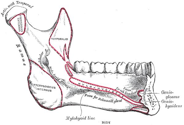 Mandible. Inner surface. Side view.
 -
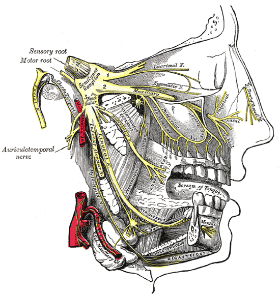 Distribution of the maxillary and mandibular nerves, and the submaxillary ganglion.
 -
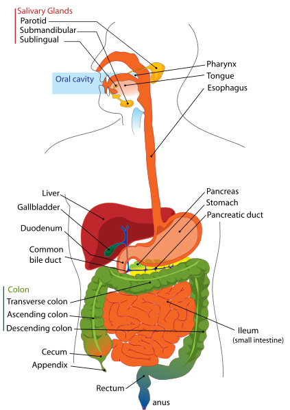 Digestive system
 -
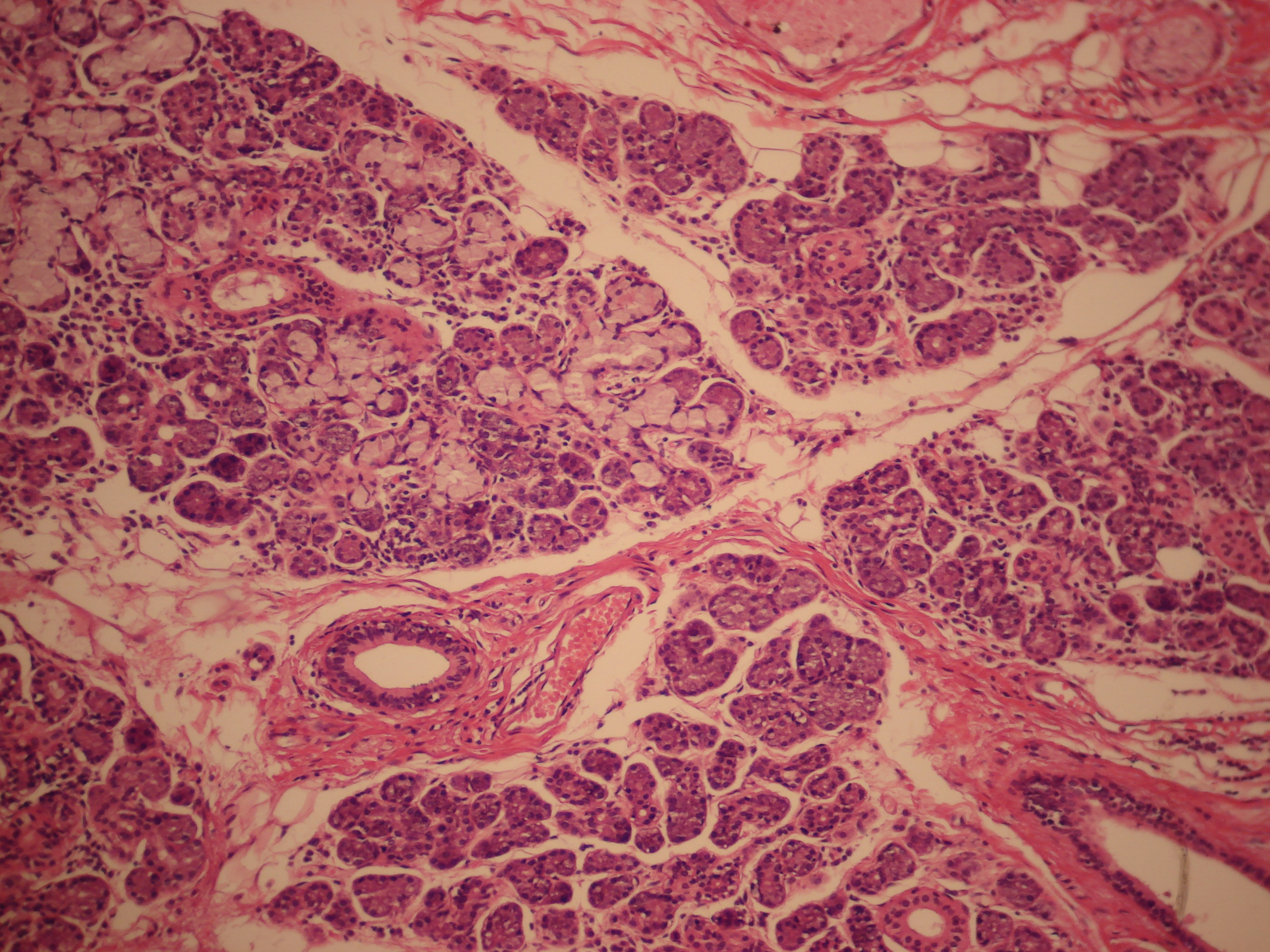 Mucus cell are identifiable by the lack of color in their cytoplasm, while serosal cells have a basophilic color.


References
References
- Douglas F. Paulsen (2000). Histology and cell biology (4th edition ed.). Stamford, Conn: Lange Medical Books/McGraw Hill. ISBN 0-8385-0593-7.
- A. R. Ten Cate (1998). Oral Histology: Development, Structure, and Function (5th edition ed.). Saint Louis: Mosby-Year Book.
External links
External links
- Histology at usc.edu
- Template:EMedicineDictionary
- Template:SUNYAnatomyLabs – “Anterior Triangle of the Neck: Nerves and Vessels of the Carotid Triangle”
- Template:SUNYAnatomyLabs – “Oral Cavity: The Submandibular Gland and Duct”
- Template:NormanAnatomy (Template:NormanAnatomyFig)
Template:Head and neck general
de:Glandula submandibularis
it:Ghiandola sottomandibolare
sr:Подвилична жлезда
Looking for the patient version?
© 2026 MyEClinic – IFTM Institut für Telematik in der Medizin GmbH