
Ebola
For patient information, click here.
Editor-In-Chief: C. Michael Gibson, M.S., M.D. [1]; Associate Editor(s)-in-Chief: Michael Maddaleni, B.S., Guillermo Rodriguez Nava, M.D. [2], Marjan Khan M.B.B.S.[3] Syed Hassan A. Kazmi BSc, MD [4]
Synonyms and keywords: Ebola hemorrhagic fever, Ebola haemorrhagic fever, Ebola HF, EHF, Zaire virus, Zaire Ebolavirus, ZEBOV, Sudan Ebolavirus, SEBOV, Reston Ebolavirus, REBOV, Cote d’Ivoire Ebolavirus, CIEBOV, Bundibugyo ebolavirus, Tai Forest ebolavirus
Overview
Editor-In-Chief: C. Michael Gibson, M.S., M.D. [1]; Associate Editor(s)-in-Chief: Guillermo Rodriguez Nava, M.D. [2] Alejandro Lemor, M.D. [3]
Overview
Ebola virus disease (EVD) is one of numerous viral hemorrhagic fevers (VHF). It is a severe, often fatal disease in human and nonhuman primates. Ebola virus is spread by direct contact with the blood or body fluids (such as urine, saliva, feces, vomit and semen) of an infected person or by being exposed to objects that have been contaminated with infected blood or body fluids. The incubation period is usually 8–10 days (rarely ranging from 2 to 21 days). Patients can transmit the virus once symptoms appear and through the later stages of disease, as well as postmortem. Ebola has caused a number of serious and highly publicized outbreaks since its discovery.[1]
Historical Perspective
The Ebola virus was named after the Ebola River Valley in the Democratic Republic of the Congo (formerly Zaïre), near the site of a 1976 outbreak at a mission run by Flemish nuns.[2] Since the initial discovery of the virus, five subtypes have subsequently been identified.
Classification
Ebola virus can be classified into 5 subtypes: Zaïre, Sudan, Reston, Tai (Ivory Coast) and Bundibugyo, according to the place of discovery.
Pathophysiology
The Ebola virus infects the mononuclear phagocyte system, but also other cells such as hepatocytes, spongiocytes, fibroblasts and endothelial cells, inducing tissue necrosis and disrupting the hematological and coagulation systems. The Ebola virus is transmitted by direct contact with infected patients or animals. The natural reservoir has not been identified.[3][4][5][6]
Causes
Ebola infection is caused by a virus that belongs to the family Filoviridae. Three viral subtypes have been reported to cause disease in humans: Ebola-Zaire virus, Ebola-Sudan virus, and Ebola-Ivory Coast virus. The human disease has so far been limited to parts of Africa. A very small number of people in the United States who were infected with the fourth type of the virus, known as Ebola Reston, did not develop any signs of disease.
Differentiating Ebola from other Diseases
Ebola must be differentiated from other diseases that cause hemorrhage and/or high fever as part of their presentation such as Marburg virus, Lassa fever, Typhoid fever and Malaria. The clinician must first rule out other more common causes of the fever before considering a viral hemorrhagic fever (VHF) such as Ebola, and the consideration of a VHF should be based upon epidemiology and demographics as well as sign and symptoms.[7] A VHF such as Ebola, should be suspected in febrile persons who, within 3 weeks before onset of fever, have either: 1) traveled in the specific local area of a country where VHF has recently occurred; 2) had direct unprotected contact with blood, other body fluids, secretions, or excretions of a person or animal with VHF; 3) if the patient had any contact with someone who was ill with fever and bleeding or who died from an unexplained illness with fever and bleeding; 4) had a possible exposure when working in a laboratory that handles hemorrhagic fever viruses; 5) If a fever continues after 3 days of empiric treatment, and if the patient has signs such as bleeding or shock, the clinician must consider a VHF; 6) if no other cause is found for the patient’s signs and symptoms, the clinician must suspect a VHF.
Epidemiology and Demographics
Outbreaks of Ebola have been generally restricted to Africa. Governments and individuals should quickly quarantine the area. Lack of roads and transportation help to contain the outbreak in remote areas. The potential for widespread Ebola virus disease epidemics is considered low due to the high case-fatality rate, the rapidity of demise of patients, and the often remote areas where infections occur.
Risk Factors
The main risk factors for Ebola virus disease are traveling to endemic areas, to be a health professional taking care of infected patients and researchers working with animal models of the Ebola virus disease.[8]
Natural History
Ebola infection rapidly progresses to death in the absence of supportive care. Ebola infection can be complicated by multiorgan failure and shock. The prognosis of Ebola infection is poor, and depends upon the Ebola virus strain. The Zaire Ebola virus has mortality rate as high as 90%.[8]
Diagnosis
History and Symptoms
Ebola causes a variety of symptoms which may include fever, chills vomiting, diarrhea, generalized pain or malaise, and sometimes internal and external bleeding, that follow an incubation period of 2-21 days. These symptoms are common to all species of Ebola virus, but the different species may present with differences in the severity of symptoms.
Physical Examination
Ebola is commonly associated with the acute onset of high fever, chills and hemorrhage as well as swollen joints, weakness, rash and red eyes.[9][10][11]
Laboratory Findings
Ebola infection is associated with nonspecific laboratory abnormalities including alterations in the white blood cell count, blood chemistry tests and liver function tests, all of which may contribute to a disruption in the clotting process and bleeding.
Other Diagnostic Studies
While the diagnosis of Ebola may be suspected based on clinical findings, the diagnosis of Ebola can be confirmed by antigen-capture enzyme-linked immunosorbent assay (ELISA) testing, IgM ELISA, polymerase chain reaction (PCR), and virus isolation, within few days of the onset of symptoms. Persons tested later in the course of the disease, or after recovery, can be tested for IgM and IgG antibodies. The disease can also be diagnosed in deceased patients by using immunohistochemistry testing, virus isolation, or PCR.[8]
Treatment
Medical Therapy
The treatment of Ebola infection is primarily supportive and includes maintaining fluids and electrolytes, homeostasis, adequate oxygen levels and blood pressure and treating any complicating superimposed infections.[8] All patients with a confirmed or suspected viral hemorrhagic fever should be put in isolation with adequate contact precautions.[12] No vaccine is currently available.
Primary Prevention
The transmission of Ebola can be limited by implementing preventive measures in both endemic and nonendemic areas which include isolation of infected patients; using gloves/masks/gowns and other standard barrier precautions; routine hand-washing; careful handling, disposal and/or maintenance of sharp objects; proper waste management and proper handling of human remains after death.
Future or Investigational Therapies
Although there is no effective human vaccine against Ebola currently available, there are promising results for antisense prevention therapies targeting the Ebola virus in monkey studies. Administration of an inhibitor of coagulation (rNAPc2) has demonstrated some benefit in monkey studies. There are non-conclusive results in human survivors from post-exposure vaccination, passive immunization with blood or serum or with recombinant human monoclonal antibodies.
References
- ↑ “Ebola Cases and Outbreaks – CDC Special Pathogens Branch”. Centers for Disease Control and Prevention. Retrieved 2007-12-08.
- ↑ Bardi, Jason Socrates (2002). “Death Called a River”. Scribbs Research Institute. 2 (1). Retrieved 2006-12-08.
- ↑ Ryabchikova E, Kolesnikova L, Smolina M, Tkachev V, Pereboeva L, Baranova S; et al. (1996). “Ebola virus infection in guinea pigs: presumable role of granulomatous inflammation in pathogenesis”. Arch Virol. 141 (5): 909–21. PMID 8678836.
- ↑ Bray M, Davis K, Geisbert T, Schmaljohn C, Huggins J (1998). “A mouse model for evaluation of prophylaxis and therapy of Ebola hemorrhagic fever”. J Infect Dis. 178 (3): 651–61. PMID 9728532.
- ↑ Connolly BM, Steele KE, Davis KJ, Geisbert TW, Kell WM, Jaax NK; et al. (1999). “Pathogenesis of experimental Ebola virus infection in guinea pigs”. J Infect Dis. 179 Suppl 1: S203–17. doi:10.1086/514305. PMID 9988186.
- ↑ Bray M, Hatfill S, Hensley L, Huggins JW (2001). “Haematological, biochemical and coagulation changes in mice, guinea-pigs and monkeys infected with a mouse-adapted variant of Ebola Zaire virus”. J Comp Pathol. 125 (4): 243–53. doi:10.1053/jcpa.2001.0503. PMID 11798241.
- ↑ “WHO Infection Control for Viral Haemorrhagic Fevers in the African Health Care Setting” (PDF).
- ↑ 8.0 8.1 8.2 8.3 “CDC Ebola Hemorrhagic Fever Information Packet” (PDF). April 2010.
- ↑ Feldmann, Heinz; Geisbert, Thomas W (2011). “Ebola haemorrhagic fever”. The Lancet. 377 (9768): 849–862. doi:10.1016/S0140-6736(10)60667-8. ISSN 0140-6736.
- ↑ Formenty, Pierre; Hatz, Christophe; Le Guenno, Bernard; Stoll, Agnés; Rogenmoser, Philipp; Widmer, Andreas (1999). “Human Infection Due to Ebola Virus, Subtype Côte d’Ivoire: Clinical and Biologic Presentation”. The Journal of Infectious Diseases. 179 (s1): S48–S53. doi:10.1086/514285. ISSN 0022-1899.
- ↑ Gradon J (2000). “An outbreak of Ebola virus: lessons for everyday activities in the intensive care unit”. Crit Care Med. 28 (1): 284–5. PMID 10667555.
- ↑ Feldmann H, Geisbert TW (2011). “Ebola haemorrhagic fever”. Lancet. 377 (9768): 849–62. doi:10.1016/S0140-6736(10)60667-8. PMC 3406178. PMID 21084112.
Historical Perspective
Editor-In-Chief: C. Michael Gibson, M.S., M.D. [1]; Associate Editor(s)-in-Chief: Michael Maddaleni, B.S.; Guillermo Rodriguez Nava, M.D. [2]; Yazan Daaboul, M.D.
Overview
Ebola virus was first discovered in 1976 when two simultaneous outbreaks occurred in Zaire and Sudan. The first description of Ebola virus was made by Ngoy Mushola in Yambuku, Zaire during the 1976 outbreak. During the outbreak, Peter Piot analyzed blood samples of an infected Belgian nun in Zaire and was the first to describe the virus morphology using electron microscopy. The Ebola virus was named after the Ebola River Valley in the Democratic Republic of the Congo (formerly Zaire).[1] Approximately 14 outbreaks of Ebola virus have been described since its discovery. The 2013-2014 outbreak marks the largest Ebola outbreak, involving Africa, Asia, Europe, and America and making the virus a worldwide disease.
Historical Perspective
Discovery
- The Ebola virus was first discovered in 1976 following two simultaneous outbreaks of Ebola hemorrhagic fever between June and November in Zaire and between August and November in Sudan.[2][3]
- Nurse Mayinga N’Seka, a nurse in Zaire, is thought to be the index case in the first recognized Ebola epidemic in 1976. She was believed to be the only patient infected via airborne transmission of the Ebola virus.
- The fist description of Ebola virus infection was made by Ngoy Mushola, who recorded the first case in Yambuku town in Zaire. In Dr. Mushola’s daily log, he stated
| “ | The illness is characterized by a high temperature of about 39 °C,hematemesis, bloody diarrhea, retrosternal abdominal pain, prostration with “heavy” articulations, and rapid evolution death after a mean of three days… | ” |
- During the outbreak, blood samples of infected Belgian nuns in Zaire were refrigerated in non-secure thermos and sent to Europe for analysis. Peter Piot was the first to analyze and describe Ebola virus morphology using electron microscopy. He noted the presence of long, worm-like agents that resemble the Marburg virus that was associated with the death of laboratory workers in Germany.
- The virus was then named after the Ebola river located in the town Yambuku, Democratic Republic of the Congo (formerly Zaire), which is the site of the first recognized Ebola outbreak.
- The first outbreaks occurred almost simultaneously in Sudan on June – November 1976 due to the so-called Sudan ebolavirus and in the Democratic Republic of Congo (formerly Zaire) on August – November 1976 due the so-called Ebola Zaire.
- Ever since the initial discovery, 5 strains of Ebola virus have been identified.
Spread
- The first two recorded outbreaks of Ebola virus occurred in 1976, followed by a third outbreak in 1979.[4]
- For 15 years, no outbreaks of Ebola virus were recorded, until Ebola re-emerged in 1994 when a Swiss ethnologist was infected during a chimpanzee autopsy in Tai National Park in Ivory Coast.[5][6] During the same period, 3 other outbreaks occurred in Mekouka, Mayibout, and Booue in Gabon between 1994 and 1997.[7][8][9]
- The period between 2000 and 2004 was remarkable for the emergence of multiple outbreaks among humans as well as among animals (gorillas and chimpanzees) in Gabon, the Republic of Congo, and Uganda.[10] At least 20 outbreaks were reported between the years 1976 (time of discovery) and 2013.
- In March 23 2014, the Ministry of Health of Guinea notified the World Health Organization (WHO) of an emerging outbreak. The outbreak rapidly evolved to become the largest Ebola outbreak since its discovery in 1976. The first case of the Ebola outbreak was reported in Guinea in December 2013. The index case was thought to be a 2-year-old boy. The 2014 outbreak then involved Liberia, Sierra Leone, Senegal, and Nigeria before the virus was spread to Europe, Asia, and America, making Ebola virus a worldwide threat. On August 8 2014, the WHO declared the Ebola epidemic to be a Public Health Emergency of International Concern (PHEIC).[11]
Major Outbreaks
Until recently, Ebola outbreaks have been restricted to Africa, with the exception of Reston ebolavirus. The International Committee on Taxonomy of Viruses currently recognizes four species of the Ebola: Zaire virus (ZEBOV), Sudan ebolavirus (SEBOV), Reston ebolavirus (REBOV), and Cote d’Ivoire ebolavirus (CIEBOV).
| Year(s) | Country | Ebola subtype | Reported number of human cases | Reported number (%) of deaths among cases | Situation |
| 1976 | Zaire (Democratic Republic of the Congo – DRC) | Ebola virus | 318 | 280 (88%) | Occurred in Yambuku and surrounding area. Disease was spread by close personal contact and by use of contaminated needles and syringes in hospitals/clinics. This outbreak was the first recognition of the disease. |
| 1976 | Sudan (South Sudan) | Sudan virus | 284 | 151 (53%) | Occurred in Nzara, Maridi and the surrounding area. Disease was spread mainly through close personal contact within hospitals. Many medical care personnel were infected. |
| 1976 | England | Sudan virus | 1 | 0 | Laboratory infection by accidental stick of contaminated needle. |
| 1977 | Zaire | Ebola virus | 1 | 1 (100%) | Noted retrospectively in the village of Tandala. |
| 1979 | Sudan (South Sudan) | Sudan virus | 34 | 22 (65%) | Occured in Nzara, Maridi. Recurrent outbreak at the same site as the 1976 Sudan epidemic. |
| 1989 | USA | Reston virus | 0 | 0 | Ebola-Reston virus was introduced into quarantine facilities in Virginia and Pennsylvania by monkeys imported from the Philippines. |
| 1990 | USA | Reston virus | 4 (asymptomatic) | 0 | Ebola-Reston virus was introduced once again into quarantine facilities in Virginia, and Texas by monkeys imported from the Philippines. Four humans developed antibodies but did not get sick. |
| 1989-1990 | Philippines | Reston virus | 3 (asymptomatic) | 0 | High mortality among cynomolgus macaques in a primate facility responsible for exporting animals in the USA. Three workers in the animal facility developed antibodies but did not get sick. |
| 1992 | Italy | Reston virus | 0 | 0 | Ebola-Reston virus was introduced into quarantine facilities in Sienna by monkeys imported from the same export facility in the Philippines that was involved in the episodes in the United States. No humans were infected. |
| 1994 | Gabon | Ebola virus | 52 | 31 (60%) | Occured in Mékouka and other gold-mining camps deep in the rain forest. Initially thought to be yellow fever; identified as Ebola hemorrhagic fever in 1995. |
| 1994 | Ivory Coast | Taï Forest virus | 1 | 0 | Scientist became ill after conducting an autopsy on a wild chimpanzee in the Tai Forest. The patient was treated in Switzerland. |
| 1995 | Democratic Republic of the Congo (formerly Zaire) | Ebola virus | 315 | 250 (81%) | Occured in Kikwit and surrounding area. Traced to index case-patient who worked in forest adjoining the city. Epidemic spread through families and hospitals. |
| 1996 (January-April) | Gabon | Ebola virus | 37 | 21 (57%) | Occured in Mayibout area. A chimpanzee found dead in the forest was eaten by people hunting for food. Nineteen people who were involved in the butchery of the animal became ill; other cases occured in family members. |
| 1996-1997 (July-January) | Gabon | Ebola virus | 60 | 45 (74%) | Occurred in Booué area with transport of patients to Libreville. Index case-patient was a hunter who lived in a forest camp. Disease was spread by close contact with infected persons. A dead chimpanzee found in the forest at the time was determined to be infected. |
| 1996 | South Africa | Ebola virus | 2 | 1 (50%) | A medical professional traveled from Gabon to Johannesburg, South Africa, after having treated Ebola virus-infected patients and thus having been exposed to the virus. He was hospitalized, and a nurse who took care of him became infected and died. |
| 1996 | USA | Reston virus | 0 | 0 | Ebola-Reston virus was introduced into a quarantine facility in Texas by monkeys imported from the Philippines. No human infections were identified. |
| 1996 | Philippines | Reston virus | 0 | 0 | Ebola-Reston virus was identified in a mokey export facility in the Philippines. No human infections were identified. |
| 1996 | Russia | Ebola virus | 1 | 1 (100%) | Laboratory contamination |
| 2000-2001 | Uganda | Sudan virus | 425 | 224 (53%) | Occurred in Gulu, Masindi, and Mbarara districts of Uganda. The three most important risks associated with Ebola virus infection were attending funerals of Ebola hemorrhagic fever case-patients, having contact with case-patients in one’s family, and providing medical care to Ebola case-patients without using adequate personal protective measures. |
| October 2001-March 2002 | Gabon | Ebola virus | 65 | 53 (82%) | Outbreak occured over the border of Gabon and the Republic of the Congo. |
| October 2001-March 2002 | Republic of Congo | Ebola virus | 57 | 43 (75%) | Outbreak occurred over the border of Gabon and the Republic of the Congo. This was the first time that Ebola hemorrhagic fever was reported in the Republic of the Congo. |
| December 2002-April 2003 | Republic of Congo | Ebola virus | 143 | 128 (89%) | Outbreak occurred in the districts of Mbomo and Kéllé in Cuvette Ouest Département. |
| November-December 2003 | Republic of Congo | Ebola virus | 35 | 29 (83%) | Outbreak occured in Mbomo and Mbandza villages located in Mbomo distric, Cuvette Ouest Département. |
| 2004 | Sudan (South Sudan) | Sudan virus | 17 | 7 (41%) | Outbreak occurred in Yambio county of southern Sudan. This outbreak was concurrent with an outbreak of measles in the same area, and several suspected EHF cases were later reclassified as measeles cases. |
| 2004 | Russia | Ebola virus | 1 | 1 (100%) | Laboratory contamination. |
| 2007 | Democratic Republic of Congo | Ebola virus | 264 | 187 (71%) | Outbreak occurred in Kasai Occidental Province. The outbreak was declared over November 20. Last confirmed case on October 4 and last death on October 10. |
| December 2007-January 2008 | Uganda | Bundibugyo virus | 149 | 37 (25%) | Outbreak occurred in Bundibugyo District in western Uganda. First reported occurance of a new strain. |
| November 2008 | Philippines | Reston virus | 6 (asymptomatic) | 0 | First known occurrence of Ebola-Reston in pigs. Strain closely similar to earlier strains. Six workers from the pig farm and slaughterhouse developed antibodies but did not become sick. |
| December 2008-February 2009 | Democratic Republic of the Congo | Ebola virus | 32 | 15 (47%) | Outbreak occurred in the Mweka and luebo health zones of the Province of Kasai Occidental. |
| May-11 | Uganda | Sudan virus | 1 | 1 (100%) | The Ugandan Ministry of Health informed the public that a patient with suspected Ebola Hemorrhagic fever died on May 6, 2011 in the Luwero district, Uganda. The quick diagnosis from a blood sample of Ebola virus was provided by the new CDC Viral Hemorrhagic Fever laboratory installed at the Uganda Viral Research Institute (UVRI). |
| June-October 2012 | Uganda | Sudan virus | 11* | 4* (36.4%) | Outbreak occurred in the Kibaale District of Uganda. Laboratory tests of blood samples were conducted by the UVRI and the U.S. Centers for Disease Control and Prevention (CDC). |
| June-November 2012 | Democratic Republic of the Congo | Bundibugyo virus | 36* | 13* (36.1%) | Outbreak occurred in DRC’s Province Orientale. Laboratory support was provided through CDC and the Public Health Agency of Canada (PHAC)’s field laboratory in Isiro, and through the CDC/UVRI lab in Uganda. The outbreak in DRC has no epidemiologic link to the near contemporaneous Ebola outbreak in the Kibaale district of Uganda. |
| November 2012-January 2013 | Uganda | Sudan virus | 6* | 3* (50%) | Outbreak occurred in the Luwero District. CDC assisted the Ministry of Health in the epidemiologic and diagnostic aspects of the outbreak. Testing of samples by CDC’s Viral Special Pathogens Branch occurred at UVRI in Entebbe. |
| March 2014-Present | Guinea, Liberia, and Sierra Leone | Ebola virus | 9936 | 4878 (49.1%) | Outbreak across Guinea, northern Liberia, and now eastern Sierra Leone. |
| May 2017 | Democratic Republic of the Congo | Not confirmed | 9 | 1 (1.1%) | In May 12, 2017, WHO declared a lab confirmed case in Bas-Uele region in the northeast Congo. Nine cases were hospitalized for hemorrhagic fever and three of them died. Only one case was confirmed to have Ebola virus.
Despite being a serious situation, it’s considered a good sign that the outbreak struck in a remote and forested region |
References
- ↑ Bardi, Jason Socrates (2002). “Death Called a River”. Scribbs Research Institute. 2 (1). Retrieved 2006-12-08.
- ↑ “Ebola haemorrhagic fever in Zaire, 1976”. Bull World Health Organ. 56 (2): 271–93. 1978. PMC 2395567. PMID 307456.
- ↑ “Ebola haemorrhagic fever in Sudan, 1976. Report of a WHO/International Study Team”. Bull World Health Organ. 56 (2): 247–70. 1978. PMC 2395561. PMID 307455.
- ↑ Baron RC, McCormick JB, Zubeir OA (1983). “Ebola virus disease in southern Sudan: hospital dissemination and intrafamilial spread”. Bull World Health Organ. 61 (6): 997–1003. PMC 2536233. PMID 6370486.
- ↑ Le Guenno B, Formenty P, Formentry P, Wyers M, Gounon P, Walker F; et al. (1995). “Isolation and partial characterisation of a new strain of Ebola virus”. Lancet. 345 (8960): 1271–4. PMID 7746057.
- ↑ Formenty P, Hatz C, Le Guenno B, Stoll A, Rogenmoser P, Widmer A (1999). “Human infection due to Ebola virus, subtype Côte d’Ivoire: clinical and biologic presentation”. J Infect Dis. 179 Suppl 1: S48–53. doi:10.1086/514285. PMID 9988164.
- ↑ Khan AS, Tshioko FK, Heymann DL, Le Guenno B, Nabeth P, Kerstiëns B; et al. (1999). “The reemergence of Ebola hemorrhagic fever, Democratic Republic of the Congo, 1995. Commission de Lutte contre les Epidémies à Kikwit”. J Infect Dis. 179 Suppl 1: S76–86. doi:10.1086/514306. PMID 9988168.
- ↑ Georges AJ, Leroy EM, Renaut AA, Benissan CT, Nabias RJ, Ngoc MT; et al. (1999). “Ebola hemorrhagic fever outbreaks in Gabon, 1994-1997: epidemiologic and health control issues”. J Infect Dis. 179 Suppl 1: S65–75. doi:10.1086/514290. PMID 9988167.
- ↑ Amblard J, Obiang P, Edzang S, Prehaud C, Bouloy M, Guenno BL (1997). “Identification of the Ebola virus in Gabon in 1994”. Lancet. 349 (9046): 181–2. doi:10.1016/S0140-6736(05)60984-1. PMID 9111553.
- ↑ Pourrut X, Kumulungui B, Wittmann T, Moussavou G, Délicat A, Yaba P; et al. (2005). “The natural history of Ebola virus in Africa”. Microbes Infect. 7 (7–8): 1005–14. doi:10.1016/j.micinf.2005.04.006. PMID 16002313.
- ↑ Briand S, Bertherat E, Cox P, Formenty P, Kieny MP, Myhre JK; et al. (2014). “The international Ebola emergency”. N Engl J Med. 371 (13): 1180–3. doi:10.1056/NEJMp1409858. PMID 25140855.
- ↑ “CDC Chronology of Ebola Hemorrhagic Fever Outbreaks”.
Classification
Editor-In-Chief: C. Michael Gibson, M.S., M.D. [1]; Associate Editor(s)-in-Chief: Guillermo Rodriguez Nava, M.D. [2]; João André Alves Silva, M.D. [3]
Overview
Ebola virus can be classified into 5 subtypes: Zaïre, Sudan, Reston, Tai (Ivory Coast) and Bundibugyo, according to the place of discovery.
Classification
The table below summarizes the ebola virus strains identified until now:
| Ebola Virus Strain | Place of Discovery | Date Identified |
|---|---|---|
| Sudan ebolavirus | Nzara and Maridi, Sudan | Between June and November 1976 |
| Zaire ebolavirus | Yambuku, Democratic Republic of the Congo | Between August and November 1976 |
| Reston ebolavirus | Reston, Virginia, US. | November 1989 |
| Ivory Coast ebolavirus | Tai Forest, Ivory Coast. | November 1994 |
| Bundibugyo ebolavirus | Bundibugyo District, Uganda | November 2007 |
Zaïre Ebolavirus
- Among the five strains, Zaïre Ebolavirus carries the highest mortality rate.
- A virus of the genus Ebolavirus is considered member of the species Zaire ebolavirus if:[1]
- It is found in the Democratic Republic of the Congo, Gabon, or the Republic of the Congo.
- It has a genome with two or three gene overlaps (VP35/VP40, GP/VP30, VP24/L).
- It has a genomic sequence that differs from the virus type by less than 30%.
Sudan Ebolavirus
- A virus of the genus Ebolavirus is considered member of the species Sudan ebolavirus if:[1]
Reston Ebolavirus
- A virus of the genus Ebolavirus is considered member of the species Reston ebolavirus if:[1]
- If its genome diverges from that of the prototype Reston virus, the Reston virus variant Pennsylvania, by ≤10% at the nucleotide level.
Tai (Ivory Coast) Ebolavirus
- A virus of the genus Ebolavirus is considered member of the species Tai Forest ebolavirus if:[1]
Bundibugyo Ebolavirus
- A virus of the genus Ebolavirus is considered member of the species Bundibugyo ebolavirus if:[1]
- It is endemic in Uganda.
- It has a genome with three gene overlaps (VP35/VP40, GP/VP30, VP24/L).
- It has a genomic sequence different from Ebola virus by ≥30%, but different from that of Bundibugyo virus by <30%.
References
- ↑ 1.0 1.1 1.2 1.3 1.4 Kuhn, Jens H.; Becker, Stephan; Ebihara, Hideki; Geisbert, Thomas W.; Johnson, Karl M.; Kawaoka, Yoshihiro; Lipkin, W. Ian; Negredo, Ana I.; Netesov, Sergey V.; Nichol, Stuart T.; Palacios, Gustavo; Peters, Clarence J.; Tenorio, Antonio; Volchkov, Viktor E.; Jahrling, Peter B. (2010). “Proposal for a revised taxonomy of the family Filoviridae: classification, names of taxa and viruses, and virus abbreviations”. Archives of Virology. 155 (12): 2083–2103. doi:10.1007/s00705-010-0814-x. ISSN 0304-8608.
Pathophysiology
Editor-In-Chief: C. Michael Gibson, M.S., M.D. [1]; Associate Editor(s)-in-Chief: Michael Maddaleni, B.S.; Guillermo Rodriguez Nava, M.D. [2]
Overview
The Ebola virus infects the mononuclear phagocyte system, but also other cells such as hepatocytes, spongiocytes, fibroblasts and endothelial cells, inducing tissue necrosis and disrupting the hematological and coagulation systems. The Ebola virus is transmitted by direct contact with infected patients or animals. The natural reservoir has not been identified.[1][2][3][4]
Pathophysiology
Tropism
- Ebola virus infects mainly the cells of the mononuclear phagocyte system, but also fibroblasts, hepatocytes, spongiocytes, adrenal cortical cells and endothelial cells.[1]
- The infection of the mononuclear phagocyte system cells plays a key role in the pathogenesis and spread of the disease as they carry the virus from of the initial infection site, through the lymphatic system and blood, to the regional lymph nodes,spleen and liver.[5]
- The next table summarizes the pathogenesis of the disease according to the virus tropism.
| Organ/Tissue | Effect |
|---|---|
| Endothelial cells | Glycoprotein (GP) on the virion envelope allows introduction of its content into the endothelial cells, which induces a cytopathic effect and damage to the endothelial barrier function that, together with effects of TNF-α released by infected mononuclear cells, leads to the loss of vascular integrity and increased leakage. |
| Liver | Causes hepatocellular necrosis which could impair the synthesis of proteins of the coagulation system[5] |
| Adrenal cortex | Affects the synthesis of enzymes responsible for the synthesis of steroids, leading to hypotension, and fluid and electrolytes disturbances.[5] |
| Lymphatic system | Necrosis of the spleen, lymph nodes and thymus; Apoptosis of lymphocytes leading to lymphopenia.[5][6] |
Immune response
- The virus activates the macrophages synthesis of interleukins (IL), which leads the Th1/Th2 balance towards a more pronounced Th1–cell mediated response.[7]
- Some inflammatory mediators produced during the ebola virus infection include: interferon (IFN)-alpha, IFN-beta, IL-2, IL-6, IL-8, IL-10, interferon-inducible protein 10; monocyte chemoattractant protein 1; regulated upon activation normal T cell expressed and secreted (RANTES); TNF-alpha; and reactive oxygen and nitrogen species.[8][9][10]
- Some viral proteins, such as VP35 and VP24, block the type I interferon response, which plays a key role of the pathogenesis of the disease.[11]
- The reactive oxygen and nitrogen species contribute to the cell and tissue damage, and therefore vascular and organ damage.[12]
- The nitric oxide is known to be an important vasodilator, therefore it plays and important role in the development of hypotension and shock.
Coagulation system
- Ebola infection is associated with hemorrhage in 50% of patients.
- Alterations of the coagulation system are induced by the ebola virus, and are thought to be mediated by the production of tissue factor:[13]
- Consumption of clotting factors
- Increased concentrations of fibrin degradation products
- Disseminated intravascular coagulopathy
Understanding the immune response to the disease
In one case study performed on an individual who had severe Ebola virus infection in Sierra Leone to further understand the immune response during all the stages of the disease.[14] The individual was provided with supportive care only without any other experimental therapies. Daily global gene expression in peripheral white blood cells was recorded and correlated with many clinical and laboratory aspects during the course of disease (Viral load, multiple organ dysfunction, coagulopathy, etc) till complete recovery after 33 days.
The study enabled us to identify the host responses (the genomic shift between increased expression of genes involved in inflammation and cell destruction to increased expression of genes promoting cell repair) that correlate with the viral clearance and recovery.
The study revealed that many changes in gene expression precede the change in the clinical status of the patient emphasizing the role of viral clearance in the cure of systemic illness.
References
- ↑ 1.0 1.1 Ryabchikova E, Kolesnikova L, Smolina M, Tkachev V, Pereboeva L, Baranova S; et al. (1996). “Ebola virus infection in guinea pigs: presumable role of granulomatous inflammation in pathogenesis”. Arch Virol. 141 (5): 909–21. PMID 8678836.
- ↑ Bray M, Davis K, Geisbert T, Schmaljohn C, Huggins J (1998). “A mouse model for evaluation of prophylaxis and therapy of Ebola hemorrhagic fever”. J Infect Dis. 178 (3): 651–61. PMID 9728532.
- ↑ Connolly BM, Steele KE, Davis KJ, Geisbert TW, Kell WM, Jaax NK; et al. (1999). “Pathogenesis of experimental Ebola virus infection in guinea pigs”. J Infect Dis. 179 Suppl 1: S203–17. doi:10.1086/514305. PMID 9988186.
- ↑ Bray M, Hatfill S, Hensley L, Huggins JW (2001). “Haematological, biochemical and coagulation changes in mice, guinea-pigs and monkeys infected with a mouse-adapted variant of Ebola Zaire virus”. J Comp Pathol. 125 (4): 243–53. doi:10.1053/jcpa.2001.0503. PMID 11798241.
- ↑ 5.0 5.1 5.2 5.3 Geisbert TW, Hensley LE, Larsen T, Young HA, Reed DS, Geisbert JB; et al. (2003). “Pathogenesis of Ebola hemorrhagic fever in cynomolgus macaques: evidence that dendritic cells are early and sustained targets of infection”. Am J Pathol. 163 (6): 2347–70. doi:10.1016/S0002-9440(10)63591-2. PMC 1892369. PMID 14633608.
- ↑ Zaki SR, Goldsmith CS (1999). “Pathologic features of filovirus infections in humans”. Curr Top Microbiol Immunol. 235: 97–116. PMID 9893381.
- ↑ Qiu X, Audet J, Wong G, Fernando L, Bello A, Pillet S; et al. (2013). “Sustained protection against Ebola virus infection following treatment of infected nonhuman primates with ZMAb”. Sci Rep. 3: 3365. doi:10.1038/srep03365. PMC 3842534. PMID 24284388.
- ↑ Villinger F, Rollin PE, Brar SS, Chikkala NF, Winter J, Sundstrom JB; et al. (1999). “Markedly elevated levels of interferon (IFN)-gamma, IFN-alpha, interleukin (IL)-2, IL-10, and tumor necrosis factor-alpha associated with fatal Ebola virus infection”. J Infect Dis. 179 Suppl 1: S188–91. doi:10.1086/514283. PMID 9988183.
- ↑ Hensley LE, Young HA, Jahrling PB, Geisbert TW (2002). “Proinflammatory response during Ebola virus infection of primate models: possible involvement of the tumor necrosis factor receptor superfamily”. Immunol Lett. 80 (3): 169–79. PMID 11803049.
- ↑ Baize S, Leroy EM, Georges AJ, Georges-Courbot MC, Capron M, Bedjabaga I; et al. (2002). “Inflammatory responses in Ebola virus-infected patients”. Clin Exp Immunol. 128 (1): 163–8. PMC 1906357. PMID 11982604.
- ↑ Basler CF, Mikulasova A, Martinez-Sobrido L, Paragas J, Mühlberger E, Bray M; et al. (2003). “The Ebola virus VP35 protein inhibits activation of interferon regulatory factor 3”. J Virol. 77 (14): 7945–56. PMC 161945. PMID 12829834.
- ↑ Sanchez A, Lukwiya M, Bausch D, Mahanty S, Sanchez AJ, Wagoner KD; et al. (2004). “Analysis of human peripheral blood samples from fatal and nonfatal cases of Ebola (Sudan) hemorrhagic fever: cellular responses, virus load, and nitric oxide levels”. J Virol. 78 (19): 10370–7. doi:10.1128/JVI.78.19.10370-10377.2004. PMC 516433. PMID 15367603.
- ↑ Geisbert TW, Young HA, Jahrling PB, Davis KJ, Kagan E, Hensley LE (2003). “Mechanisms underlying coagulation abnormalities in ebola hemorrhagic fever: overexpression of tissue factor in primate monocytes/macrophages is a key event”. J Infect Dis. 188 (11): 1618–29. doi:10.1086/379724. PMID 14639531.
- ↑ “Longitudinal peripheral blood transcriptional analysis of a patient with severe Ebola virus disease | Science Translational Medicine”.
Causes
Editor-In-Chief: C. Michael Gibson, M.S., M.D. [1]
Overview
Ebola infection is caused by the Ebola virus that belongs to the family Filoviridae. Four viral subtypes have been reported to cause clinical illness in humans: Bundibugyo ebolavirus, Sudan ebolavirus, Tai Forest ebolavirus, and Zaire ebolavirus.
Taxonomy
Viruses; ssRNA viruses; ssRNA negative-strand viruses; Mononegavirales; Filoviridae; Ebolavirus[1]
- Ebolavirus
- Bundibugyo ebolavirus
- Reston ebolavirus
- Reston ebolavirus – Reston
- Reston ebolavirus – Reston (1989)
- Reston ebolavirus – Siena/Philippine-92
- Sudan ebolavirus
- Sudan ebolavirus – Boniface (1976)
- Sudan ebolavirus – Maleo (1979)
- Sudan ebolavirus – Nakisamata
- Sudan ebolavirus – Uganda (2000)
- Tai Forest ebolavirus
- Tai Forest virus – Côte d’Ivoire, Côte d’Ivoire, 1994
- Zaire ebolavirus
- Ebola virus – Mayinga, Zaire, 1976
- Zaire ebolavirus – Eckron (Zaire, 1976)
- Zaire ebolavirus – Gabon (1994-1997)
- Zaire ebolavirus – Zaire (1995)
- Unclassified Ebolavirus
- Ebola virus Yambio0401
- Ebola virus Yambio0402
- Ebola virus Yambio0403
- Ebola virus sp.
Virology
Structure
Electron micrographs of Ebola virus demonstrates the characteristic thread-like structure of a filovirus.[2] EBOV VP30 is around 288 amino acids long.[2] The virions are tubular and variable in shape and may resemble a “U”, “6”, coiled, circular, or branched shape. It is important to note that laboratory purification techniques, such as centrifugation, may contribute to the various shapes.[2] Virions are generally 80 nm in diameter.[2] They are variable in length, and can be up to 1400 nm long. On average, however, the length of a typical Ebola virus is closer to 1000 nm. In the center of the virion is a structure called nucleocapsid, which is formed of the helically wound viral genomic RNA complexed to the proteins NP, VP35, VP30 and L. The virus has an average diameter of 40-50 nm and contains a central channel of 20–30 nm in diameter. Virally encoded glycoprotein (GP) spikes 10 nm long and 10 nm apart are present on the outer viral envelope of the virion, which is derived from the host cell membrane. Between the envelope and the nucleocapsid exists a matrix space that contains the viral proteins VP40 and VP24.
Genome
Each virion contains one minor molecule of linear, single-stranded, negative-sense RNA, totaling 18959 to 18961 nucleotides in length. The 3′ terminus is not polyadenylated and the 5′ end is not capped. Only 472 nucleotides from the 3′ end and 731 nucleotides from the 5′ end are sufficient for replication.[2] The genome codes for seven structural proteins and one non-structural protein. The gene order is as follows: 3′ – leader – NP – VP35 – VP40 – GP/sGP – VP30 – VP24 – L – trailer – 5′. Leader and trailer regions are non-transcribed regions that carry important signals to control transcription, replication, and packaging of the viral genomes into new virions. The genomic material by itself is not infectious, because viral proteins such as RNA-dependent RNA polymerase are necessary for viral transcription and replication.
Life Cycle
- The virus attaches to host receptors through the GP (glycoprotein) surface peplomer and is endocytosed into vesicles in the host cell.
- Fusion of virus membrane with the vesicle membrane occurs; nucleocapsid is released into the cytoplasm.
- The encapsidated, negative-sense genomic ssRNA is used as a template for the synthesis (3′-5′) of polyadenylated, monocistronic mRNAs using the viral RNA-dependent RNA polymerase.
- Translation of the mRNA into viral proteins occurs using the host cell’s machinery.
- Post-translational processing of viral proteins occurs. GP0 (glycoprotein precursor) is cleaved to GP1 and GP2, which are heavily glycosylated. These two molecules assemble, first into heterodimers, and then into trimers to give the surface peplomers. SGP (secreted glycoprotein) precursor is cleaved to SGP and delta peptide, both of which are released from the cell.
- As viral protein levels rise, a switch occurs from translation to replication. Using the negative-sense genomic RNA as a template, a complementary positive-sense ssRNA is synthesized; this is then used as a template for the synthesis of new genomic negative-sense ssRNA, which is rapidly encapsidated.
- The newly-formed nucleocapsides and envelope proteins associate at the host cell’s plasma membrane; budding occurs, and the virions are released.
Transmission
- Among humans, the virus is transmitted by direct contact with infected body fluids, or to a lesser extent, skin or mucous membrane contact. The incubation period can be anywhere from 2 to 21 days, but is generally between 5 and 10 days.
- Human-to-human airborne transmission has not been reported in any reported epidemics.
- The infection of human cases with Ebola virus has been documented through the handling of infected chimpanzees, gorillas, and forest antelopes – both dead and alive – as was documented in Côte d’Ivoire, the Republic of Congo and Gabon.
- So far, all epidemics of Ebola have occurred in sub-optimal hospital conditions, where practices of basic hygiene and sanitation are often either luxuries or unknown to caretakers and where disposable needles and autoclaves are unavailable or too expensive. In modern hospitals with disposable needles and knowledge of basic hygiene and barrier nursing techniques, Ebola has never spread on such a large scale.
- In the early stages, particularly when the patient is asymptomatic, Ebola is not generally contagious. Contact with someone in early stages very rarely transmits the disease. As the illness progresses, bodily fluids from diarrhea, vomiting, and bleeding represent an extreme biohazard.
- Other than samples that are grossly contaminated with body fluids, Ebola virus is rarely detected on environmental surfaces and thus transmission by fomites is not very common. However, given that the infectious dose of the virus is low, and that current evaluation techniques such as cell culture and RT-PCR have not been well validated for environmental detection (unknown sensitivity and specificity), the true risk of transmission by contaminated surfaces is unknown.[3]
Transmission in Recovering Patients and Continued Shedding
- Ebola virus has been isolated in the semen of recovering patients 40 days after the onset of illness. This stresses the risk of sexual transmission during the recovery phase. The Zaire Ebola virus in particular has a higher risk of continued shedding in the semen. It has been detected in samples for up to 90 days after the onset of illness.
- Continued shedding may be detected in many bodily fluids with varying durations. The only bodily fluid that does not contain Ebola virus, even during the symptomatic phase of the infection, is urine. This is probably related to the inability of the kidney to filter the Ebola virus particles.
- It is generally recommended to abstain from sex or to use condoms for up to 3 months after the onset of illness. It is also recommended to avoid breastfeeding and contact with the mucous membranes of the eye for the same amount of time.[3]
Viral Reservoirs
Despite numerous studies, the wildlife reservoir of Ebolavirus has not yet been identified. Between 1976 and 1998, 30,000 mammals, birds, reptiles, amphibians, and arthropods were sampled from outbreak regions with no virus detected.[4] Ebolavirus has been detected in the carcasses of gorillas, chimpanzees, and duikers during outbreaks in 2001 and 2003 and these carcasses were deemed to be the source of initial human infection. However given the high mortality in these species, they are unlikely to be able to act as reservoirs.[4] Plants, arthropods, and birds have also been considered as reservoirs, however bats are considered the most likely candidate[5].
Bats
Bats were known to reside in the cotton factory in which the index cases for the 1976 and 1979 outbreaks were employed.[4] Of 24 plant species and 19 vertebrate species experimentally inoculated with Ebolavirus, only bats became infected.[6] The absence of clinical signs in these bats is characteristic of a reservoir species. In 2002-03, a survey of 1,030 animals from Gabon and the Republic of the Congo including 679 bats found Ebolavirus RNA in 13fruit bats (Hyspignathus monstrosus, Epomops franquetti and Myonycteris torquata).[7] Bats are also known to be the reservoirs for a number of related viruses including Nipah virus, Hendra virus andlyssaviruses.
Microscopic Pathology
The images below display key features of the Ebola virus.
-
![This transmission electron micrograph (TEM) demonstrates the ultrastructural morphology displayed by an Ebola virus. Source: CDC microbiologist Frederick A. Murphy.Adapted from Public Health Image Library (PHIL), Centers for Disease Control and Prevention.[8]](https://www.wikidoc.org/images/9/9d/Ebola_virus2.png) This transmission electron micrograph (TEM) demonstrates the ultrastructural morphology displayed by an Ebola virus. Source: CDC microbiologist Frederick A. Murphy.Adapted from Public Health Image Library (PHIL), Centers for Disease Control and Prevention.[8]
This transmission electron micrograph (TEM) demonstrates the ultrastructural morphology displayed by an Ebola virus. Source: CDC microbiologist Frederick A. Murphy.Adapted from Public Health Image Library (PHIL), Centers for Disease Control and Prevention.[8] -
![This transmission electron micrograph (TEM) demonstrates the ultrastructural morphologic changes in this tissue sample isolate.Adapted from Public Health Image Library (PHIL), Centers for Disease Control and Prevention.[8]](https://www.wikidoc.org/images/7/78/Ebola_virus1.png) This transmission electron micrograph (TEM) demonstrates the ultrastructural morphologic changes in this tissue sample isolate.Adapted from Public Health Image Library (PHIL), Centers for Disease Control and Prevention.[8]
This transmission electron micrograph (TEM) demonstrates the ultrastructural morphologic changes in this tissue sample isolate.Adapted from Public Health Image Library (PHIL), Centers for Disease Control and Prevention.[8] -
![Scanning electron micrograph (SEM) revealing ultrastructural morphologic features of the Ebola virus from the Ivory Coast of Africa.Adapted from Public Health Image Library (PHIL), Centers for Disease Control and Prevention.[8]](https://www.wikidoc.org/images/c/c4/Ebola_virus3.png) Scanning electron micrograph (SEM) revealing ultrastructural morphologic features of the Ebola virus from the Ivory Coast of Africa.Adapted from Public Health Image Library (PHIL), Centers for Disease Control and Prevention.[8]
Scanning electron micrograph (SEM) revealing ultrastructural morphologic features of the Ebola virus from the Ivory Coast of Africa.Adapted from Public Health Image Library (PHIL), Centers for Disease Control and Prevention.[8] -
![Negatively-stained transmission electron micrograph (TEM) demonstrating the ultrastructural curvilinear morphologic features displayed by the Ebola virus from the Ivory Coast of Africa.Adapted from Public Health Image Library (PHIL), Centers for Disease Control and Prevention.[8]](https://www.wikidoc.org/images/2/2e/Ebola_virus4.png) Negatively-stained transmission electron micrograph (TEM) demonstrating the ultrastructural curvilinear morphologic features displayed by the Ebola virus from the Ivory Coast of Africa.Adapted from Public Health Image Library (PHIL), Centers for Disease Control and Prevention.[8]
Negatively-stained transmission electron micrograph (TEM) demonstrating the ultrastructural curvilinear morphologic features displayed by the Ebola virus from the Ivory Coast of Africa.Adapted from Public Health Image Library (PHIL), Centers for Disease Control and Prevention.[8]
![This transmission electron micrograph (TEM) demonstrates the ultrastructural morphology displayed by an Ebola virus. Source: CDC microbiologist Frederick A. Murphy.Adapted from Public Health Image Library (PHIL), Centers for Disease Control and Prevention.[8]](https://www.wikidoc.org/index.php/File%3AEbola_virus2.png)
![This transmission electron micrograph (TEM) demonstrates the ultrastructural morphologic changes in this tissue sample isolate.Adapted from Public Health Image Library (PHIL), Centers for Disease Control and Prevention.[8]](https://www.wikidoc.org/index.php/File%3AEbola_virus1.png)
![Scanning electron micrograph (SEM) revealing ultrastructural morphologic features of the Ebola virus from the Ivory Coast of Africa.Adapted from Public Health Image Library (PHIL), Centers for Disease Control and Prevention.[8]](https://www.wikidoc.org/index.php/File%3AEbola_virus3.png)
![Negatively-stained transmission electron micrograph (TEM) demonstrating the ultrastructural curvilinear morphologic features displayed by the Ebola virus from the Ivory Coast of Africa.Adapted from Public Health Image Library (PHIL), Centers for Disease Control and Prevention.[8]](https://www.wikidoc.org/index.php/File%3AEbola_virus4.png)
Gallery
-
![Produced by the National Institute of Allergy and Infectious Diseases (NIAID), this digitally-colorized scanning electron micrograph (SEM) depicts numerous string-like Ebola virus particles as they were in the process of being shed from an infected cell. From Public Health Image Library (PHIL). [8]](https://www.wikidoc.org/images/3/3e/Ebolavirus10.jpeg) Produced by the National Institute of Allergy and Infectious Diseases (NIAID), this digitally-colorized scanning electron micrograph (SEM) depicts numerous string-like Ebola virus particles as they were in the process of being shed from an infected cell. From Public Health Image Library (PHIL). [8]
Produced by the National Institute of Allergy and Infectious Diseases (NIAID), this digitally-colorized scanning electron micrograph (SEM) depicts numerous string-like Ebola virus particles as they were in the process of being shed from an infected cell. From Public Health Image Library (PHIL). [8] -
![Produced by the National Institute of Allergy and Infectious Diseases (NIAID), this digitally-colorized scanning electron micrograph (SEM) depicts numerous filamentous Ebola virus particles (blue) budding from a chronically-infected VERO E6 cell (yellow-green). From Public Health Image Library (PHIL). [8]](https://www.wikidoc.org/images/1/1d/Ebolavirus09.jpeg) Produced by the National Institute of Allergy and Infectious Diseases (NIAID), this digitally-colorized scanning electron micrograph (SEM) depicts numerous filamentous Ebola virus particles (blue) budding from a chronically-infected VERO E6 cell (yellow-green). From Public Health Image Library (PHIL). [8]
Produced by the National Institute of Allergy and Infectious Diseases (NIAID), this digitally-colorized scanning electron micrograph (SEM) depicts numerous filamentous Ebola virus particles (blue) budding from a chronically-infected VERO E6 cell (yellow-green). From Public Health Image Library (PHIL). [8] -
![Produced by the National Institute of Allergy and Infectious Diseases (NIAID), under a magnification of 25,000X, this digitally-colorized scanning electron micrograph (SEM) depicts numerous filamentous Ebola virus particles (green) budding from a chronically-infected VERO E6 cell (orange). From Public Health Image Library (PHIL). [8]](https://www.wikidoc.org/images/6/68/Ebolavirus08.jpeg) Produced by the National Institute of Allergy and Infectious Diseases (NIAID), under a magnification of 25,000X, this digitally-colorized scanning electron micrograph (SEM) depicts numerous filamentous Ebola virus particles (green) budding from a chronically-infected VERO E6 cell (orange). From Public Health Image Library (PHIL). [8]
Produced by the National Institute of Allergy and Infectious Diseases (NIAID), under a magnification of 25,000X, this digitally-colorized scanning electron micrograph (SEM) depicts numerous filamentous Ebola virus particles (green) budding from a chronically-infected VERO E6 cell (orange). From Public Health Image Library (PHIL). [8] -
![Produced by the National Institute of Allergy and Infectious Diseases (NIAID), under a magnification of 15,000X, this scanning electron photomicrograph (SEM) depicts numerous filamentous Ebola virus particles attached and budding from a chronically-infected VERO E6 cell. From Public Health Image Library (PHIL). [8]](https://www.wikidoc.org/images/1/1d/Ebolavirus07.jpeg) Produced by the National Institute of Allergy and Infectious Diseases (NIAID), under a magnification of 15,000X, this scanning electron photomicrograph (SEM) depicts numerous filamentous Ebola virus particles attached and budding from a chronically-infected VERO E6 cell. From Public Health Image Library (PHIL). [8]
Produced by the National Institute of Allergy and Infectious Diseases (NIAID), under a magnification of 15,000X, this scanning electron photomicrograph (SEM) depicts numerous filamentous Ebola virus particles attached and budding from a chronically-infected VERO E6 cell. From Public Health Image Library (PHIL). [8] -
![Produced by the National Institute of Allergy and Infectious Diseases (NIAID), under a magnification of 50,000X, this scanning electron micrograph (SEM) depicts numerous filamentous Ebola virus particles replicating from an infected VERO E6 cell. From Public Health Image Library (PHIL). [8]](https://www.wikidoc.org/images/8/8c/Ebolavirus06.jpeg) Produced by the National Institute of Allergy and Infectious Diseases (NIAID), under a magnification of 50,000X, this scanning electron micrograph (SEM) depicts numerous filamentous Ebola virus particles replicating from an infected VERO E6 cell. From Public Health Image Library (PHIL). [8]
Produced by the National Institute of Allergy and Infectious Diseases (NIAID), under a magnification of 50,000X, this scanning electron micrograph (SEM) depicts numerous filamentous Ebola virus particles replicating from an infected VERO E6 cell. From Public Health Image Library (PHIL). [8] -
![Produced by the National Institute of Allergy and Infectious Diseases (NIAID), under a magnification of 50,000X, this scanning electron micrograph (SEM) depicts numerous filamentous Ebola virus particles replicating from an infected VERO E6 cell. From Public Health Image Library (PHIL). [8]](https://www.wikidoc.org/images/f/f4/Ebolavirus05.jpeg) Produced by the National Institute of Allergy and Infectious Diseases (NIAID), under a magnification of 50,000X, this scanning electron micrograph (SEM) depicts numerous filamentous Ebola virus particles replicating from an infected VERO E6 cell. From Public Health Image Library (PHIL). [8]
Produced by the National Institute of Allergy and Infectious Diseases (NIAID), under a magnification of 50,000X, this scanning electron micrograph (SEM) depicts numerous filamentous Ebola virus particles replicating from an infected VERO E6 cell. From Public Health Image Library (PHIL). [8] -
![Produced by the National Institute of Allergy and Infectious Diseases (NIAID), under a very-high magnification, this digitally-colorized scanning electron micrograph (SEM) depicts a single filamentous Ebola virus particle that had budded from the surface of a VERO cell of the African green monkey kidney epithelial cell line. From Public Health Image Library (PHIL). [8]](https://www.wikidoc.org/images/1/13/Ebolavirus04.jpeg) Produced by the National Institute of Allergy and Infectious Diseases (NIAID), under a very-high magnification, this digitally-colorized scanning electron micrograph (SEM) depicts a single filamentous Ebola virus particle that had budded from the surface of a VERO cell of the African green monkey kidney epithelial cell line. From Public Health Image Library (PHIL). [8]
Produced by the National Institute of Allergy and Infectious Diseases (NIAID), under a very-high magnification, this digitally-colorized scanning electron micrograph (SEM) depicts a single filamentous Ebola virus particle that had budded from the surface of a VERO cell of the African green monkey kidney epithelial cell line. From Public Health Image Library (PHIL). [8]
![Produced by the National Institute of Allergy and Infectious Diseases (NIAID), this digitally-colorized scanning electron micrograph (SEM) depicts numerous string-like Ebola virus particles as they were in the process of being shed from an infected cell. From Public Health Image Library (PHIL). [8]](https://www.wikidoc.org/index.php/File%3AEbolavirus10.jpeg)
![Produced by the National Institute of Allergy and Infectious Diseases (NIAID), this digitally-colorized scanning electron micrograph (SEM) depicts numerous filamentous Ebola virus particles (blue) budding from a chronically-infected VERO E6 cell (yellow-green). From Public Health Image Library (PHIL). [8]](https://www.wikidoc.org/index.php/File%3AEbolavirus09.jpeg)
![Produced by the National Institute of Allergy and Infectious Diseases (NIAID), under a magnification of 25,000X, this digitally-colorized scanning electron micrograph (SEM) depicts numerous filamentous Ebola virus particles (green) budding from a chronically-infected VERO E6 cell (orange). From Public Health Image Library (PHIL). [8]](https://www.wikidoc.org/index.php/File%3AEbolavirus08.jpeg)
![Produced by the National Institute of Allergy and Infectious Diseases (NIAID), under a magnification of 15,000X, this scanning electron photomicrograph (SEM) depicts numerous filamentous Ebola virus particles attached and budding from a chronically-infected VERO E6 cell. From Public Health Image Library (PHIL). [8]](https://www.wikidoc.org/index.php/File%3AEbolavirus07.jpeg)
![Produced by the National Institute of Allergy and Infectious Diseases (NIAID), under a magnification of 50,000X, this scanning electron micrograph (SEM) depicts numerous filamentous Ebola virus particles replicating from an infected VERO E6 cell. From Public Health Image Library (PHIL). [8]](https://www.wikidoc.org/index.php/File%3AEbolavirus06.jpeg)
![Produced by the National Institute of Allergy and Infectious Diseases (NIAID), under a magnification of 50,000X, this scanning electron micrograph (SEM) depicts numerous filamentous Ebola virus particles replicating from an infected VERO E6 cell. From Public Health Image Library (PHIL). [8]](https://www.wikidoc.org/index.php/File%3AEbolavirus05.jpeg)
![Produced by the National Institute of Allergy and Infectious Diseases (NIAID), under a very-high magnification, this digitally-colorized scanning electron micrograph (SEM) depicts a single filamentous Ebola virus particle that had budded from the surface of a VERO cell of the African green monkey kidney epithelial cell line. From Public Health Image Library (PHIL). [8]](https://www.wikidoc.org/index.php/File%3AEbolavirus04.jpeg)
References
- ↑ “Taxonomy browser (Ebolavirus)”.
- ↑ 2.0 2.1 2.2 2.3 2.4 Klenk, Hans-Dieter (2004). Ebola and Marburg Viruses, Molecular and Cellular Biology. Wymondham, Norfolk: Horizon Bioscience. ISBN 0954523237. Unknown parameter
|coauthors=ignored (help) - ↑ 3.0 3.1 Bausch DG, Towner JS, Dowell SF, Kaducu F, Lukwiya M, Sanchez A; et al. (2007). “Assessment of the risk of Ebola virus transmission from bodily fluids and fomites”. J Infect Dis. 196 Suppl 2: S142–7. doi:10.1086/520545. PMID 17940942.
- ↑ 4.0 4.1 4.2 Pourrut, Xavier (2005). “The natural history of Ebola virus in Africa”. Microbes and Infection. 7 (7–8): 1005–1014. doi:10.1016/j.micinf.2005.04.006. Unknown parameter
|coauthors=ignored (help) - ↑ “Fruit bats may carry Ebola virus”. BBC News. 2005-12-11. Retrieved 2008-02-25.
- ↑ Swanepoel, R (1996). “Experimental inoculation of plants and animals with Ebola virus”. Emerging Infectious Diseases. 2: 321–325. Unknown parameter
|coauthors=ignored (help) - ↑ Leroy, Eric (2005). “Fruit bats as reservoirs of Ebola virus”. Nature. 438: 575–576. doi:10.1038/438575a. Unknown parameter
|coauthors=ignored (help) - ↑ 8.00 8.01 8.02 8.03 8.04 8.05 8.06 8.07 8.08 8.09 8.10 “Public Health Image Library (PHIL), Centers for Disease Control and Prevention Lassa fever history”.
Differentiating Ebola from other Diseases
Editor-In-Chief: C. Michael Gibson, M.S., M.D. [1]; Associate Editor(s)-in-Chief: Alejandro Lemor, M.D. [2]; Guillermo Rodriguez Nava, M.D. [3]
Overview
Ebola must be differentiated from other diseases that cause hemorrhage and/or high fever as part of their presentation such as Marburg virus, Lassa fever, Typhoid fever and Malaria. The clinician must first rule out other more common causes of the fever before considering a viral hemorrhagic fever (VHF) such as Ebola, and the consideration of a VHF should be based upon epidemiology and demographics as well as sign and symptoms.[1] A VHF such as Ebola, should be suspected in febrile persons who, within 3 weeks before onset of fever, have either: 1) traveled in the specific local area of a country where VHF has recently occurred; 2) had direct unprotected contact with blood, other body fluids, secretions, or excretions of a person or animal with VHF; 3) if the patient had any contact with someone who was ill with fever and bleeding or who died from an unexplained illness with fever and bleeding; 4) had a possible exposure when working in a laboratory that handles hemorrhagic fever viruses; 5) If a fever continues after 3 days of empiric treatment, and if the patient has signs such as bleeding or shock, the clinician must consider a VHF; 6) if no other cause is found for the patient’s signs and symptoms, the clinician must suspect a VHF.
Differentiating Ebola from other Diseases
The table below summarizes the findings that differentiate Ebola from other conditions that cause fever and hemorrhage:
| Disease | Findings |
|---|---|
| Shigellosis & other bacterial enteric infections | Presents with diarrhea, possibly bloody, accompanied by fever, nausea, and sometimes toxemia, vomiting, cramps, and tenesmus. Stools contain blood and mucous in a typical case. A search for possible sites of bacterial infection, together with cultures and blood smears, should be made. Presence of leucocytosis distinguishes bacterial infections from viral infections. |
| Typhoid fever | Presents with fever, headache, rash, gastrointestinal symptoms, with lymphadenopathy, relative bradycardia, cough and leucopenia and sometimes sore throat. Blood and stool culture can confirm the presence of the causative bacteria. |
| Malaria | Presents with acute fever, headache and sometimes diarrhea (children). A blood smears must be examined for malaria parasites. The presence of parasites does not exclude a concurrent viral infection. An antimalarial should be prescribed as an empiric therapy. Although both Malaria and Ebola virus may present with constitutional symptoms and similar lab abnormalities, Malaria is more likely to involve paroxysms of fever, hypoglycemia, acute respiratory distress syndrome (ARDS), whereas Ebola virus is more likely to involve hemorrhagic sequelae.[2] |
| Lassa fever | Disease onset is usually gradual, with fever, sore throat, cough, pharyngitis, and facial edema in the later stages. Inflammation and exudation of the pharynx and conjunctiva are common. |
| Yellow fever and other Flaviviridae | Present with hemorrhagic complications. Epidemiological investigation may reveal a pattern of disease transmission by an insect vector. Virus isolation and serological investigation serves to distinguish these viruses. Confirmed history of previous yellow fever vaccination will rule out yellow fever. |
| Others | Viral hepatitis, leptospirosis, dengue fever, rheumatic fever, typhus, acute leukemia, systemic lupus erythematosus, idiopathic thrombocytopenic purpura, thrombotic thrombocytopenic purpura, hemolytic uremic syndrome, and mononucleosis |
| Table adapted from WHO Guidelines For Epidemic Preparedness And Response: Ebola Haemorrhagic Fever [3] | |
Differentiating Ebola from Influenza
Ebola virus should be differentiated from influenza virus since both occur in outbreaks and may present with similar signs and symptoms. The following table provided by the Centers for Disease Control and Prevention (CDC) demonstrates the key differences in transmission, risk factors, and clinical features between Ebola and influenza virus.[4]
| Influenza (Flu) | Ebola | |
|---|---|---|
| Cause | The flu is a common, contagious mild/severe respiratory illness caused by infection with influenza virus. | Ebola is a rare and deadly disease caused by infection with Ebola virus |
| Transmission | Transmission of influenza virus is mainly by droplets during cough, sneeze, or talk. Less commonly, the influenza virus can also spread on surfaces. | Transmission of Ebola is mainly by direct contact with blood or body fluids from:
Ebola cannot spread in the air or by water or food. |
| Susceptible patients | Anyone can be infected by influenza virus. Patients at higher risk include young children, elderly, and individuals with co-morbidities | At-risk individuals include healthcare providers taking care of Ebola patients and friends/family who have had unprotected direct contact with blood or body fluids of a person sick with Ebola |
| Signs and Symptoms | Signs and symptoms usually develop within 2 days following exposure. Symptoms often develop rapidly and simultaneously.
|
Signs and symptoms of Ebola can appear 2 to 21 days following exposure, with an average time of 8-10 days. Symptoms of Ebola often develop over several days and progressively worsen. Early symptoms include:
Late signs and symptoms include:
|
Table adapted from the Centers for Disease Control and Prevention (CDC) – Is it Flu or Ebola?[4]
References
- ↑ “WHO Infection Control for Viral Haemorrhagic Fevers in the African Health Care Setting” (PDF).
- ↑ “Ebola virus outbreak 2014: clinical review for emergency physicians”. Annals of Emergency Medicine. 2014. Invalid parameter “physicians” in
<ref>tag. The supported parameters are: dir, follow, group, name. - ↑ “WHO Guidelines For Epidemic Preparedness And Response: Ebola Haemorrhagic Fever”.
- ↑ 4.0 4.1 “Is it Flu or Ebola?” (PDF).
Epidemiology and Demographics
Editor-In-Chief: C. Michael Gibson, M.S., M.D. [1]
Michael Maddaleni, B.S.;
Guillermo Rodriguez Nava, M.D. [2]
Ahmed Younes M.B.B.CH [3]
Syed Hassan A. Kazmi BSc, MD [4]
Overview
Outbreaks of Ebola have been generally restricted to Africa. Governments and individuals should quickly quarantine the area. Lack of roads and transportation help to contain the outbreak in remote areas. The potential for widespread Ebola virus disease epidemics is considered low due to the high case-fatality rate, the rapidity of the demise of patients, and the often remote areas where infections occur.
2020 Outbreak Updates
- On 31 May 2020, the World Health Organization (WHO) received information that between 18 and 30 May, 4 deaths were reported from Mbandaka Health Zone, Mbandaka city, Equateur Province, the Democratic Republic of the Congo.
- As of 2 June 2020, 8 cases, including 2 confirmed alive cases, 2 suspected cases and 4 deaths (1 confirmed and 3 probable deaths), have been reported. On June 1, 2020, the Ministry of Health (MOH) officially declared the outbreak in Equateur Province.
- This is the 11th outbreak of Ebola virus disease reported in the Democratic Republic of the Congo since the discovery of the virus in 1976.
2019 Outbreak Updates
On June 11, 2019, the Ministry of Health and the World Health Organization (WHO) confirmed a case of Ebola Virus Disease in Uganda. Although there have been numerous previous alerts, this is the first confirmed case in Uganda during the Ebola outbreak on-going in neighboring Democratic Republic of the Congo. A 5-year-old Congolese boy was diagnosed with the virus in Uganda. This is the 10th outbreak in Congo since 1976.
- An increase in the incidence of new cases has been reported from Mabalako Health Zone in recent weeks, and high infection rates continue within Butembo metropolitan. Times between detecting, reporting and admission of cases at Ebola treatment/transit centres (ETCs) remains too long (median 6 days, interquartile range 4–9 days in the past 3 weeks), with about a third (34% in the past 3 weeks) of cases dying outside of ETCs.
- In the 21 days between 15 May to 4 June 2019, 80 health areas within 12 health zones reported new cases, representing 12% of the 664 health areas within North Kivu and Ituri provinces. During this period, a total of 280 confirmed cases were reported, the majority of which were from the Mabalako (27%, n=75), Butembo (23%, n=63), Katwa (16%, n=44), Beni (11%, n=30), Kalunguta (8%, n=23), Mandima (7%, n=19) and Musienene (5%, n=14) health zones.
- As of 4 June 2019, a total of 2025 EVD cases, including 1931 confirmed and 94 probable cases, were reported. A total of 1357 deaths were reported (overall case fatality ratio 67%), including 1263 deaths among confirmed cases. Of the 2025 confirmed and probable cases with known age and sex, 58% (1170) were female, and 29% (589) were children aged less than 18 years. The number of healthcare workers affected has risen to 110 (5% of total cases).

2018 Outbreak Updates
In early May 2018, there was an outbreak of Ebola virus disease (EVD) in the Bikoro region of Equateur Province in the northwestern part of the Democratic Republic of Congo (DRC). The DRC government declared the outbreak on May 8 after two cases were confirmed by laboratory testing at the Institut National de Recherche Biomédicale in Kinshasa. CDC is assisting the DRC government and local and international partners, including the World Health Organization (WHO) to address the outbreak.[1]
- On August 1, 2018, the Ministry of Health of the Democratic Republic of Congo reported an outbreak of Ebola Virus Disease in North Kivu Province. The current outbreak is located in the Mabalako, Beni, Oicha, and Mandima health zones of North Kivu and Ituri provinces. The area is about 780 miles away from Equateur province, where an Ebola outbreak was reported in May 2018. Although the ebolavirus species associated with the current outbreak is the same species that caused the earlier outbreak (Zaire ebolavirus), genetic differences between the viruses suggest the two outbreaks are not linked.
- This is the 10th Ebola outbreak in the Democratic Republic of Congo since the virus was discovered in 1976 in DRC.
Map of DR Congo Affected Regions

2017 Outbreak updates
| Country | Date | Cases (confirmed and suspected) | Deaths | More details |
| The Democratic Republic of Congo (DRC) | May 12th, 2017 | 9 | 1 (11.1%) |
|
| May 14th, 2017 | 11 | 3 (27%)[2] |
| |
| May 17th, 2017 | 11 | 3 (27%) |
| |
| May 18th, 2017 | 18 | 3 (17%) |
| |
| May 19th, 2017 | 29 | 3 (10%) |
| |
| May 21st, 2017 | 32 | 4 (13%) |
| |
| May 22nd, 2017 | 34 | 4 (12%) |
| |
| May 22nd, 2017 | 37 | 4 (11%) |
| |
| May 22nd, 2017 | 38 | 4 (11%) | ||
| May 25th, 2017 | 43 | 4 (9%) |
| |
| May 28th, 2017 | 43 | 4 (9%) | ||
| May 30th, 2017 | 52 | 4 (7%) |
| |
| June 3rd, 2017 | 52 | 4 (7%) |
| |
| July 2nd, 2017 | 8 (confirmed cases) | 4 (50%) |
{{#ev:youtube|tNZi8MLsMWM}} |
Map of DR Congo Affected Regions
2014 Outbreak
The first reported case of the recent outbreak was diagnosed in Guinea in December 2013. From there it spread to Liberia, Sierra Leone, Nigeria, and Senegal. The first case in the United States was confirmed on September 30, 2014.
| Country | Case definition | Cases | Deaths |
| Guinea | Confirmed | 2762 | 1704 |
| Probable | 387 | 387 | |
| Suspected | 6 | – | |
| All | 3155 | 2091 | |
| Liberia | Confirmed | 3153 | – |
| Probable | 1888 | – | |
| Suspected | 4197 | – | |
| All | 9238 | 4037 | |
| Sierra Leone | Confirmed | 8289 | 3095 |
| Probable | 287 | 208 | |
| Suspected | 2725 | 158 | |
| All | 11,301 | 3461 | |
| Nigeria | Confirmed | 19 | 7 |
| Probable | 1 | 1 | |
| Suspected | 0 | 0 | |
| All | 20 | 8 | |
| Senegal | Confirmed | 1 | 0 |
| Probable | 0 | 0 | |
| Suspected | 0 | 0 | |
| All | 1 | 0 | |
| United States | Confirmed | 3 | 1 |
| Probable | 0 | 0 | |
| Suspected | 0 | 0 | |
| All | 3 | 1 | |
| Spain | Confirmed | 1 | 0 |
| Probable | 0 | 0 | |
| Suspected | 0 | 0 | |
| All | 1 | 0 | |
| Total | 23,719 | 9,598 |
As of February 25, 2015. Data from the World Health Organization
Cases of Ebola in Africa
|
 |
Major Historical Outbreaks
Until recently, Ebola outbreaks have been restricted to Africa, with the exception of Reston ebolavirus. The International Committee on Taxonomy of Viruses currently recognizes four species of the Ebola: Zaire virus (ZEBOV), Sudan ebolavirus (SEBOV), Reston ebolavirus (REBOV), and Cote d’Ivoire ebolavirus (CIEBOV).
| Year(s) | Country | Ebola subtype | Reported number of human cases | Reported number (%) of deaths among cases | Situation |
| 1976 | Zaire (Democratic Republic of the Congo – DRC) | Ebola virus | 318 | 280 (88%) | Occurred in Yambuku and surrounding area. Disease was spread by close personal contact and by use of contaminated needles and syringes in hospitals/clinics. This outbreak was the first recognition of the disease. |
| 1976 | Sudan (South Sudan) | Sudan virus | 284 | 151 (53%) | Occurred in Nzara, Maridi and the surrounding area. Disease was spread mainly through close personal contact within hospitals. Many medical care personnel were infected. |
| 1976 | England | Sudan virus | 1 | 0 | Laboratory infection by accidental stick of contaminated needle. |
| 1977 | Zaire | Ebola virus | 1 | 1 (100%) | Noted retrospectively in the village of Tandala. |
| 1979 | Sudan (South Sudan) | Sudan virus | 34 | 22 (65%) | Occured in Nzara, Maridi. Recurrent outbreak at the same site as the 1976 Sudan epidemic. |
| 1989 | USA | Reston virus | 0 | 0 | Ebola-Reston virus was introduced into quarantine facilities in Virginia and Pennsylvania by monkeys imported from the Philippines. |
| 1990 | USA | Reston virus | 4 (asymptomatic) | 0 | Ebola-Reston virus was introduced once again into quarantine facilities in Virginia, and Texas by monkeys imported from the Philippines. Four humans developed antibodies but did not get sick. |
| 1989-1990 | Philippines | Reston virus | 3 (asymptomatic) | 0 | High mortality among cynomolgus macaques in a primate facility responsible for exporting animals in the USA. Three workers in the animal facility developed antibodies but did not get sick. |
| 1992 | Italy | Reston virus | 0 | 0 | Ebola-Reston virus was introduced into quarantine facilities in Sienna by monkeys imported from the same export facility in the Philippines that was involved in the episodes in the United States. No humans were infected. |
| 1994 | Gabon | Ebola virus | 52 | 31 (60%) | Occured in Mékouka and other gold-mining camps deep in the rain forest. Initially thought to be yellow fever; identified as Ebola hemorrhagic fever in 1995. |
| 1994 | Ivory Coast | Taï Forest virus | 1 | 0 | Scientist became ill after conducting an autopsy on a wild chimpanzee in the Tai Forest. The patient was treated in Switzerland. |
| 1995 | Democratic Republic of the Congo (formerly Zaire) | Ebola virus | 315 | 250 (81%) | Occured in Kikwit and surrounding area. Traced to index case-patient who worked in forest adjoining the city. Epidemic spread through families and hospitals. |
| 1996 (January-April) | Gabon | Ebola virus | 37 | 21 (57%) | Occured in Mayibout area. A chimpanzee found dead in the forest was eaten by people hunting for food. Nineteen people who were involved in the butchery of the animal became ill; other cases occured in family members. |
| 1996-1997 (July-January) | Gabon | Ebola virus | 60 | 45 (74%) | Occurred in Booué area with transport of patients to Libreville. Index case-patient was a hunter who lived in a forest camp. Disease was spread by close contact with infected persons. A dead chimpanzee found in the forest at the time was determined to be infected. |
| 1996 | South Africa | Ebola virus | 2 | 1 (50%) | A medical professional traveled from Gabon to Johannesburg, South Africa, after having treated Ebola virus-infected patients and thus having been exposed to the virus. He was hospitalized, and a nurse who took care of him became infected and died. |
| 1996 | USA | Reston virus | 0 | 0 | Ebola-Reston virus was introduced into a quarantine facility in Texas by monkeys imported from the Philippines. No human infections were identified. |
| 1996 | Philippines | Reston virus | 0 | 0 | Ebola-Reston virus was identified in a mokey export facility in the Philippines. No human infections were identified. |
| 1996 | Russia | Ebola virus | 1 | 1 (100%) | Laboratory contamination |
| 2000-2001 | Uganda | Sudan virus | 425 | 224 (53%) | Occurred in Gulu, Masindi, and Mbarara districts of Uganda. The three most important risks associated with Ebola virus infection were attending funerals of Ebola hemorrhagic fever case-patients, having contact with case-patients in one’s family, and providing medical care to Ebola case-patients without using adequate personal protective measures. |
| October 2001-March 2002 | Gabon | Ebola virus | 65 | 53 (82%) | Outbreak occured over the border of Gabon and the Republic of the Congo. |
| October 2001-March 2002 | Republic of Congo | Ebola virus | 57 | 43 (75%) | Outbreak occurred over the border of Gabon and the Republic of the Congo. This was the first time that Ebola hemorrhagic fever was reported in the Republic of the Congo. |
| December 2002-April 2003 | Republic of Congo | Ebola virus | 143 | 128 (89%) | Outbreak occurred in the districts of Mbomo and Kéllé in Cuvette Ouest Département. |
| November-December 2003 | Republic of Congo | Ebola virus | 35 | 29 (83%) | Outbreak occured in Mbomo and Mbandza villages located in Mbomo distric, Cuvette Ouest Département. |
| 2004 | Sudan (South Sudan) | Sudan virus | 17 | 7 (41%) | Outbreak occurred in Yambio county of southern Sudan. This outbreak was concurrent with an outbreak of measles in the same area, and several suspected EHF cases were later reclassified as measeles cases. |
| 2004 | Russia | Ebola virus | 1 | 1 (100%) | Laboratory contamination. |
| 2007 | Democratic Republic of Congo | Ebola virus | 264 | 187 (71%) | Outbreak occurred in Kasai Occidental Province. The outbreak was declared over November 20. Last confirmed case on October 4 and last death on October 10. |
| December 2007-January 2008 | Uganda | Bundibugyo virus | 149 | 37 (25%) | Outbreak occurred in Bundibugyo District in western Uganda. First reported occurance of a new strain. |
| November 2008 | Philippines | Reston virus | 6 (asymptomatic) | 0 | First known occurrence of Ebola-Reston in pigs. Strain closely similar to earlier strains. Six workers from the pig farm and slaughterhouse developed antibodies but did not become sick. |
| December 2008-February 2009 | Democratic Republic of the Congo | Ebola virus | 32 | 15 (47%) | Outbreak occurred in the Mweka and luebo health zones of the Province of Kasai Occidental. |
| May-11 | Uganda | Sudan virus | 1 | 1 (100%) | The Ugandan Ministry of Health informed the public that a patient with suspected Ebola Hemorrhagic fever died on May 6, 2011 in the Luwero district, Uganda. The quick diagnosis from a blood sample of Ebola virus was provided by the new CDC Viral Hemorrhagic Fever laboratory installed at the Uganda Viral Research Institute (UVRI). |
| June-October 2012 | Uganda | Sudan virus | 11* | 4* (36.4%) | Outbreak occurred in the Kibaale District of Uganda. Laboratory tests of blood samples were conducted by the UVRI and the U.S. Centers for Disease Control and Prevention (CDC). |
| June-November 2012 | Democratic Republic of the Congo | Bundibugyo virus | 36* | 13* (36.1%) | Outbreak occurred in DRC’s Province Orientale. Laboratory support was provided through CDC and the Public Health Agency of Canada (PHAC)’s field laboratory in Isiro, and through the CDC/UVRI lab in Uganda. The outbreak in DRC has no epidemiologic link to the near contemporaneous Ebola outbreak in the Kibaale district of Uganda. |
| November 2012-January 2013 | Uganda | Sudan virus | 6* | 3* (50%) | Outbreak occurred in the Luwero District. CDC assisted the Ministry of Health in the epidemiologic and diagnostic aspects of the outbreak. Testing of samples by CDC’s Viral Special Pathogens Branch occurred at UVRI in Entebbe. |
| March 2014-Present | Guinea, Liberia, and Sierra Leone | Ebola virus | 9936 | 4878 (49.1%) | Outbreak across Guinea, northern Liberia, and now eastern Sierra Leone. |
West Africa outbreak (2013-2014)

Image extracted from WHO: Ebola Hemorrhagic Fever in Guinea[8]

Image extracted from WHO: Ebola Virus Disease (EVD) in West Africa (situation as of 16 June 2014)[9]
On March 23, 2014, the Ministry of Health of Guinea notified the World Health Organization (WHO) of a rapidly evolving outbreak of Ebola virus disease (EVD) in forested areas south eastern Guinea: Guekedou, Macenta, Nzerekore and Kissidougou districts. As of 22 March 2014, a total of 49 cases including 29 deaths (case fatality ratio: 59%) were reported. Four health care workers were among the victims. At the same time, suspected cases in border areas of Liberia and Sierra Leone were being investigated. Six blood samples were tested at Institut Pasteur in Lyon, France, resulting positive for Ebola virus by PCR, confirming the first Ebola virus disease outbreak in Guinea. Preliminary results from sequencing of a part of the L gene showed strong homology with Zaire Ebolavirus. The ministry of health together with the WHO and other partners initiated measures to control the outbreak and prevent further spread. Médecins Sans Frontières, Switzerland (MSF-CH) started working in the affected areas and assisted with the establishment of isolation facilities, and also supported transport of the biological samples from suspected cases and contacts to international reference laboratories for urgent testing. The Emerging and Dangerous Pathogens Laboratory Network (EDPLN) worked with the Guinean VHF Laboratory in Donka, the Institut Pasteur in Lyon, the Institut Pasteur in Dakar, and the Kenema Lassa fever laboratory in Sierra Leone to make available appropriate Filovirus diagnostic capacity in Guinea and Sierra Leone.[8]
On 30 March, 2014, the Ministry of Health of Liberia provided updated details on the suspected and confirmed cases of Ebola virus disease in Liberia. As of 29 March, seven clinical samples, all from adult patients from Foya District, Lofa County, were tested by PCR using Ebola Zaire virus primers by the mobile laboratory of the Institut Pasteur (IP) Dakar in Conakry. Two of those samples tested positive for the ebolavirus. There were 2 deaths among the suspected cases; a 35-year-old woman who died on 21 March tested positive for ebolavirus while a male patient who died on 27 March tested negative. At that time, Foya was the only district in Liberia that reported confirmed or suspected cases of Ebola Hemorrhagic Fever. As of 26 March, Liberia had 27 contacts under medical follow-up. Liberia established a high-level National Task Force to lead the response. Response partners include WHO, the International Red Cross (IRC), Samaritan’s Purse (SP) Liberia, Pentecostal Mission Unlimited (PMU)-Liberia, CHF-WASH Liberia, PLAN-Liberia, UNFPA and UNICEF.[10]
On 3 April, 2014, the outbreak was confirmed to be caused by a strain of ebolavirus with very close homology (98%) to the Zaire ebolavirus. This is the first time the disease has been detected in West Africa.[11] [12]
Democratic Republic of the Congo (2007)
As of August 30, 2007, 103 people (100 adults and three children) were infected by a suspected hemorrhagic fever outbreak in the village of Mweka, Democratic Republic of the Congo (DRC). The outbreak started after the funerals of two village chiefs, and 217 people in four villages fell ill. The World Health Organization sent a team to take blood samples for analysis and confirmed that many of the cases are the result of the Ebola virus [13]. The Congo’s last major Ebola epidemic killed 245 people in 1995 inKikwit, about 200 miles from the source of the Aug. 2007 outbreak.[14]
On November 30, 2007, the Uganda Ministry of Health confirmed an outbreak of Ebola in the Bundibugyo District. After confirmation of samples tested by the United States National Reference Laboratories and the Centers for Disease Control, the World Health Organization confirmed the presence of a new species of the Ebola virus.[15] The epidemic came to an official end on February 20, 2008. 149 cases of this new strain were reported and 37 of those led to deaths.
Uganda Outbreak (2000)
On October 8, 2000, an outbreak of an unusual febrile illness with occasional hemorrhage and significant mortality was reported to the Ministry of Health (MoH) in Kampala by the superintendent of St. Mary’s Hospital in Lacor, and the District Director of Health Services in the Gulu District. A preliminary assessment conducted by MoH found additional cases in Gulu District and in Gulu Hospital, the regional referral hospital. On October 15, suspicion of Ebola hemorrhagic fever (EHF) was confirmed when the National Institute of Virology (NIV), Johannesburg, South Africa, identified Ebola virus infection among specimens from patients, including health-care workers at St. Mary’s Hospital. This report describes surveillance and control activities related to the EHF outbreak and presents preliminary clinical and epidemiologic findings.
Control activities were organized around surveillance and epidemiology, clinical case management, social education and mobilization, and coordination and logistic support. An active EHF surveillance system was initiated to determine the extent and magnitude of the outbreak, identify foci of disease activity, and detect cases early. Ill persons were encouraged to be assessed at a hospital and, if indicated, to be hospitalized to reduce further community transmission. Targeted prevention activities included follow-up of contacts of identified cases for 21 days; establishment of trained burial teams for all potential and confirmed EHF deaths; community education; cessation of traditional healing and burial practices; cessation of large public gatherings; and updates of hospital infection-control measures, including isolation wards. Laboratory testing was performed at a field laboratory established at St. Mary’s Hospital by CDC and supplemented by additional testing at CDC and NIV. Sequence analysis revealed that the virus associated with this outbreak was Ebola-Sudan and differed at the nucleotide sequence level from earlier Ebola-Sudan isolates by 3.3% and 4.2% in the polymerase (362 nucleotides sequenced) and nucleocapsid (146 nucleotides sequenced) protein encoding genes, respectively.
During the third week of October, active surveillance was established and included three case notification categories: alert, suspect, and probable. The alert category comprised persons with sudden onset of high fever, sudden death, or hemorrhage, and was used by community members to alert health-care personnel. The suspect category comprised persons with fever and contact with a potential case-patient; persons with unexplained bleeding; persons with fever and three or more specified symptoms (i.e., headache, vomiting, anorexia, diarrhea, weakness or severe fatigue, abdominal pain, body aches or joint pains, difficulty swallowing, difficulty breathing, and hiccups), and all unexplained deaths. The suspect category was used by mobile surveillance teams to determine whether a patient required transport to an isolation ward. The probable category included persons who met these criteria and were assessed and reported by a physician. Laboratory tests included virus antigen detection and antibody ELISA tests and reverse transcriptase polymerase chain reaction. Laboratory-confirmed case-patients were defined as patients who met the surveillance case definitions and were either positive for Ebola virus antigen or Ebola IgG antibody.
During October 5–November 27, among 62 persons with laboratory-confirmed EHF admitted to Gulu Hospital, symptoms included diarrhea (66%), asthenia(64%),anorexia (61%), headache (63%), nausea and vomiting (60%), abdominal pain (55%), and chest pain (48%). Patients presented for care a mean of 8 days (range: 2–20 days) after symptom onset. Bleeding occurred in 12 (20%) patients and primarily involved the gastrointestinal tract. Among the 62 confirmed case-patients, 36 (58%) died; among patients aged <15 years, four of five died (case fatality: 80%). Spontaneous abortions were reported among pregnant women infected with EHF. Patients who died usually exhibited a rapid progression of shock, increasing coagulopathy, and loss ofconsciousness.
As of January 23, 2001, 425 presumptive* case-patients with 224 (53%) deaths attributed to EHF were recorded from three districts in Uganda: 393 (93%) from Gulu, 27 (6%) from Masindi, and five (1%) from Mbarara. The combined area comprises approximately 11,700 square miles (31,000 square kilometers; 2000 combined population: 1.8 million) (See the map of Uganda below) (1). Although the cluster of cases in early October triggered identification of the outbreak and response measures, investigations (i.e., case-record review and interviews with surviving patients or their surrogates) identified cases occurring in the community and patients hospitalized several weeks earlier. The onset of illness of the earliest presumptive case was August 30, 2000, and onset of last presumptive case was January 9, 2001 (See the graph below the map of Uganda). The ages of presumptive case-patients ranged from 3 days–72 years (median: 28 years); 269 (63%) were women. Mean time from symptom onset to death was 8 days (95% confidence interval=±5 days); 218 (51%) presumptive cases were laboratory confirmed.
Epidemiologic investigations identified the three most important means of transmission as attending funerals of presumptive EHF case-patients where ritual contact with the deceased occurred, and intrafamilial or nosocomial transmission. Fourteen (64%) of 22 health-care workers in Gulu were infected after establishing the isolation wards; these incidences led to the reinforcement of infection-control measures. Two distant focal outbreaks were initiated by movement of infected contacts of EHF cases from Gulu to Mbarara and Masindi districts. National notification and surveillance efforts led to the rapid identification of these foci and effective containment.
 |
 |
References
- ↑ “2018 Democratic Republic of the Congo, Bikoro | Democratic Republic of Congo | Ebola (Ebola Virus Disease) | CDC”.
- ↑ 2.0 2.1 “Ebola kills 3 in Democratic Republic of Congo, WHO says – CNN.com”.
- ↑ “Congo’s Ebola outbreak threatens CAR – WHO – Vanguard News”.
- ↑ “apps.who.int” (PDF).
- ↑ “WHO declares an end to the Ebola outbreak in the Democratic Republic of the Congo – WHO | Regional Office for Africa”.
- ↑ 6.0 6.1 “CDC Ebola Hemorrhagic Fever Distribution Map”.
- ↑ “CDC Chronology of Ebola Hemorrhagic Fever Outbreaks”.
- ↑ 8.0 8.1 “Ebola Hemorrhagic Fever in Guinea”.
- ↑ “Ebola Virus Disease (EVD) in West Africa (situation as of 16 June 2014)”.
- ↑ “30 March 2014 Ebola virus disease in Liberia”.
- ↑ “3 April 2014 Ebola virus disease: background and summary”.
- ↑ “Ebola virus disease, West Africa (Situation as of 17 June 2014)”.
- ↑ “Ebola Outbreak Confirmed in Congo”. NewScientist.com. 2007-09-11. Retrieved 2008-02-25.
- ↑ “Mystery DR Congo fever kills 100”. BBC News. 2007-08-31. Retrieved 2008-02-25.
- ↑ “Uganda: Deadly Ebola Outbreak Confirmed -UN”. UN News Service. 2007-11-30. Retrieved 2008-02-25.
- ↑ 16.0 16.1 “Public Health Image Library (PHIL), Centers for Disease Control and Prevention”.
Risk Factors
Editor-In-Chief: C. Michael Gibson, M.S., M.D. [1]: Associate Editor(s)-in-Chief: Michael Maddaleni, B.S.;Guillermo Rodriguez Nava, M.D. [2]; Rim Halaby, M.D. [3]; Yazan Daaboul, M.D.
Overview
The main risk factors for Ebola virus disease (EVD) include a recent travel to endemic regions, provision of direct care or exposure/processing of blood or body fluids of a symptomatic patient with Ebola virus disease, and direct contact with a dead body in an endemic region without personal protective equipment (PPE).[1]
Exposure Risk Levels
Levels of exposure risk are defined as follows[1]:
- High risk
- Some risk
- Low (but not zero) risk
- No identifiable risk
Risk Factors
The following epidemiologic risk factors should be considered when evaluating a person for Ebola virus disease (EVD), classifying contacts, or considering public health actions such as monitoring and movement restrictions based on exposure.[1]
High Risk
High risk includes any of the following[1]:
- Percutaneous (e.g., needle stick) or mucous membrane exposure to blood or body fluids of EVD patient while the patient was symptomatic
- Exposure to the blood or body fluids (including but not limited to feces, saliva, sweat, urine, vomit, and semen) of a person with Ebola while the person was symptomatic without appropriate personal protective equipment (PPE)
- Processing blood or body fluids of a person with Ebola while the person was symptomatic without appropriate PPE or standard biosafety precautions
- Direct contact with a dead body without appropriate PPE in a country with widespread Ebola virus transmission
- Having lived in the immediate household and provided direct care to a person with Ebola while the person was symptomatic
Some Risk
Some risk includes any of the following[1]:
- In countries with widespread Ebola virus transmission: direct contact while using appropriate PPE with a person with Ebola while the person was symptomatic
- Close contact in households, health care facilities, or community settings with a person with Ebola while the person was symptomatic. Close contact is defined as being for a prolonged period of time while not wearing appropriate PPE within approximately 3 feet (1 meter) of a person with Ebola while the person was symptomatic
Low Risk
Low (but not zero) risk exposure includes any of the following[1]:
- Having been in a country with widespread Ebola virus transmission within the past 21 days and having had no known exposures
- Having brief direct contact (e.g., shaking hands) while not wearing appropriate PPE, with a person with Ebola while the person was in the early stage of disease
- Brief proximity, such as being in the same room for a brief period of time, with a person with Ebola while the person was symptomatic
- In countries without widespread Ebola virus transmission: direct contact while using appropriate PPE with a person with Ebola while the person was symptomatic
- Traveled on an aircraft with a person with Ebola while the person was symptomatic
No Identifiable Risk
No identifiable risk includes[1]:
- Contact with an asymptomatic person who had contact with person with Ebola
- Contact with a person with Ebola before the person developed symptoms
- Having been more than 21 days previously in a country with widespread Ebola virus transmission
- Having been in a country without widespread Ebola virus transmission and not having any other exposures as defined above
References
Screening
Editor-In-Chief: C. Michael Gibson, M.S., M.D. [2]
Overview
The CDC has developed a screening tool for patients with suspected Ebola virus disease that can be used to triage patients urgently to the appropriate level of care. The screening tool below requires that the patient meets both the symptom criteria as well as a travel history to be placed in isolation (a private room with standard, contact, and droplet precautions).[1]
Screening
Click on the picture below to access the PDF format of this poster and print it. The PDF format is interactive; you can click on the blue fields to enter information about the hospital and local and state public health authorities to which person under investigation (PUI) for Ebola should be reported.
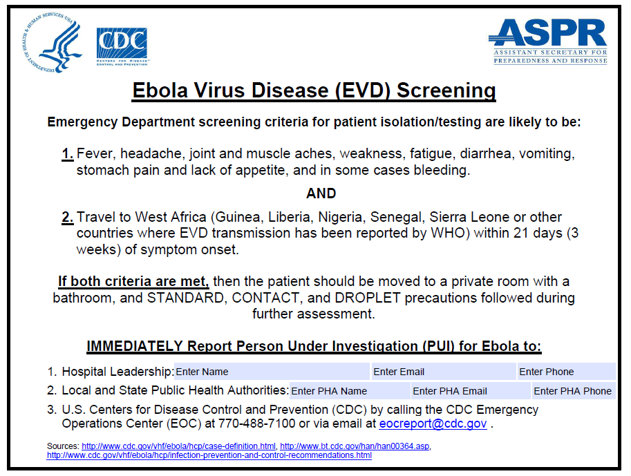
Screening Travelers at Airports
Entry screening in the United States
Because of the Ebola outbreak spreading to the US, CDC and Customs and Border Protection (CBP) have set in place enhanced entry screening of individuals who have traveled from or through Guinea, Liberia, and Sierra Leone. By doing enhanced entry screening at 5 U.S. airports (JFK, Washington-Dulles, Newark, Chicago-O’Hare, and Atlanta international), the CDC hopes to cover over 94% of travelers from the affected countries.
For each arriving traveler who has been in Guinea, Liberia, or Sierra Leone:
1. CBP will give each traveler health information that includes
- Information about Ebola
- Symptoms to look for and what to do if symptoms develop
- Information for doctors if travelers need to seek medical attention
2. Travelers will undergo screening measures to include:
- Answer questions to determine potential risk
- Have their temperature taken
- Be observed for other symptoms of Ebola
3. If a traveler has a fever or other symptoms or has been exposed to Ebola, CBP will refer to CDC to further evaluate the traveler to determine whether the traveler:
- can continue to travel
- should be taken to a hospital for evaluation, testing, and treatment
- should referred to a local health department for further monitoring and support
References
Natural History, Complications and Prognosis
Editor-In-Chief: C. Michael Gibson, M.S., M.D. [1] Associate Editor(s)-in-Chief: Michael Maddaleni, B.S.; Guillermo Rodriguez Nava, M.D. [2]; João André Alves Silva, M.D. [3]; Yazan Daaboul, M.D.
Overview
The natural history of Ebola hemorrhagic fever is highly dependent on the Ebola virus species and the host immunity. The symptoms of Ebola hemorrhagic fever usually develop 2 to 21 days following exposure to Ebola virus. The clinical course of Ebola hemorrhagic fever has 2 phases, which may possibly be separated by a phase of “pseudoremission”. The majority of patients develop severe symptoms that gradually worsen with time. Patients with non-fatal disease typically develop isolated high-grade fever that resolves within 7 to 10 days of disease onset. In contrast, fatal disease is associated with early clinical signs and symptoms, and these patients typically experience rapid clinical deterioration, including hemorrhagic, infectious, and neurological complications, and die 6 to 16 days following the onset of symptoms. Recovery from Ebola depends on good supportive care and the patient’s immune response. People who recover from Ebola infection develop antibodies that last for at least 10 years, possibly longer. It is not known if people who recover are immune for life or if they can become infected with a different species of Ebola. Patients who survive are susceptible to late complications that are not related to the acute illness. The most common long-term complication of Ebola virus among survivors is arthralgia of the large joints that is usually not accompanied by findings on physical examination.
Natural History
The natural history of Ebola hemorrhagic fever is highly dependent on the Ebola virus species and the host immunity. The symptoms of Ebola hemorrhagic fever usually develop 2 to 21 days following exposure to Ebola virus. The clinical course of Ebola hemorrhagic fever has 2 phases, which may possibly be separated by a phase of “pseudoremission”.[1] The clinical course of Ebola virus is a spectrum of manifestations. If left untreated, patients may remain asymptomatic or develop mild non-fatal disease, but the majority of patients develop severe symptoms that gradually worsen with time. Patients with non-fatal disease typically develop isolated high-grade fever that resolves within 7 to 10 days of disease onset. In contrast, fatal disease is associated with early clinical signs and symptoms, and these patients typically experience rapid clinical deterioration and die 6 to 16 days following the onset of symptoms.[2][3]
Phase 1: Early, Non-specific Signs and Symptoms
- Early symptoms include high-grade fever, chills, myalgias, arthritis, and generalized fatigue that typically develop within 6 to 13 days of viral incubation (incubation period ranges from 2 to 21 days). Early symptoms typically persist for approximately one week.[4]
- Without treatment, patients subsequently develop non-specific multisystem symptoms, including constitutional (asthenia and anorexia), respiratory (cough and nasal discharge), and gastrointestinal (abdominal pain, nausea, and vomiting) manifestations.
- During this phase, work-up is usually remarkable for leucopenia with lymphopenia.
- At day 5-7 following onset of symptoms, patients may develop cutaneous flushing or a characteristic desquematous, maculopapular, non-pruritic, erythematous rash with a centripetal distribution.
- Patients with non-fatal disease usually develop non-life-threatening symptoms that self-resolve approximately 7 to 10 days following onset of symptoms. When no improvement is observed within one week of phase 1 symptoms, patients are more likely to deteriorate into the potentially fatal phase 2 symptoms.[5]
Pseudoremission Phase
- A phase of pseudoremission, that typically occurs at day 7-8 of symptoms onset and lasts for 1 to 2 days, may be observed prior to the development of neurological and hemorrhagic manifestations.[1][4]
- Patients often report significant improvement in clinical symptoms with adequate food intake and mobility.[4]
- In the minority of cases, patients recover during this phase and survive. However, the majority of cases progress into developing life-threatening complications.
Phase 2: Life-threatening Signs and Symptoms
- A second phase of manifestations, characterized by hemorrhagic and neurological manifestations, typically develops during the peak of the illness.
- Approximately 50% of patients develop mucosal and visceral hemorrhage.
- At advanced stages, patients’ symptoms worsen; and patients develop multisystem failure (renal failure, hepatic failure, and pancreatitis), dyspnea, convulsions, encephalopathy, diffuse coagulopathy (disseminated intravascular coagulopathy), hypovolemic shock, and eventually death.[6][7][3]
- In contrast to early leucopenia, the late course of the disease is often characterized by prolonged prothrombin time (PT) and partial thromboplastin time (PTT), neutrophilia with left shift and atypical lymphocytes, thrombocytopenia, elevated liver enzymes, hyperproteinemia, and proteinuria.[2][3]
- Patients typically experience rapid clinical deterioration and perish within 6 to 16 days following the onset of symptoms.
Complications
Acute Complications of Advanced Disease
Ebola hemorrhagic fever usually leads to death by multiorgan failure and systemic complications. Infectious (overwhelming sepsis) and hemorrhagic (visceral bleeding and disseminated intravascular coagulopathy) complications are the most significant life-threatening causes of death associated with Ebola virus disease.[8] Generally, the following complications have been reported in patients with advanced Ebola virus disease[9]:
ENT
- Tinnitus
- Hearing loss
- Sudden bilateral blindness
Cardiovascular system
- Hypovolemic shock
- Disseminated intravascular coagulopathy (DIC)
- Acute heart failure
- Myocarditis
- Serous pericardial effusion
Respiratory system
Gastrointestinal system
Genitourinary system
Neurological system
- Hiccups (may herald further neurological symptoms and death)
- Dysesthesia and burning skin sensation
- Convulsions
- Meningitis
- Encephalopathy
- Coma
Late Complications Among Survivors
Survivors of Ebola hemoarrhagic fever may present with late complications of the disease that are not related to the acute illness. However, the majority of these symptoms seem to resolve by 1-2 years.[10] The most common long-term complication of Ebola virus among survivors is arthralgias of the large joints that are not accompanied by physical exam findings. Other unexplained reported complications that have occurred between 2 weeks to 2 months[9], at 6-months, and 21-month follow-up of the acute Ebola infection are listed below[10]:
Constitutional signs and symptoms
- Persistent weight loss
- Extreme fatigue
- Anorexia
- Fever
- Headache
ENT
Ocular disease
Musculoskeletal system
- Migratory arthralgias. Notably, arthralgias are the most common non-acute complication of Ebola virus. Arthralgias usually involve the large joints (knees, back, hips). Joint pain may be symmetric and is typically worse in the morning and following exertion. Arthralgias are typically not accompanied by signs on physical exam.
- Myalgias
Cardiovascular system
Gastrointestinal system
Genitourinary system
- Unilateral orchitis
- Amenorrhea
Neuropsychiatric system
Prognosis
- Ebola infection is associated with poor survival. Case-fatality rates is highly dependent on the species of virus:[3]
- Zaire Ebola virus species: case-fatality rate of 60 – 90%
- Sudan Ebola virus species: case-fatality rate 40 – 60%
- Bundibugyo Ebola virus species: case-fatality rate 25%
- Côte d’Ivoire Ebola virus: case-fatality rate 0% (only 1 case reported)
- Reston Ebola virus: case-fatality rate 0% (Reston virus seems to be relatively harmless to humans)
- Patients who survived Ebola infection for two weeks are usually able to recover slowly, despite potential complications.[3]
- Survival for 11 days is generally associated with recovery.
- Desquamation of the maculopapular rash by the 5th or 7th day is often associated with survival.[3]
- Tachypnea is the strongest correlate of fatal outcome. It often appears a few hours before death. Other correlates of fatal outcome are hypotension, tachycardia and anuria.
- Severe hemorrhagic complications such as hematemesis, melena, epistaxis, ear bleeding and hematuria are associated with a poorer prognosis and are often associated with death within a week.[11]
- In pregnant women, there is an increased risk of miscarriage.[3]
References
- ↑ 1.0 1.1 Ndambi R, Akamituna P, Bonnet MJ, Tukadila AM, Muyembe-Tamfum JJ, Colebunders R (1999). “Epidemiologic and clinical aspects of the Ebola virus epidemic in Mosango, Democratic Republic of the Congo, 1995”. J Infect Dis. 179 Suppl 1: S8–10. doi:10.1086/514297. PMID 9988156.
- ↑ 2.0 2.1 Ksiazek TG, West CP, Rollin PE, Jahrling PB, Peters CJ (1999). “ELISA for the detection of antibodies to Ebola viruses”. J Infect Dis. 179 Suppl 1: S192–8. doi:10.1086/514313. PMID 9988184.
- ↑ 3.0 3.1 3.2 3.3 3.4 3.5 3.6 Feldmann H, Geisbert TW (2011). “Ebola haemorrhagic fever”. Lancet. 377 (9768): 849–62. doi:10.1016/S0140-6736(10)60667-8. PMC 3406178. PMID 21084112.
- ↑ 4.0 4.1 4.2 Muyembe-Tamfum JJ, Mulangu S, Masumu J, Kayembe JM, Kemp A, Paweska JT (2012). “Ebola virus outbreaks in Africa: past and present”. Onderstepoort J Vet Res. 79 (2): 451. doi:10.4102/ojvr.v79i2.451. PMID 23327370.
- ↑ Casillas AM, Nyamathi AM, Sosa A, Wilder CL, Sands H (2003). “A current review of Ebola virus: pathogenesis, clinical presentation, and diagnostic assessment”. Biol Res Nurs. 4 (4): 268–75. PMID 12698919.
- ↑ Peters CJ, LeDuc JW (1999). “An introduction to Ebola: the virus and the disease”. J Infect Dis. 179 Suppl 1: ix–xvi. doi:10.1086/514322. PMID 9988154.
- ↑ Feldmann H, Geisbert T, Kawaoka Y (2007). “Filoviruses: recent advances and future challenges”. J Infect Dis. 196 Suppl 2: S129–30. doi:10.1086/520550. PMID 17940939.
- ↑ Parkes-Ratanshi R, Ssekabira U, Crozier I (2014). “Ebola in West Africa: be aware and prepare”. Intensive Care Med. 40 (11): 1742–5. doi:10.1007/s00134-014-3497-z. PMID 25253023.
- ↑ 9.0 9.1 Bwaka MA, Bonnet MJ, Calain P, Colebunders R, De Roo A, Guimard Y; et al. (1999). “Ebola hemorrhagic fever in Kikwit, Democratic Republic of the Congo: clinical observations in 103 patients”. J Infect Dis. 179 Suppl 1: S1–7. doi:10.1086/514308. PMID 9988155.
- ↑ 10.0 10.1 Rowe AK, Bertolli J, Khan AS, Mukunu R, Muyembe-Tamfum JJ, Bressler D; et al. (1999). “Clinical, virologic, and immunologic follow-up of convalescent Ebola hemorrhagic fever patients and their household contacts, Kikwit, Democratic Republic of the Congo. Commission de Lutte contre les Epidémies à Kikwit”. J Infect Dis. 179 Suppl 1: S28–35. doi:10.1086/514318. PMID 9988162.
- ↑ Sureau PH (1989). “Firsthand clinical observations of hemorrhagic manifestations in Ebola hemorrhagic fever in Zaire”. Rev Infect Dis. 11 Suppl 4: S790–3. PMID 2749110.
Diagnosis
Diagnosis
Algorithm for the Evaluation of the Returned Traveler | Emergency Department Evaluation | Case Definition | History and Symptoms | Physical Examination | Laboratory Findings | Other Diagnostic Studies
Looking for the patient version?
© 2026 MyEClinic – IFTM Institut für Telematik in der Medizin GmbH