
Orbital cellulitis
Editor-In-Chief: C. Michael Gibson, M.S., M.D. [1]; Associate Editor(s)-in-Chief: Prashanth Saddala M.B.B.S, Tarek Nafee, M.D. [2]
Synonyms and Keywords: Retroseptal cellulitis; Postseptal cellulitis; Retrobulbar cellulitis
For patient information, click here
Overview
Editor-In-Chief: C. Michael Gibson, M.S., M.D. [1]; Associate Editor(s)-in-Chief: Tarek Nafee, M.D. [2]
Overview
Orbital cellulitis is an inflammation of the deep, soft tissue cells surrounding the eye, posterior to the orbital septum. This inflammation can occur by means of extension of an adjacent infection, by direct inoculation through traumatic or iatrogenic pathways, or by hematogenous spread from a distant infected site. Orbital cellulitis most commonly occurs from typical bacterial infections. In some cases, mycobacteria or mycosis may be implicated. The most common underlying condition is ethmoid sinusitis. Children are more commonly affected by orbital cellulitis than adults. Orbital cellulitis has a higher incidence in the winter months and follows the seasonal patterns of sinusitis and upper respiratory tract infections. The most commonly reported pathogens are Staphylococcus aureus, Streptococcus spp. and Haemophilus influenzae. MRSA must be considered as a potential cause and is correlated with geographic prevalence. A positive history of fever, proptosis, eyelid edema and erythema, diplopia, and painful or restricted ocular movements is highly suggestive of orbital cellulitis. Orbital cellulitis must be differentiated from various conditions which affect the eye and present similarly, such as periorbital cellulitis, cavernous sinus thrombosis, and orbital or periorbital neoplasia. Both CT and MRI of the head may be used as gold standard diagnostic imaging modalities. The most common risk factors for orbital cellulitis include acute or chronic ethmoid sinusitis, upper respiratory tract infection, and recent ocular or periocular procedures or infections. If left untreated, orbital cellulitis may result in blindness in 20% of patients and death in 17%. Serious complications can develop as a result of orbital cellulitis, such as abscess, orbital compartment syndrome, intracranial extension of infection, and septic shock. Although orbital cellulitis is an ophthalmologic emergency, the prognosis is good if prompt medical treatment is received. The mainstay of therapy for orbital cellulitis involves prompt intravenous antimicrobial therapy. Surgery may be necessary if medical therapy is ineffective, if there is intracranial extension of the disease, or in the presence of abscesses. Effective measures for the primary prevention of orbital cellulitis include prompt, appropriate, and aggressive treatment of adjacent infections.
Historical Perspective
Orbital cellulitis first appears in the literature as early as 1882. Hypotheses regarding the etiology and management approaches were developed by various physicians and scientists. In 1937, Dr. Davis reported a review of 54 successfully treated patients, and suggested a management guideline for orbital cellulitis.[1][2][3]
Classification
Orbital cellulitis may be classified according to the microbial family of the offending pathogen (bacterial vs. fungal), or by the management protocol (medical vs. surgical therapy). In 1970, Chandler’s classification was created to describe five groups of complications of sinusitis by invading the surrounding tissue. This system is often used to describe the extent of orbital and periorbital disease.[4]
Pathophysiology
Orbital cellulitis occurs secondary to microbial infiltration of the deep soft tissue cells surrounding the eye, posterior to the orbital septum. The infiltration can occur by means of extension of an adjacent infection (e.g. rhinosinusitis), by direct inoculation through traumatic or iatrogenic pathways, or by hematogenous spread from a distant infected site. Regardless of the mode of infiltration of the retroseptal orbital space, symptoms may arise due to the immune system triggering a regional or systemic acute inflammatory response.[5][6][7][8][9]
Causes
Orbital cellulitis occurs most commonly from typical bacterial infections. In some cases, mycobacteria or mycosis may also be implicated. By far, the most common underlying condition is ethmoid sinusitis. It has been reported as the cause in 90-98% of orbital cellulitis cases. Thus, the most commonly reported pathogens were Staphylococcus aureus, Streptococcus spp., and Haemophilus influenzae. With the rise of microbial resistance in more recent years, methicillin-resistant Staphylococcus aureus (MRSA) must be considered as a potential cause and correlated with geographic prevalence.Though some causes may be uncommon, orbital cellulitis is a medical emergency. Thus, it is pertinent to consider all possible etiologies and the most common pathogens according to the clinical scenario.[5][6][10][11][12][13]
Differentiating Orbital cellulitis from other Diseases
Orbital cellulitis must be differentiated from various conditions which affect the eye and present with similar symptoms, and include periorbital cellulitis, cavernous sinus thrombosis, and neoplasia.
Epidemiology and Demographics
Children are more affected by orbital cellulitis than adults. In childhood, males are more likely to contract the disease than females. Orbital cellulitis has a higher incidence in the winter months and follows the seasonal patterns of sinusitis and upper respiratory tract infections.[13][14][15]
Risk Factors
The most common risk factors for orbital cellulitis include acute or chronic ethmoid sinusitis, pansinusitis, upper respiratory tract infection and recent ocular or periocular procedures or infections.[12][16]
Screening
There are no recommended screening guidelines for orbital cellulitis.[17]
Natural History, Complications, and Prognosis
If left untreated, orbital cellulitis may result in death in 17% of patients, and blindness in 20%. Complications that can develop as a result of orbital cellulitis include abscess, orbital compartment syndrome, intracranial extension of infection, and septic shock. Although orbital cellulitis is considered an ophthalmic emergency, the prognosis is good if prompt medical treatment is received. Depending on the extent of complications at the time of diagnosis, prognosis may vary. The most feared complications include septic shock, cavernous sinus thrombosis, and meningitis. These complications carry a much poorer prognosis.[18][19][20][21][22]
Diagnosis
History and Symptoms
A positive history of fever, proptosis, eyelid edema and erythema, and painful or restricted ocular movements is suggestive of orbital cellulitis. Additionally, patients with orbital cellulitis often have a recent history of rhinosinusitis, upper respiratory tract infection, facial insect bite, orbital surgery or trauma, tooth abscess, or otitis media.[5][6][10][11][23][24]
Physical Examination
Patients with orbital cellulitis usually appear ill. Physical examination of patients with orbital cellulitis is usually remarkable for fever, proptosis, ophthalmoplegia, and impaired visual acuity. Patients should undergo a complete physical examination, paying particular attention to general appearance, vital signs, visual acuity, visual field, orbital positioning, ocular movmements, oropharynx, and nasopharynx examinations.[5][6][10][11][23][24]
Laboratory Findings
There are no diagnostic lab findings associated with orbital cellulitis. Some patients with orbital cellulitis may have elevated ESR, CRP, and white blood cells with a left shift. These are non-specific findings associated with infections, inflammatory conditions and some neoplasia.[10] Blood and nasal mucosal cultures are often ordered to help guide medical therapy, though they have low positive and negative predictive values, and thus do not contribute to diagnosis of orbital cellulitis.
X Ray
There are no diagnostic x ray findings associated with orbital cellulitis. In cases where CT scan is contraindicated, plain radiograph (x ray) is indicated prior to MRI in patients with a suspected history of metallic object ocular trauma.[25][26]
CT
Computed tomography of the orbit is the imaging modality of choice for patients with orbital cellulitis. On CT scan of the head, orbital cellulitis is characterized by hyperdensity within low density periorbital fat and generalized elevation of the periobital space. CT is also diagnostic of subperiosteal and orbital abscesses associated with orbital cellulitis.[6][11][12][27][28][29]
MRI
MRI has demonstrated equivalence to CT in diagnosing orbital disease and is also accepted as a gold standard diagnostic imaging modality.[11][30] On MRI scan of the head, orbital cellulitis is characterized by hypointense signal on T1-weighted fat-suppressed images, and hyperintense signal on T2-weighted fat-suppressed images.[28] Although an MRI scan is safer in children since there is no risk of radiation exposure, the long acquisition time and the need for prolonged sedation make CT scan the imaging modality of choice.[12][30]
Ultrasound
There are no ultrasound findings specifically associated with orbital cellulitis. Ultrasound can detect an abscess in the anterior orbit or medial orbital wall with high sensitivity.[6][25] On orbital ultrasound, orbital abscess may show an anechoic mass with low internal reflectivity.[6]
Other Imaging Findings
There are no other diagnostic imaging findings associated with orbital cellulitis.
Other Diagnostic Studies
There are no other diagnostic studies associated with orbital cellulitis.
Treatment
Medical Therapy
Orbital cellulitis is considered an ophthalmologic emergency. The mainstay of therapy for orbital cellulitis involves prompt intravenous antimicrobial therapy with either beta-lactams or Clindamycin. Patients suspected to have MRSA-induced orbital cellulitis require more extensive antimicrobial therapy.
Surgery
An abscess can threaten the vision or neurological status of a patient with orbital cellulitis, therefore sometimes surgical intervention is necessary. Surgery typically requires drainage of the sinuses and if a subperiosteal abscess is present in the medial orbit, drainage can be performed endoscopically. Post-operatively, patients must follow up regularly with their surgeon and remain under close observation.[22]
Primary Prevention
Effective measures for the primary prevention of orbital cellulitis include prompt, appropriate and aggressive treatment of adjacent infections. Cases of acute bacterial rhinosinusitis should be managed effectively.[31]
Secondary Prevention
Secondary Prevention strategies following orbital cellulitis include obtaining blood cultures and immediately starting aggressive empirical antimicrobial therapy, diagnostic imaging studies to monitor for abscess formation and evaluation of necessity of surgical intervention. If cultures are positive, tailor antimicrobial therapy to the pathogen.[22]
References
- ↑ Holt EE (1892). “Orbital Cellulitis, the inflammation spreading to the temporal region, to the neck, obstructing deglutition, and to the brain, causing death, with reports of five other cases”. Trans Am Ophthalmol Soc. 6: 295–306. PMC 1326882. PMID 25259124.
- ↑ Layton TB (1935). “Four Cases of Orbital Cellulitis Secondary to Nasal Disease Treated by Simple Incision”. Proc R Soc Med. 28 (12): 1569–71. PMC 2205946. PMID 19990471.
- ↑ “Orbital Cellulitis due to Sinus Infection, and its Treatment: (Section of Laryngology and Section of Otology)”. Proc R Soc Med. 30 (11): 1397–407. 1937. PMC 2076505. PMID 19991275.
- ↑ Martin-Hirsch DP, Habashi S, Hinton AH, Kotecha B (1992). “Orbital cellulitis”. Arch Emerg Med. 9 (2): 143–8. PMC 1285851. PMID 1388488.
- ↑ 5.0 5.1 5.2 5.3 Hasanee K, Sharma S (2004). “Ophthaproblem. Orbital cellulitis”. Can Fam Physician. 50: 359, 365, 367. PMC 2214559. PMID 15318671.
- ↑ 6.0 6.1 6.2 6.3 6.4 6.5 6.6 Chaudhry IA, Al-Rashed W, Arat YO (2012). “The hot orbit: orbital cellulitis”. Middle East Afr J Ophthalmol. 19 (1): 34–42. doi:10.4103/0974-9233.92114. PMC 3277022. PMID 22346113.
- ↑ Turvey TA, Golden BA (2012). “Orbital anatomy for the surgeon”. Oral Maxillofac Surg Clin North Am. 24 (4): 525–36. doi:10.1016/j.coms.2012.08.003. PMC 3566239. PMID 23107426.
- ↑ U.S. National Library of Medicine Medlineplus(2014) https://medlineplus.gov/ency/article/000821.htm
- ↑ Zhang J, Stringer MD (2010). “Ophthalmic and facial veins are not valveless”. Clin Experiment Ophthalmol. 38 (5): 502–10. doi:10.1111/j.1442-9071.2010.02325.x. PMID 20491800.
- ↑ 10.0 10.1 10.2 10.3 Lam Choi VB, Yuen HK, Biswas J, Yanoff M (2011). “Update in pathological diagnosis of orbital infections and inflammations”. Middle East Afr J Ophthalmol. 18 (4): 268–76. doi:10.4103/0974-9233.90127. PMC 3249811. PMID 22224014.
- ↑ 11.0 11.1 11.2 11.3 11.4 Merck Manual Professional Version (2016)https://www.merckmanuals.com/professional/eye-disorders/orbital-diseases/preseptal-and-orbital-cellulitis
- ↑ 12.0 12.1 12.2 12.3 American Academy of Ophthalmology Eyewiki (2015)http://eyewiki.aao.org/Orbital_Cellulitis#Etiology
- ↑ 13.0 13.1 Nageswaran S, Woods CR, Benjamin DK, Givner LB, Shetty AK (2006). “Orbital cellulitis in children”. Pediatr Infect Dis J. 25 (8): 695–9. doi:10.1097/01.inf.0000227820.36036.f1. PMID 16874168.
- ↑ American Association of Family Physicians (2003)http://www.aafp.org/afp/2003/0315/p1349a.html
- ↑ Bergin DJ, Wright JE (1986). “Orbital cellulitis”. Br J Ophthalmol. 70 (3): 174–8. PMC 1040961. PMID 3954974.
- ↑ Torretta S, Marchisio P, Gaffuri M, Capaccio P, Esposito S, Pignataro L (2014). “Step-by-step iconographic description of a prolonged but still favourable course of orbital cellulitis in a child with acute rhinosinusitis: an iconographic case study”. Ital J Pediatr. 40 (1): 25. doi:10.1186/1824-7288-40-25. PMC 3995968. PMID 24594215.
- ↑ United States Preventative Services Task Force Recommendations (2016) http://www.uspreventiveservicestaskforce.org/Page/Name/recommendations
- ↑ Healy GB (1997). “Chandler et al.: “The pathogenesis of orbital complications in acute sinusitis.” (Laryngoscope 1970;80:1414-1428)”. Laryngoscope. 107 (4): 441–6. PMID 9111370.
- ↑ Ryan JT, Preciado DA, Bauman N, Pena M, Bose S, Zalzal GH; et al. (2009). “Management of pediatric orbital cellulitis in patients with radiographic findings of subperiosteal abscess”. Otolaryngol Head Neck Surg. 140 (6): 907–11. doi:10.1016/j.otohns.2009.02.014. PMID 19467413.
- ↑ “Reorganized text”. JAMA Otolaryngol Head Neck Surg. 141 (5): 428. 2015. doi:10.1001/jamaoto.2015.0540. PMID 25996397.
- ↑ Bedwell J, Bauman NM (2011). “Management of pediatric orbital cellulitis and abscess”. Curr Opin Otolaryngol Head Neck Surg. 19 (6): 467–73. doi:10.1097/MOO.0b013e32834cd54a. PMID 22001661.
- ↑ 22.0 22.1 22.2 Lee S, Yen MT (2011). “Management of preseptal and orbital cellulitis”. Saudi J Ophthalmol. 25 (1): 21–9. doi:10.1016/j.sjopt.2010.10.004. PMC 3729811. PMID 23960899.
- ↑ 23.0 23.1 American Academy of Ophthalmology EyeWiki (2015)http://eyewiki.aao.org/Orbital_Cellulitis
- ↑ 24.0 24.1 Stanford Orbital Cellulitis Summary (2011)http://peds.stanford.edu/Rotations/blue_team/documents/Periorbital_and_Orbital_Cellulitis_Summary.pdf
- ↑ 25.0 25.1 Mair MH, Geley T, Judmaier W, Gassner I (2002). “Using orbital sonography to diagnose and monitor treatment of acute swelling of the eyelids in pediatric patients”. AJR Am J Roentgenol. 179 (6): 1529–34. doi:10.2214/ajr.179.6.1791529. PMID 12438049.
- ↑ Murphy KJ, Brunberg JA (1996). “Orbital plain films as a prerequisite for MR imaging: is a known history of injury a sufficient screening criterion?”. AJR Am J Roentgenol. 167 (4): 1053–5. doi:10.2214/ajr.167.4.8819411. PMID 8819411.
- ↑ LeBedis CA, Sakai O (2008). “Nontraumatic orbital conditions: diagnosis with CT and MR imaging in the emergent setting”. Radiographics. 28 (6): 1741–53. doi:10.1148/rg.286085515. PMID 18936033.
- ↑ 28.0 28.1 Radiopaedia (2016)http://radiopaedia.org/articles/orbital-infection
- ↑ Yousem DM (1993). “Imaging of sinonasal inflammatory disease”. Radiology. 188 (2): 303–14. doi:10.1148/radiology.188.2.8327669. PMID 8327669.
- ↑ 30.0 30.1 Sepahdari AR, Aakalu VK, Kapur R, Michals EA, Saran N, French A; et al. (2009). “MRI of orbital cellulitis and orbital abscess: the role of diffusion-weighted imaging”. AJR Am J Roentgenol. 193 (3): W244–50. doi:10.2214/AJR.08.1838. PMID 19696266.
- ↑ Brooks I, Gooch WM, Jenkins SG, Pichichero ME, Reiner SA, Sher L; et al. (2000). “Medical management of acute bacterial sinusitis. Recommendations of a clinical advisory committee on pediatric and adult sinusitis”. Ann Otol Rhinol Laryngol Suppl. 182: 2–20. PMID 10823486.
Historical Perspective
Editor-In-Chief: C. Michael Gibson, M.S., M.D. [1]; Associate Editor(s)-in-Chief: Tarek Nafee, M.D. [2]
Overview
In 1882, Dr. Nettleship, a British surgeon, described orbital cellulitis in a case study.[1] In 1937, Dr. Davis reported a review of 54 successfully treated patients and suggested a management guideline for orbital cellulitis.
Historical Perspective
- In 1882, Dr. Nettleship, a British surgeon, described orbital cellulitis in a case study.[1]
- In 1892, Dr. E.E. Holt first described orbital cellulitis as a case study in the literature. He hypothesized that orbital cellulitis was a result of extension of an adjacent infection. In his work, Dr. Holt references the work of Dr. Nettleship.[1]
- In 1935, Dr. T.B. Layton outlines a surgical approach to treatment by simple incision, drainage, and irrigation of the retro-orbital space.[2]
- In 1937, the procedure created by Dr. Layton was confirmed in a review of 54 successfully treated cases of orbital cellulitis by Dr. Davis.[3]
References
- ↑ 1.0 1.1 1.2 Holt EE (1892). “Orbital Cellulitis, the inflammation spreading to the temporal region, to the neck, obstructing deglutition, and to the brain, causing death, with reports of five other cases”. Trans Am Ophthalmol Soc. 6: 295–306. PMC 1326882. PMID 25259124.
- ↑ Layton TB (1935). “Four Cases of Orbital Cellulitis Secondary to Nasal Disease Treated by Simple Incision”. Proc R Soc Med. 28 (12): 1569–71. PMC 2205946. PMID 19990471.
- ↑ “Orbital Cellulitis due to Sinus Infection, and its Treatment: (Section of Laryngology and Section of Otology)”. Proc R Soc Med. 30 (11): 1397–407. 1937. PMC 2076505. PMID 19991275.
Classification
Editor-In-Chief: C. Michael Gibson, M.S., M.D. [1]; Associate Editor(s)-in-Chief: Tarek Nafee, M.D. [2]
Overview
Orbital cellulitis may be classified according to the microbial family of the offending pathogen (bacterial vs. fungal), or by the management protocol (medical vs. surgical therapy). In 1970, Chandler’s classification was created to describe five groups of orbital complications of sinusitis. This system is often used to describe the extent of orbital and periorbital disease.[1]
Classification
Orbital cellulitis may be classified as follows:
- Bacterial vs. non-bacterial infection: This classification determines the course of the disease and the type of antibiotics to be used.
- Abscess vs. no abscess: This classification determines the need for surgical therapy.
- Chandler’s Classification of Orbital Complications of Sinusitis:[1]
- Stage I: Preseptal Cellulitis – Also known as periorbital cellulitis; this condition is not classified as true orbital cellulitis. It rarely extends to become true postseptal orbital cellulitis. It is characterized by lid edema, lack of proptosis, changes in visual acuity, pain with ocular movements, and opthalmoplegia.
- Stage II: Orbital cellulitis – Also known as postseptal cellulitis or true orbital cellulitis; diffuse orbital inflammation without abscess formation.
- Stage III: Subperiosteal abscess
- Stage IV: Orbital abscess
- Stage V: Intracranial involvement / cavernous sinus thrombosis
References
Pathophysiology
Editor-In-Chief: C. Michael Gibson, M.S., M.D. [1]; Associate Editor(s)-in-Chief: Tarek Nafee, M.D. [2]
Overview
Orbital cellulitis occurs secondary to microbial infiltration of the deep soft tissue cells surrounding the eye, posterior to the orbital septum. The infiltration can occur by means of extension of an adjacent infection (e.g., rhinosinusitis), by direct inoculation through traumatic or iatrogenic pathways, or by hematogenous spread from a distant infected site. Regardless of the mode of infiltration of the retroseptal orbital space, symptoms may arise due to the immune system triggering a regional acute inflammatory response.[1][2][3][4][5]
Pathogenesis
Orbital cellulitis occurs secondary to microbial infiltration of the deep soft tissue cells surrounding the eye, posterior to the orbital septum.[1] Damage to the cells triggers an acute inflammatory response resulting in vasodilation, increased vascular permeability, and induction of a cascade of inflammatory markers and white blood cell chemoattractants.[4]
Extension of adjacent infection
Orbital cellulitis may spread through direct extension of acute or chronic adjacent infections. This is due to the fragility of the medial and inferior orbital walls, the presence of natural foramina and defects in these structures, and the medial check ligaments extending from extraocular structures and muscle sheaths to the thin medial orbital wall, which separates the orbital cavity from the paranasal sinuses. Some infections that may affect these structures include:[1][2][3]
- Rhinosinusitis (ethmoid sinusitis and pansinusitis)
- Dacryocystitis/Dacryoadenitis
- Panophthalmitis
- Infected tumor
- Otitis media
- Mucormycosis
- Dental abscess
Direct Inoculation
Traumatic Inoculation
Orbital cellulitis may occur as a result of microbial inoculation to the orbital space due to trauma. Examples of this include:[1]
Iatrogenic Inoculation
Orbital cellulitis may also occur as a result of direct inoculation during surgical procedures, such as:[1][2]
- Ocular or periocular surgeries
- Paranasal sinus surgeries
- Other ENT surgeries
Hematogenous Seeding
In some cases, infections from a distant source may seed to the retroseptal orbital soft tissue by means of hematogenous spread in patients with bacteremia. This highly vascularized space, coupled with a valveless inferior ophthalmic vein, has been implicated in facilitating this mode of infection.[1][2][5]
Associated Conditions
The following conditions are associated with orbital cellulitis:[2]
- Chronic sinusitis
- Upper respiratory tract infection
- Subperiosteal abscess
Gross Pathology
The following are gross pathology images of orbital cellulitis:[6][7]
-
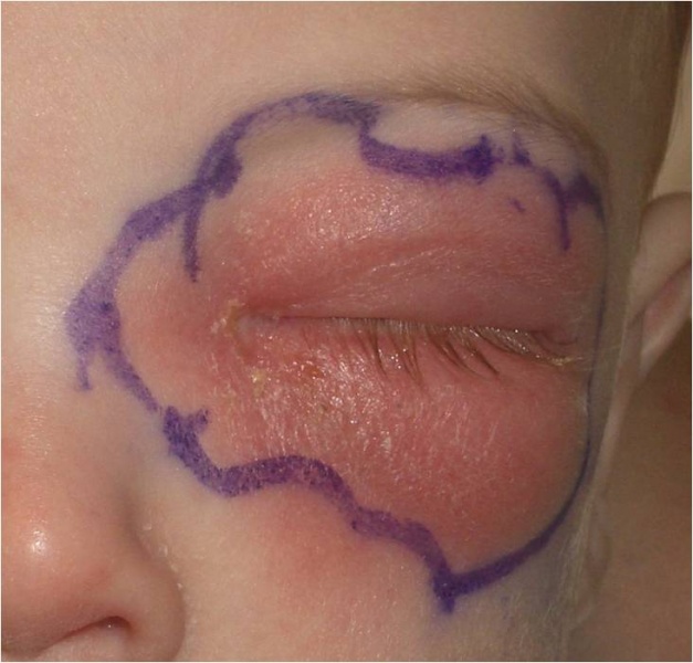 Orbital cellulitis in the left eye of a child
 -
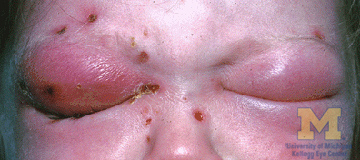 Bilateral orbital cellulitis gross pathology


References
- ↑ 1.0 1.1 1.2 1.3 1.4 1.5 Hasanee K, Sharma S (2004). “Ophthaproblem. Orbital cellulitis”. Can Fam Physician. 50: 359, 365, 367. PMC 2214559. PMID 15318671.
- ↑ 2.0 2.1 2.2 2.3 2.4 Chaudhry IA, Al-Rashed W, Arat YO (2012). “The hot orbit: orbital cellulitis”. Middle East Afr J Ophthalmol. 19 (1): 34–42. doi:10.4103/0974-9233.92114. PMC 3277022. PMID 22346113.
- ↑ 3.0 3.1 Turvey TA, Golden BA (2012). “Orbital anatomy for the surgeon”. Oral Maxillofac Surg Clin North Am. 24 (4): 525–36. doi:10.1016/j.coms.2012.08.003. PMC 3566239. PMID 23107426.
- ↑ 4.0 4.1 U.S. National Library of Medicine Medlineplus(2014) https://medlineplus.gov/ency/article/000821.htm
- ↑ 5.0 5.1 Zhang J, Stringer MD (2010). “Ophthalmic and facial veins are not valveless”. Clin Experiment Ophthalmol. 38 (5): 502–10. doi:10.1111/j.1442-9071.2010.02325.x. PMID 20491800.
- ↑ American Academy of Ophthalmology EyeWiki(2010)http://eyewiki.org/File%3AOrbital_cellulitis1.jpg
- ↑ University of Michigan Eyes Have It(2009)http://eyewiki.org/File%3AOrbital_cellulitis1.jpg
Causes
Editor-In-Chief: C. Michael Gibson, M.S., M.D. [1]; Associate Editor(s)-in-Chief: Ogheneochuko Ajari, MB.BS, MS [2]; Tarek Nafee, M.D. [3]
Overview
Orbital cellulitis occurs most commonly from typical bacterial infections. In some cases, mycobacteria or mycosis may also be implicated. The most common underlying condition is ethmoid sinusitis, which has been reported in 90-98% of orbital cellulitis cases. The most commonly reported pathogens are Staphylococcus aureus, Streptococcus spp., and Haemophilus influenzae. With the rise of microbial resistance, methicillian-resistant Staphylococcus aureus (MRSA) must be considered as a potential cause and correlated with geographic prevalence. Though some causes may be uncommon, orbital cellulitis is a medical emergency and all etiologies must be considered.[1][2][3][4][5][6]
Causes
Orbital cellulitis occurs most commonly from bacterial infection. In some cases, mycobacterial or fungal infections are observed.[1][2][3][4] Difficulty arises in identifying a specific organism due to challenges in culturing the retroseptal orbital region.[7]
Cause by Pathogen
Common bacterial causes of orbital cellulitis include:[1][2][3]
- Staphylococcus aureus
- Streptococcus spp.
- Haemophilus influenzae
- With the dissemination of Haemophilus influenza type b (Hib) vaccine, the incidence of Haemophilus influenzae-caused orbital cellulitis has decreased significantly.
- Methicillin-resistant Staphylococcus aureus (MRSA)
- Rise in microbial resistance, MRSA must be considered as a potential cause and correlated with geographic prevalence.
- Aerobic and anaerobic bacteria
In immunocompromised patients, additional causes may include:[2]
- Fungal infection
- Mycobacterial infection
Cause by Etiology
Based on etiology, the most common underlying condition causing orbital cellulitis is ethmoid sinusitis, which occurs in 90-98% of cases.[1][5][6] Additional causes based on etiology include:[2][7]
- Sinusitis:
- Dacryocystitis, dacryoadenitis, and other lacrimal duct abnormalities
- Traumatic/Foreign body:
- Spread from superficial infections of the face or adjacent soft tissue:
- Dental caries/Tooth abscess:
- Iatrogenic/post-surgical procedures:
- Immunocompromised patient:
- Diabetic patients with or without history of diabetic ketoacidosis
Cause by Organ System
Causes in Alphabetical Order
- Aeromonas hydrophila
- Anaerobes
- Arcanobacterium
- Aspergillosis
- Aspergillus
- Bacterial rhinosinusitis
- Bacteroides
- Beta-hemolytic streptococci
- Blepharoplasty
- Dacryocystitis
- Dacryocystorhinostomy
- Dental infection
- Eikenella corrodens
- Enterococcus
- Ethmoid sinusitis
- Eyelid surgery
- Foreign body
- Haemophilus influenzae
- Haemophilus parainfluenzae
- Infected mucocele
- Klebsiella pneumoniae
- Moraxella catarrhalis
- MRSA
- MSSA
- Mucor
- Mucorales
- Mucormycosis
- Mycobacterium tuberculosis
- Neisseria gonorrhea
- Ophthalmic surgery
- Orbital decompression
- Orbital fracture
- Osteomyelitis of the orbital bones
- Otitis media
- Peptostreptococcus
- Peribulbar anesthesia
- Phlebitis of the facial veins
- Pseudomonas aeruginosa
- Radial keratotomy
- Retinal surgery
- Rothia mucilaginosa
- Sinusitis
- Staphylococcus aureus
- Strabismus surgery
- Streptococcus
- Streptococcus anginosus
- Streptococcus milleri
- Streptococcus pneumoniae
- Streptococcus pyogenes
- Surgical trauma
- Tooth abscess
- Trauma
- Varicella
References
- ↑ 1.0 1.1 1.2 1.3 Hasanee K, Sharma S (2004). “Ophthaproblem. Orbital cellulitis”. Can Fam Physician. 50: 359, 365, 367. PMC 2214559. PMID 15318671.
- ↑ 2.0 2.1 2.2 2.3 2.4 Lam Choi VB, Yuen HK, Biswas J, Yanoff M (2011). “Update in pathological diagnosis of orbital infections and inflammations”. Middle East Afr J Ophthalmol. 18 (4): 268–76. doi:10.4103/0974-9233.90127. PMC 3249811. PMID 22224014.
- ↑ 3.0 3.1 3.2 Merck Manual Professional Version (2016)https://www.merckmanuals.com/professional/eye-disorders/orbital-diseases/preseptal-and-orbital-cellulitis
- ↑ 4.0 4.1 American Academy of Ophthalmology Eyewiki (2015)http://eyewiki.aao.org/Orbital_Cellulitis#Etiology
- ↑ 5.0 5.1 Nageswaran S, Woods CR, Benjamin DK, Givner LB, Shetty AK (2006). “Orbital cellulitis in children”. Pediatr Infect Dis J. 25 (8): 695–9. doi:10.1097/01.inf.0000227820.36036.f1. PMID 16874168.
- ↑ 6.0 6.1 Chaudhry IA, Al-Rashed W, Arat YO (2012). “The hot orbit: orbital cellulitis”. Middle East Afr J Ophthalmol. 19 (1): 34–42. doi:10.4103/0974-9233.92114. PMC 3277022. PMID 22346113.
- ↑ 7.0 7.1 Baring DE, Hilmi OJ (2011). “An evidence based review of periorbital cellulitis”. Clin Otolaryngol. 36 (1): 57–64. doi:10.1111/j.1749-4486.2011.02258.x. PMID 21232022.
Differentiating Orbital cellulitis from other Diseases
Editor-In-Chief: C. Michael Gibson, M.S., M.D. [1]; Associate Editor(s)-in-Chief: Tarek Nafee, M.D. [2]
Overview
Orbital cellulitis must be differentiated from various conditions which affect the eye and present with similar symptoms, and include periorbital cellulitis, cavernous sinus thrombosis, and neoplasia.
Differential Diagnosis
Orbital cellulitis may share common features or present similarly to other eye diseases with significantly varying management protocols. Orbital cellulitis must be differentiated from the following:[1][2]
Infectious diseases
Orbital cellulitis must be differentiated from other diseases that may cause erythema, fever, and edema, such as:
- Periorbital cellulitis
- Endophthalmitis
- Severe conjunctivitis
- Herpes Zoster Ophthalmicus
- Posterior Scleritis
- Mucormycosis,
- Aspergillosis of the globe
- M.tuberculosis of the globe
Vascular
Orbital cellulitis must be differentiated from other diseases that may cause proptosis, edema, and visual disturbance, such as:
Autoimmune/Inflammatory
Orbital cellulitis must be differentiated from other diseases that may cause inflammation, proptosis, and visual disturbance, such as:
- Exophthalmos
- Wegener’s granulomatosis
- Sarcoidosis granuloma
- Orbital pseudotumor
Structural/Trauma
Orbital cellulitis must be differentiated from other diseases that may cause edema, visual disturbance, and proptosis, such as:
Toxicity
Orbital cellulitis must be differentiated from other diseases that may cause proptosis such as:
- Bisphosphonate use
Neoplastic
Orbital cellulitis must be differentiated from other diseases that may cause erythema, edema, fever, visual disturbance, and proptosis, such as:
- Metastatic carcinoma
- Neurofibroma
- Schwannoma
- Glioma
- Retinoblastoma
- Rhabdomyosarcoma
- Neuroblastoma
- Lymphoma
- Leukemia
- Histiocytosis
References
- ↑ American Academy of Ophthalmology EyeWiki (2015)http://eyewiki.aao.org/Orbital_Cellulitis#Differential_Diagnosis-
- ↑ Lam Choi VB, Yuen HK, Biswas J, Yanoff M (2011). “Update in pathological diagnosis of orbital infections and inflammations”. Middle East Afr J Ophthalmol. 18 (4): 268–76. doi:10.4103/0974-9233.90127. PMC 3249811. PMID 22224014.
Epidemiology and Demographics
Editor-In-Chief: C. Michael Gibson, M.S., M.D. [1]; Associate Editor(s)-in-Chief: Tarek Nafee, M.D. [2]
Overview
Children are more affected by orbital cellulitis than adults. In childhood, males are more likely to contract the disease than females. Orbital cellulitis has a higher incidence in the winter months and follows the seasonal patterns of sinusitis and upper respiratory tract infections.[1][2][3]
Epidemiology and demographics
Age
Children are more affected by orbital cellulitis than adults. The mean age at diagnosis is 12 years among the entire population. Among children, the mean age at diagnosis is 7.5 years.[1][2]
Gender
In childhood, males are 2.7 times as likely to be affected by orbital cellulitis than females. In adulthood the incidence is the same among both sexes.[2]
Seasonality
Orbital cellulitis occurs more commonly in the winter months and follows the seasonal patterns of sinusitis and upper respiratory tract infections.[3]
References
- ↑ 1.0 1.1 American Association of Family Physicians (2003)http://www.aafp.org/afp/2003/0315/p1349a.html
- ↑ 2.0 2.1 2.2 Nageswaran S, Woods CR, Benjamin DK, Givner LB, Shetty AK (2006). “Orbital cellulitis in children”. Pediatr Infect Dis J. 25 (8): 695–9. doi:10.1097/01.inf.0000227820.36036.f1. PMID 16874168.
- ↑ 3.0 3.1 Bergin DJ, Wright JE (1986). “Orbital cellulitis”. Br J Ophthalmol. 70 (3): 174–8. PMC 1040961. PMID 3954974.
Risk Factors
Editor-In-Chief: C. Michael Gibson, M.S., M.D. [1]; Associate Editor(s)-in-Chief: Tarek Nafee, M.D. [2]
Overview
The most common risk factors for orbital cellulitis include acute or chronic ethmoid sinusitis, pansinusitis, upper respiratory tract infection, and recent ocular or periocular procedures or infections.[1][2]
Risk Factors
Risk factors in the development of orbital cellulitis include:[1][2]
- Acute or chronic sinusitis
- Upper respiratory tract infection
- Recent paranasal sinus procedures
- Recent ocular or periocular procedures or infections
- Recent trauma to the eye
- Immunodeficiency
- Systemic infection
- Age (children)
References
- ↑ 1.0 1.1 American Academy of Ophthalmology EyeWiki (2015)http://eyewiki.aao.org/Orbital_Cellulitis
- ↑ 2.0 2.1 Torretta S, Marchisio P, Gaffuri M, Capaccio P, Esposito S, Pignataro L (2014). “Step-by-step iconographic description of a prolonged but still favourable course of orbital cellulitis in a child with acute rhinosinusitis: an iconographic case study”. Ital J Pediatr. 40 (1): 25. doi:10.1186/1824-7288-40-25. PMC 3995968. PMID 24594215.
Screening
Editor-In-Chief: C. Michael Gibson, M.S., M.D. [1]; Associate Editor(s)-in-Chief: Tarek Nafee, M.D. [2]
Overview
There are no recommended screening guidelines for orbital cellulitis.[1]
Screening
There are no recommended screening guidelines for orbital cellulitis.[1]
References
- ↑ 1.0 1.1 United States Preventative Services Task Force Recommendations (2016) http://www.uspreventiveservicestaskforce.org/Page/Name/recommendations
Natural History, Complications and Prognosis
Editor-In-Chief: C. Michael Gibson, M.S., M.D. [1]; Associate Editor(s)-in-Chief: Tarek Nafee, M.D. [2]
Overview
If left untreated, orbital cellulitis may result in blindness in 20% of patients and death in 17%. Complications that can develop as a result of orbital cellulitis include abscess, orbital compartment syndrome, intracranial extension of infection, and septic shock. Although orbital cellulitis is considered an ophthalmologic emergency, the prognosis is good if prompt medical treatment is received. Depending on the extent of complications at the time of diagnosis, prognosis may vary. The most serious complications include septic shock, cavernous sinus thrombosis, and meningitis. These complications carry a poor prognosis.[1][2][3][4][5]
Natural History
If left untreated, orbital cellulitis may result in death in blindness in 20% of patients, and death in 17%.[1]
Complications
Complications that can develop as a result of orbital cellulitis are:[2][3][4]
Abscesses are a common complication of orbital cellulitis. The most commonly encountered abscesses in this scenario are:
- Subperiosteal abscess
- Orbital abscess
Orbital compartment syndrome is among the most serious complications of orbital cellulitis and has a high risk of causing permanent vision loss. In orbital cellulitis cases, it occurs due to compression of the ophthalmic vasculature or optic nerve by a growing abscess, or uncontrolled infection in the retrobulbar region.
Intracranial extension
Intracranial extension of orbital cellulitis is among the most serious complications; it may result in encephalitis, meningitis, or sepsis.
Cavernous sinus thrombosis
Cavernous sinus thrombosis is among the most common causes of death due to orbital cellulitis; it occurs secondary to bacterial colonization causing venous stasis.
Septic shock
Bacteremia and sepsis may result from a sustained infection of orbital cellulitis. Sepsis carries a high risk of mortality.
Prognosis
Depending on the time of diagnosis, the prognosis may vary. Although orbital cellulitis is considered an ophthalmic emergency, the prognosis is good if prompt medical treatment is received.[5] In some cases, surgical intervention is required to mitigate serious complications. Undergoing surgical intervention for orbital cellulitis is associated with longer hospital admissions.[4]
The most serious complications include septic shock, cavernous sinus thrombosis, and meningitis. These complications carry a poor prognosis. Abscess formation is another complication that may require surgery. Complications secondary to abscess formation include bacteremia and optic nerve damage. which may lead to permanent vision loss. [2][3]
References
- ↑ 1.0 1.1 “Reorganized text”. JAMA Otolaryngol Head Neck Surg. 141 (5): 428. 2015. doi:10.1001/jamaoto.2015.0540. PMID 25996397.
- ↑ 2.0 2.1 2.2 Bedwell J, Bauman NM (2011). “Management of pediatric orbital cellulitis and abscess”. Curr Opin Otolaryngol Head Neck Surg. 19 (6): 467–73. doi:10.1097/MOO.0b013e32834cd54a. PMID 22001661.
- ↑ 3.0 3.1 3.2 Healy GB (1997). “Chandler et al.: “The pathogenesis of orbital complications in acute sinusitis.” (Laryngoscope 1970;80:1414-1428)”. Laryngoscope. 107 (4): 441–6. PMID 9111370.
- ↑ 4.0 4.1 4.2 Ryan JT, Preciado DA, Bauman N, Pena M, Bose S, Zalzal GH; et al. (2009). “Management of pediatric orbital cellulitis in patients with radiographic findings of subperiosteal abscess”. Otolaryngol Head Neck Surg. 140 (6): 907–11. doi:10.1016/j.otohns.2009.02.014. PMID 19467413.
- ↑ 5.0 5.1 Lee S, Yen MT (2011). “Management of preseptal and orbital cellulitis”. Saudi J Ophthalmol. 25 (1): 21–9. doi:10.1016/j.sjopt.2010.10.004. PMC 3729811. PMID 23960899.
Diagnosis
Diagnosis
History and Symptoms | Physical Examination | Laboratory Findings | X Ray | CT | MRI | Ultrasound | Other Imaging Findings | Other Diagnostic Studies
Treatment
Treatment
Medical Therapy | Surgery | Primary Prevention | Secondary Prevention | Cost-Effectiveness of Therapy | Future or Investigational Therapies
Looking for the patient version?
© 2026 MyEClinic – IFTM Institut für Telematik in der Medizin GmbH