
Right ventricular myocardial infarction
For patient information click here
Editor-In-Chief: C. Michael Gibson, M.S., M.D. [1]; Associate Editor-In-Chief: Cafer Zorkun, M.D., Ph.D. [2]; Priyamvada Singh, M.D. [3]
Overview
Editor-In-Chief: C. Michael Gibson, M.S., M.D. [1]
Overview
Acute myocardial infarction involving only the free wall of the right ventricle is a rare event. [1] More commonly, right ventricular infarction is associated with infarction of the inferior wall of the left ventricle, occurring in more than one-third of such cases,[2] [3] [4] [5] [6] [7] [8] [9] [10] even when a thrombolytic therapy is given. [11]
One study of 113 patients with a first acute inferior wall infarction reported that the presence of preinfarction angina within 72 hours of the infarction was associated with a reduction in the incidence of right ventricular infarction (odds ratio 0.2) and combined hypotension and shock (odds ratio 0.1). [12] This is possibly the result of ischemic preconditioning.
- Ninety percent of right ventricular infarcts result from occlusion of the proximal right coronary artery, while another 5 to 10 percent arise after occlusion of the left anterior descending artery [13] [14] [15] [16]
Although more than one-third of cases are clinically silent [17], the presence of right ventricular infarction often has important implications for both management and prognosis.
References
- ↑ Anderson, HR, Falk, E, Nielsen, D. Right ventricular infarction: Frequency, size, and topography in coronary heart disease. J Am Coll Cardiol 1987; 10:1223. PMID 3680789
- ↑ Anderson, HR, Falk, E, Nielsen, D. Right ventricular infarction: Frequency, size, and topography in coronary heart disease. J Am Coll Cardiol 1987; 10:1223. PMID 3680789
- ↑ Isner, JM, Roberts, WC. Right ventricular infarction complicating left ventricular infarction secondary to coronary artery disease: frequency, location, associated findings and significance from analysis of 236 necropsy patients with acute or healed myocardial infarction. Am J Cardiol 1978; 42:885. PMID 153103
- ↑ Isner, JM. Right ventricular myocardial infarction. JAMA 1988; 259:712. PMID 3275819
- ↑ Williams, JF. Right ventricular infarction. Clin Cardiol 1990; 13:309. PMID 2189611
- ↑ Cabin, HS, Clubb, S, Wackers FJ, et al. Right ventricular myocardial infarction with anterior wall left ventricular infarction: an autopsy study. Am Heart J 1987; 113:16. PMID 3799430
- ↑ Kinch, JW, Ryan, TJ. Right ventricular infarction. N Engl J Med 1994; 330:1211. PMID 8139631
- ↑ Setaro, JF, Cabin, HS. Right ventricular infarction. Cardiol Clin 1992; 10:69. PMID 1739961
- ↑ Cohn, JN. Right ventricular infarction revisited. Am J Cardiol 1979; 43:666. PMID 420117
- ↑ Wackers, FJ, Lie, KI, Sokole, EB, et al. Prevalence of right ventricular involvement in inferior wall infarction associated with myocardial imaging with thallium-201 and technetium-99m pyrophosphate. Am J Cardiol 1978; 42:358. PMID 210648
- ↑ Zeymer, U, Neuhaus, K-L, Wegscheider, K, et al. Effects of thrombolytic therapy in acute inferior myocardial infarction with and without right ventricular involvement. J Am Coll Cardiol 1998; 32:876. PMID 9768705
- ↑ Shiraki, H, Yoshikawa, T, Anzai, T, et al. Association between preinfarction angina and a lower risk of right ventricular infarction. N Engl J Med 1998; 338:941. PMID 9521981
- ↑ Isner, JM, Roberts, WC. Right ventricular infarction complicating left ventricular infarction secondary to coronary artery disease: frequency, location, associated findings and significance from analysis of 236 necropsy patients with acute or healed myocardial infarction. Am J Cardiol 1978; 42:885.PMID 153103
- ↑ Isner, JM. Right ventricular myocardial infarction. JAMA 1988; 259:712. PMID 3275819
- ↑ Williams, JF. Right ventricular infarction. Clin Cardiol 1990; 13:309. PMID 2189611
- ↑ Cabin, HS, Clubb, S, Wackers FJ, et al. Right ventricular myocardial infarction with anterior wall left ventricular infarction: an autopsy study. Am Heart J 1987; 113:16. PMID 3799430
- ↑ Wackers, FJ, Lie, KI, Sokole, EB, et al. Prevalence of right ventricular involvement in inferior wall infarction associated with myocardial imaging with thallium-201 and technetium-99m pyrophosphate. Am J Cardiol 1978; 42:358. PMID 210648
Pathophysiology
Pathophysiology of Reperfusion | Gross Pathology | Histopathology
Editor-In-Chief: C. Michael Gibson, M.S., M.D. [1]; Associate Editor(s)-in-Chief: Cafer Zorkun, M.D., Ph.D. [2]
Overview
ST elevation myocardial infarction is largely influenced by the role of plaque rupture.
The Role of Plaque Rupture in ST Elevation Myocardial Infarction
Atherosclerosis, or hardening of the arteries, is the gradual buildup of cholesterol and fibrous tissue (collagen and smooth muscle cells) throughout the vascular tree. When there is localized accumulation of lipids and scar tissue, this is called a “plaque”. Somewhat paradoxically, it is not the most severe plaque narrowing that leads to ST elevation MI. Pathological studies indicate that it is often mild-to-moderate, lipid-laden, inflamed plaques that are the ones most likely to rupture and cause an ST elevation MI (STEMI) or a non ST elevation MI (NSTEMI). [1] The role of plaque rupture in STEMI and NSTEMI is supported by studies demonstrating that plaque rupture is present in about 70% and superficial erosion is present in 30% of patients who die suddenly in whom there is documented coronary artery disease. [2] Exposure of the blood stream to the thrombogenic components of the plaque leads to activation of the coagulation cascade and thrombus formation. In STEMI, the clot completely occludes the epicardial artery, and there is a complete lack of blood flow to the involved territory. This causes transmural injury and ST elevation. In NSTEMI, there is partial obstruction with embolization. This causes ischemia and subendocardial injury that are manifested by ST depression.


Pathophysiology of and Risk Factors for Plaque Rupture
- Macrophage accumulation has been shown to be present to a greater degree in patients with acute coronary syndromes than in those patients with chronic stable angina [3] [4] These activated macrophages can release enzymes such as metalloproteinases, interstitial collagenase, gelatinase, and stromelysin that degrade collagen, elastin, and proteoglycans. [5] This enzymatic degradation in turn leads to breakdown of the fibrous cap. The thin shoulders or edges of the fibrous cap appear to be particularly vulnerable to erosion and breakdown.
- Neovascularization of the plaque Moreno et have shown that microvessel density was increased in ruptured plaques when compared with nonruptured plaques (P=0.0001). Furthermore, among lesions with severe macrophage infiltration at the fibrous cap, microvessel density was increased (P=0.0001) was well as at the edges or shoulders of the plaque (P=0.0001). Intraplaque hemorrhage was also associated with an increase in microvessel density (P=0.04) as was the presence of thin-cap fibroatheromas (P=0.038). Microvessel density at the base of the plaque was identified as an independent (P=0.003) correlate of plaque rupture. [6]
- High oscillatory shear stress
- Vasoconstriction
- Spontaneous coronary dissection
Pathophysiology of and Risk Factors for Thrombosis Following Plaque Rupture
There are numerous systemic risk factors associated with thrombus formation following plaque rupture:
- Smoking: Smoking increases platelet aggregation and plasma epinephrine levels [7]
- Fibrinogen: Elevated levels of fibrinogen have been associated with thrombosis including abnormal levels of fibrinogen [8]
- Von Willebrand factor antigen [8]
- Tissue plasminogen activator [8]
- Anticardiolipin antibodies [9]
- Cross-linked fibrin-degradation products [10]
- Polymorphisms of a platelet glycoprotein receptor [11]
Gross Pathology Findings in Plaque Rupture
-
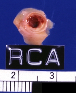
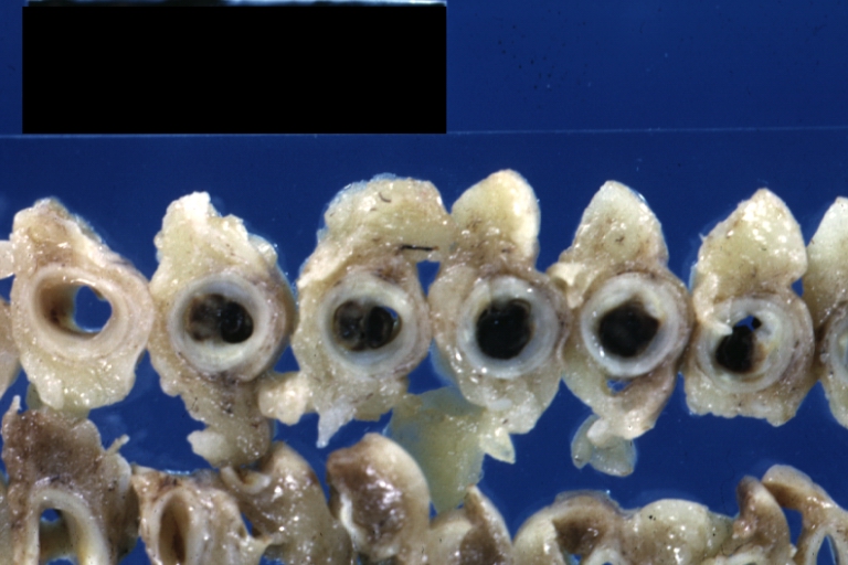
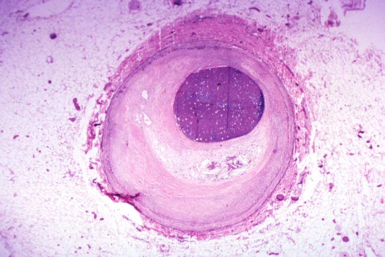
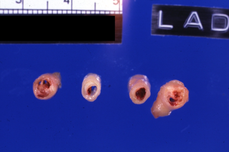 Left anterior descending coronary artery: Atherosclerosis Plaque Ruptured with Thrombosis: Gross; natural color; four cross sections, close-up view (acute anterior myocardial infarction with rupture)
 -
 Coronary artery: Atherosclerotic Plaque: Gross natural color close-up view of a typical plaque


-
 Coronary Atherosclerosis: Gross, natural color, close-up view of large atherosclerotic plaque with soft atheroma (a quite good example in 54yo male. Smoker with hypertension). This slide shows the left main artery
 -
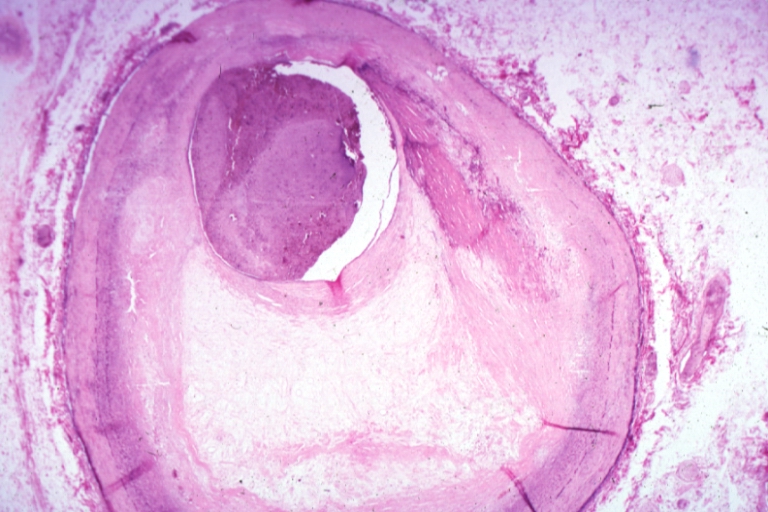
 Coronary artery: Atherosclerotic Plaque: Gross, natural color, close-up view of plaque with atheroma core causing more than 90% lumen occlusion (an excellent example)


-
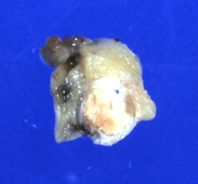
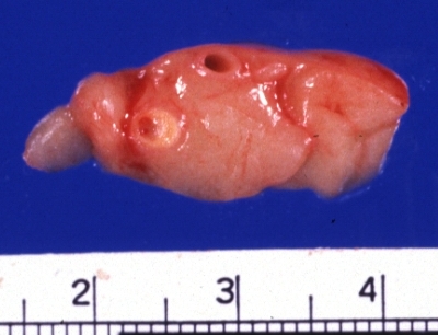
 Coronary artery: Atherosclerotic Plaque with Hemorrhage and Thrombosis: Gross, natural color, cross section, close-up, an excellent example of right coronary artery in 71yo female.
 -
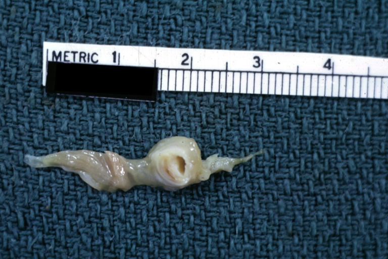
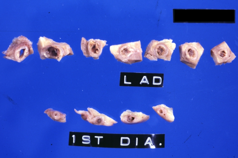
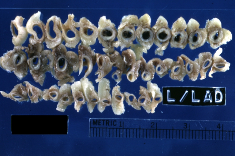
 Coronary artery: Atherosclerotic Plaque with Hemorrhage and Thrombosis: Gross, natural color, cross sections; there is excellent example of hemorrhagic plaque and thrombus at and just below the origin of first diagonal artery. Another one (a more acute one) was in the right coronary artery.


-
 Coronary artery: Atherosclerotic Plaque with Thrombus: Gross natural color, close-up of cross section.
 -
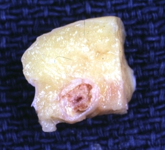
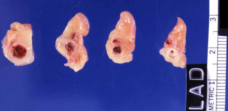
 Coronary artery: Atherosclerotic Plaque with Hemorrhage: Gross fixed tissue, cross sections. LAD and 1st diagonal with large plaques and several apparent areas of hemorrhage.


-
 Coronary artery: Atherosclerosis: Gross, an excellent close-up atherosclerosis with hemorrhage into plaque.
 -
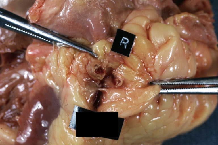
 Coronary artery: Atherosclerosis: Gross, cross sections coronary artery with hemorrhage into plaque (image shows full length of the artery).


-
 Coronary artery: Atherosclerosis: Gross, cross sections of artery showing plaques (an excellent example)
 -
 Coronary artery: Atherosclerosis: Gross natural color in situ cross section with large fibrocalcific plaque with hemorrhage (an excellent example)


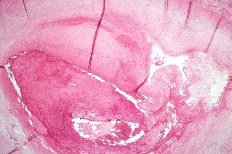
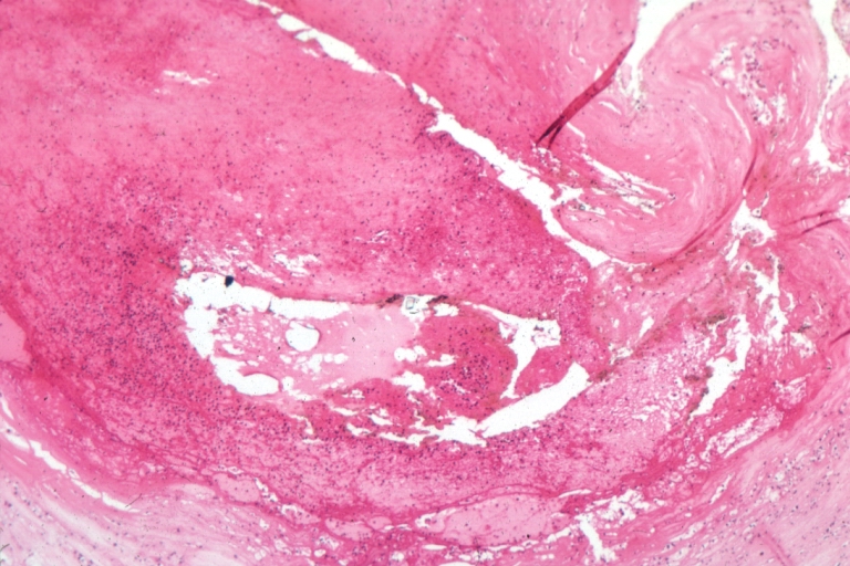
Plaque Rupture Histopathological Findings
-
 Coronary artery: Atherosclerosis: Micro H&E med mag; A good example of plaque rupture with thrombosis.
 -
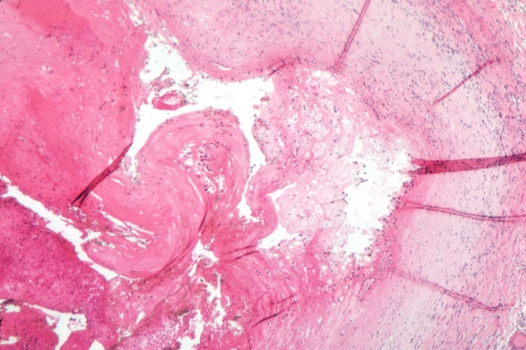 Right coronary artery: Ruptured Plaque: Micro low mag H&E; Ruptured plaque with foam cell lesion (near rupture site).


-
 Right coronary artery: Atherosclerosis Plaque Ruptured with Thrombus: Micro low mag H&E; an excellent view of ruptured plaque with thrombus and some old fibrin in it.
 -
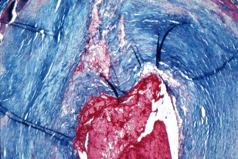 Right coronary artery: Atherosclerosis Plaque Ruptured with Thrombus: Micro low mag trichrome.


-
 Right coronary artery: Atherosclerosis Plaque Ruptured: Micro low mag H&E; large plaque with hemorrhage; (an excellent example of hemorrhage).
 -
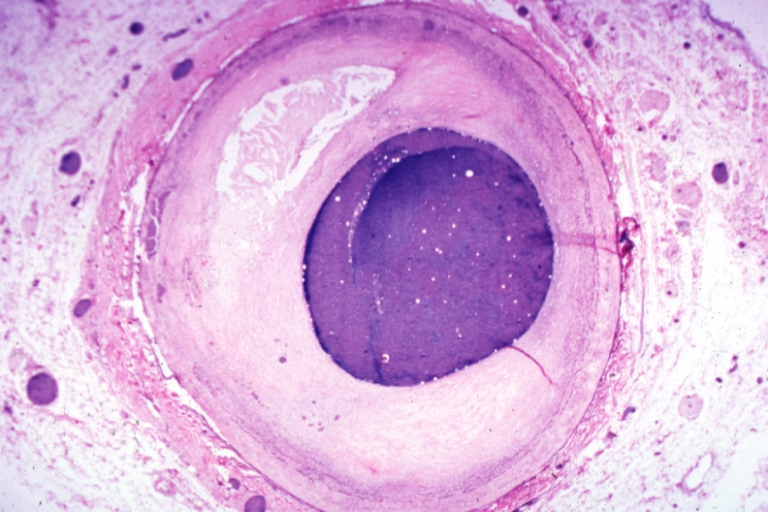 Coronary artery: Atherosclerosis: Micro H&E low mag injected artery fairly typical uncomplicated atheromatous plaque


-
 Coronary artery: Atherosclerosis: Micro H&E low mag, injected artery has typical fibrous plaque with small hemorrhage in atheroma.
 -
 Coronary artery: Atherosclerosis: Micro H&E low mag, injected artery is a very good example of marked lumen stenosis due to typical fibrous plaque with calcification


The Consequence of Plaque Rupture and Vessel Occlusion: The Time Dependent Wavefront of Necrosis

In 1940, Blumgart ligated or tied off the coronary artery in dogs and cats and for the first time demonstrated a wavefront of cell death folllowing vessel occlusion [12] [13] [14] [15] [16]
Irreversible injury of ischemic myocytes occurs first in the subendocardial zone. With more extended ischemia, a wavefront of cell death moves through the myocardium to involve progressively more of the transmural thickness of the ischemic zone. The precise location, size, and specific morphologic features of an acute myocardial infarction depend on:
- The location, severity, and rate of development of coronary atherosclerotic obstructions,
- The size of the vascular bed perfused by the obstructed vessels
- The duration of the coronary artery occlusion
- The metabolic / oxygen needs of the myocardium at risk,
- The extent of collateral blood vessels
Decrease of ATP levels in myocytes in reaction to ischemia starts within seconds and causes loss of contractility in first two minutes. If ischemia persists, ATP levels reduced to its half level within 10 minutes and to 1/10 within 40 minutes. Irreversible cell injury occurs between 20-40 minutes and microvascular level injury starts if ischemia lasts more than an hour.[17]
If impaired blood flow to the heart lasts long enough, it triggers a process called the ischemic cascade; the heart cells die (chiefly through necrosis) and do not grow back. A collagen scar forms in its place. Recent studies indicate that another form of cell death called apoptosis also plays a role in the process of tissue damage subsequent to myocardial infarction.[18] As a result, the patient’s heart can be permanently damaged. This scar tissue also puts the patient at risk for potentially life threatening arrhythmias.
Pathophysiology of ST segment elevation on the electrocardiogram
In ST segment myocaridal infarction (STEMI), the ST segments on the ECG are by definition elevated and there is myonecrosis (death of myocytes) as reflected by elevation of biomarkers such as creatine kinase MB fraction (CK-MB) or troponin T or I (tn). The ST segments are elevated due to full thickness injury of the myocardium.
Videos of STEMI pathophysiology
The following are excellent videos demonstrating the underlying pathophysiology. {{#ev:youtube|L6EiPLli5x8}} {{#ev:youtube|cOMzh2hf_Vw}} {{#ev:youtube|a8Idk4EUYTs}}
References
- ↑ Falk E, Shah PK, Fuster V (1995). “Coronary plaque disruption”. Circulation. 92 (3): 657–71. PMID 7634481. Unknown parameter
|month=ignored (help) - ↑ Burke AP, Farb A, Malcom GT, Liang YH, Smialek J, Virmani R (1997). “Coronary risk factors and plaque morphology in men with coronary disease who died suddenly”. N. Engl. J. Med. 336 (18): 1276–82. PMID 9113930. Unknown parameter
|month=ignored (help) - ↑ Moreno PR, Falk E, Palacios IF, Newell JB, Fuster V, Fallon JT (1994). “Macrophage infiltration in acute coronary syndromes. Implications for plaque rupture”. Circulation. 90 (2): 775–8. PMID 8044947. Unknown parameter
|month=ignored (help) - ↑ van der Wal AC, Becker AE, van der Loos CM, Das PK (1994). “Site of intimal rupture or erosion of thrombosed coronary atherosclerotic plaques is characterized by an inflammatory process irrespective of the dominant plaque morphology”. Circulation. 89 (1): 36–44. PMID 8281670. Unknown parameter
|month=ignored (help) - ↑ Shah PK, Falk E, Badimon JJ; et al. (1995). “Human monocyte-derived macrophages induce collagen breakdown in fibrous caps of atherosclerotic plaques. Potential role of matrix-degrading metalloproteinases and implications for plaque rupture”. Circulation. 92 (6): 1565–9. PMID 7664441. Unknown parameter
|month=ignored (help) - ↑ Moreno PR, Purushothaman KR, Fuster V; et al. (2004). “Plaque neovascularization is increased in ruptured atherosclerotic lesions of human aorta: implications for plaque vulnerability”. Circulation. 110 (14): 2032–8. doi:10.1161/01.CIR.0000143233.87854.23. PMID 15451780. Unknown parameter
|month=ignored (help) - ↑ Hung J, Lam JY, Lacoste L, Letchacovski G (1995). “Cigarette smoking acutely increases platelet thrombus formation in patients with coronary artery disease taking aspirin”. Circulation. 92 (9): 2432–6. PMID 7586342. Unknown parameter
|month=ignored (help) - ↑ 8.0 8.1 8.2 Thompson SG, Kienast J, Pyke SD, Haverkate F, van de Loo JC (1995). “Hemostatic factors and the risk of myocardial infarction or sudden death in patients with angina pectoris. European Concerted Action on Thrombosis and Disabilities Angina Pectoris Study Group”. N. Engl. J. Med. 332 (10): 635–41. PMID 7845427. Unknown parameter
|month=ignored (help) - ↑ Vaarala O, Mänttäri M, Manninen V; et al. (1995). “Anti-cardiolipin antibodies and risk of myocardial infarction in a prospective cohort of middle-aged men”. Circulation. 91 (1): 23–7. PMID 7805207. Unknown parameter
|month=ignored (help) - ↑ Ridker PM, Hennekens CH, Cerskus A, Stampfer MJ (1994). “Plasma concentration of cross-linked fibrin degradation product (D-dimer) and the risk of future myocardial infarction among apparently healthy men”. Circulation. 90 (5): 2236–40. PMID 7955179. Unknown parameter
|month=ignored (help) - ↑ Weiss EJ, Bray PF, Tayback M; et al. (1996). “A polymorphism of a platelet glycoprotein receptor as an inherited risk factor for coronary thrombosis”. N. Engl. J. Med. 334 (17): 1090–4. PMID 8598867. Unknown parameter
|month=ignored (help) - ↑ Blumgart HL, Schlesinge MJ, Davis D: Studies on the relation of the clinical manifestations of angina pectoris, coronary thrombosis, and myocardial infarction to the pathologic findings, with particular reference to the significance of collateral circulation. Amer Heart J 19: 1, 1940
- ↑ Blumgart HL, Zoll PM, Freedberg AS, Gilligan DR: The experimental production of intercoronary arterial anastomoses and their functional significance. Circulation 1: 10, 1950 PMID 15401193
- ↑ Blumgart HL, Zoll PM, Kurland CS: Discussion of direct relief of coronary occlusion. Arch Intern Med (Chicago) 104: 862, 1959 PMID 13801751
- ↑ Blumgart HL, Zoll PM. Pathologic physiology of angina pectoris and acute myocardial infarction. Circulation. 1960 Aug;22:301-7. PMID 13801752
- ↑ Blumgart HL, Zoll PM, Clinical Pathologic Correlations in Coronary Artery Disease, Circulation, Volume XLVII, No 6, June 1973, 1139-43 PMID 4575525
- ↑ Robbins Pathologic Basis of Disease, Kumar V, 7th ed
- ↑ Krijnen PA, Nijmeijer R, Meijer CJ, Visser CA, Hack CE, Niessen HW. (2002). “Apoptosis in myocardial ischaemia and infarction”. J Clin Pathol. 55 (11): 801–11. PMID 12401816.
Additional Resources
- Reimer KA, Jennings RB. The “wavefront phenomenon” of myocardial ischemic cell death. II. Transmural progression of necrosis within the framework of ischemic bed size (myocardium at risk) and collateral flow. Lab Invest. 1979 Jun 40(6): 633-44. PMID 449273
- Hasche ET, Fernandes C, Freedman SB, Jeremy RW. Relation between ischemia time, infarct size, and left ventricular function in humans. Circulation. 1995 Aug 15; 92(4): 710-9. PMID 7641348
- Gibson CM, Kirtane AJ, Morrow DA, Palabrica TM, Murphy SA, Stone PH, Scirica BM, Jennings LK, Herrmann HC, Cohen DJ, McCabe CH, Braunwald E; TIMI Study Group. Association between thrombolysis in myocardial infarction myocardial perfusion grade, biomarkers, and clinical outcomes among patients with moderate- to high-risk acute coronary syndromes: observations from the randomized trial to evaluate the relative PROTECTion against post-PCI microvascular dysfunction and post-PCI ischemia among antiplatelet and antithrombotic agents-Thrombolysis In Myocardial Infarction 30 (PROTECT-TIMI 30). Am Heart J. 2006 Oct; 152 (4): 756-61. PMID 16996854
- Christian TF, Schwartz RS, Gibbons RJ. Determinants of infarct size in reperfusion therapy for acute myocardial infarction. Circulation. 1992 Jul; 86(1): 81-90. PMID 1617793
- Gibson CM, Cannon CP, Murphy SA, Marble SJ, Barron HV, Braunwald E; TIMI Study Group. Relationship of the TIMI myocardial perfusion grades, flow grades, frame count, and percutaneous coronary intervention to long-term outcomes after thrombolytic administration in acute myocardial infarction. Circulation 2002 Apr 23; 105 (16): 1909-13. PMID 11997276
- Kandzari DE, Tcheng JE, Gersh BJ, Cox DA, Stuckey T, Turco M, Mehran R, Garcia E, Zimetbaum P, McGlaughlin MG, Lansky AJ, Costantini CO, Grines CL, Stone GW; CADILLAC Investigators. Relationship between infarct artery location, epicardial flow, and myocardial perfusion after primary percutaneous revascularization in acute myocardial infarction. Am Heart J. 2006 Jun; 151(6): 1288-95. PMID 16781238
- Elsman P, van ‘t Hof AW, de Boer MJ, Hoorntje JC, Suryapranata H, Dambrink JH, Zijlstra F; Zwolle Myocardial Infarction Study Group. Role of collateral circulation in the acute phase of ST-segment-elevation myocardial infarction treated with primary coronary intervention. Eur Heart J. 2004 May; 25(10): 854-8. PMID 15140533
- Ortiz-Pérez JT, Meyers SN, Lee DC, Kansal P, Klocke FJ, Holly TA, Davidson CJ, Bonow RO, Wu E. Angiographic estimates of myocardium at risk during acute myocardial infarction: validation study using cardiac magnetic resonance imaging. Eur Heart J. 2007 Jul;28(14):1670-2. Epub 2007 Jun 22 PMID 17586811
- Maehara A, Mintz GS, Bui AB, Walter OR, Castagna MT, Canos D, Pichard AD, Satler LF, Waksman R, Suddath WO, Laird JR Jr, Kent KM, Weissman NJ. Morphologic and angiographic features of coronary plaque rupture detected by intravascular ultrasound. J Am Coll Cardiol. 2002 Sep 4;40 (5): 904-10. PMID 12225714
- Gibson CM, Murphy SA, Kirtane AJ, Giugliano RP, Cannon CP, Antman EM, Braunwald E; TIMI Study Group. Association of duration of symptoms at presentation with angiographic and clinical outcomes after fibrinolytic therapy in patients with ST-segment elevation myocardial infarction. J Am Coll Cardiol. 2004 Sep 1; 44 (5): 980-7. PMID 15337207
- D. Garcia-Dorado, P. Theroux, M. Desco, J. Solares, J. Elizaga, F. Fernandez-Aviles, J. Alonso and J. Soriano, Cell-to-cell interaction: a mechanism to explain wave-front progression of myocardial necrosis. Am J Physiol Heart Circ Physiol 256: H1266-H1273, 1989; 0363-6135/89 $5.00 PMID 2719127
- Sorajja P, Gersh BJ, Cox DA, McLaughlin MG, Zimetbaum P, Costantini C, Stuckey T, Tcheng JE, Mehran R, Lansky AJ, Grines CL, Stone GW. Impact of multivessel disease on reperfusion success and clinical outcomes in patients undergoing primary percutaneous coronary intervention for acute myocardial infarction. Eur Heart J. 2007 Jul; 28(14): 1709-16. Epub 2007 Jun 7. PMID 17556348
- Brener S. Insights into the pathophysiology of ST-elevation myocardial infarction. American Heart Journal, Volume 151, Issue 6, Pages S4 – S10, 2006 PMID 16777509
- Biasucci LM, Leo M, De Maria GL. Local and Systemic Mechanisms of Plaque Rupture. Angiology. 2008 Jun 10. [Epub ahead of print] PMID 1854458
- El-Menyar AA. Cytokines and coronary artery disease: the state of the art. Crit Pathw Cardiol. 2008 Jun; 7(2): 139-51. PMID 18520532
- Kaneda H. Coronary plaque rupture and vessel remodeling. Am J Cardiol 2008 May 15; 101 (10): 1519; PMID 18471472
- Sorajja P, Gersh BJ, Mehran R, Lansky AJ, Krucoff MW, Webb J, Cox DA, Brodie BR, Stone GW. Impact of collateral flow on myocardial reperfusion and infarct size in patients undergoing primary angioplasty for acute myocardial infarction. Am Heart J. 2007 Aug;154(2):379-84. PMID 17643592
- Kitabata H, Kubo T, Akasaka T.Identification of multiple plaque ruptures by optical coherence tomography in a patient with acute myocardial infarction: a three-vessel study. Heart 2008; 94: 544; doi:10.1136/hrt.2007.124339 PMID 18411345
- Hong MK, Mintz GS, Lee CW, Park KM, Lee BK, Kim YH, Kang DH, Cheong SS, Song JK, Kim JJ, Park SW, Park SJ. Plaque ruptures in stable angina pectoris compared with acute coronary syndrome. Int J Cardiol. 2007 Jan 2; 114(1): 78-82. Epub 2006 May 18. PMID 1671298
- Kubo T, Imanishi T, Takarada S, Kuroi A, Ueno S, Yamano T, Tanimoto T, Matsuo Y, Masho T, Kitabata H, Tsuda K, Tomobuchi Y, Akasaka T. Assessment of culprit lesion morphology in acute myocardial infarction: ability of optical coherence tomography compared with intravascular ultrasound and coronary angioscopy. J Am Coll Cardiol. 2007 Sep 4;50(10):933-9. Epub 2007 Aug 20. PMID 17765119
- Rioufol G, Finet G, Ginon I, André-Fouët X, Rossi R, Vialle E, Desjoyaux E, Convert G, Huret JF, Tabib A. Multiple atherosclerotic plaque rupture in acute coronary syndrome: a three-vessel intravascular ultrasound study. Circulation. 2002 Aug 13; 106(7): 804-8. PMID 12176951
- Hong MK, Mintz GS, Lee CW, Lee BK, Yang TH, Kim YH, Song JM, Han KH, Kang DH, Cheong SS, Song JK, Kim JJ, Park SW, Park SJ. The site of plaque rupture in native coronary arteries: a three-vessel intravascular ultrasound analysis. J Am Coll Cardiol. 2005 Jul 19; 46 (2): 261-5. PMID 16022952
- Kusama I, Hibi K, Kosuge M, Nozawa N, Ozaki H, Yano H, Sumita S, Tsukahara K, Okuda J, Ebina T, Umemura S, Kimura K. Impact of plaque rupture on infarct size in ST-segment elevation anterior acute myocardial infarction. J Am Coll Cardiol. 2007 Sep 25;50(13):1230-7. Epub 2007 Sep 10. PMID 17888839
- Tanaka N, Ehara M, Surmely JF, Matsubara T, Terashima M, Tsuchikane E, Katoh O, Suzuki T. Images in cardiovascular medicine. Sixty-four-multislice computed tomography image of a ruptured coronary plaque. Circulation. 2006 Oct 3; 114 (14): e519-20. PMID 17015797
- Fujii K, Mintz GS, Carlier SG, Costa JR Jr, Kimura M, Sano K, Tanaka K, Costa RA, Lui J, Stone GW, Moses JW, Leon MB. Intravascular ultrasound profile analysis of ruptured coronary plaques. Am J Cardiol. 2006 Aug 15;98(4):429-35. Epub 2006 Jun 19. PMID 16893692
- Gilard M, Rioufol G, Zeller M, Cottin Y, Rochette L, Finet G. Reliability and limitations of angiography in the diagnosis of coronary plaque rupture: an intravascular ultrasound study Arch Cardiovasc Dis. 2008 Feb;101(2):114-20. PMID 18398396
- Appelbaum E, Kirtane AJ, Clark A, Pride YB, Gelfand EV, Harrigan CJ, Kissinger KV, Manning WJ, Gibson CM. Association of TIMI Myocardial Perfusion Grade and ST-segment resolution with cardiovascular magnetic resonance measures of microvascular obstruction and infarct size following ST-segment elevation myocardial infarction. J Thromb Thrombolysis. 2008 Feb 2. [Epub ahead of print] PMID 18246410
- Leshnower BG, Sakamoto H, Hamamoto H, Zeeshan A, Gorman JH 3rd, Gorman RC. Progression of myocardial injury during coronary occlusion in the collateral-deficient heart: a non-wavefront phenomenon. Am J Physiol Heart Circ Physiol. 2007 Sep;293(3):H1799-804. Epub 2007 Jul 20. PMID 17644569
Causes
Editor-In-Chief: C. Michael Gibson, M.S., M.D. [1]
Overview
Causes
- Acute myocardial infarction involving only the free wall of the right ventricle is a rare event. [1] More commonly, right ventricular infarction is associated with infarction of the inferior wall of the left ventricle, occurring in more than one-third of such cases. [2] [3] [4] [5] [6] [7] [8] [9] [10], even when thrombolytic therapy is given [11]
- Ninety percent of right ventricular infarcts result from occlusion of the proximal right coronary artery.
- Another 5 to 10 percent arise after occlusion of the left anterior descending artery. [12] [13] [14] [15]
References
- ↑ Anderson, HR, Falk, E, Nielsen, D. Right ventricular infarction: Frequency, size, and topography in coronary heart disease. J Am Coll Cardiol 1987; 10:1223. PMID 3680789
- ↑ Anderson, HR, Falk, E, Nielsen, D. Right ventricular infarction: Frequency, size, and topography in coronary heart disease. J Am Coll Cardiol 1987; 10:1223. PMID 3680789
- ↑ Isner, JM, Roberts, WC. Right ventricular infarction complicating left ventricular infarction secondary to coronary artery disease: frequency, location, associated findings and significance from analysis of 236 necropsy patients with acute or healed myocardial infarction. Am J Cardiol 1978; 42:885. PMID 153103
- ↑ Isner, JM. Right ventricular myocardial infarction. JAMA 1988; 259:712. PMID 3275819
- ↑ Williams, JF. Right ventricular infarction. Clin Cardiol 1990; 13:309. PMID 2189611
- ↑ Cabin, HS, Clubb, S, Wackers FJ, et al. Right ventricular myocardial infarction with anterior wall left ventricular infarction: an autopsy study. Am Heart J 1987; 113:16. PMID 3799430
- ↑ Kinch, JW, Ryan, TJ. Right ventricular infarction. N Engl J Med 1994; 330:1211. PMID 8139631
- ↑ Setaro, JF, Cabin, HS. Right ventricular infarction. Cardiol Clin 1992; 10:69. PMID 1739961
- ↑ Cohn, JN. Right ventricular infarction revisited. Am J Cardiol 1979; 43:666. PMID 420117
- ↑ Wackers, FJ, Lie, KI, Sokole, EB, et al. Prevalence of right ventricular involvement in inferior wall infarction associated with myocardial imaging with thallium-201 and technetium-99m pyrophosphate. Am J Cardiol 1978; 42:358. PMID 210648
- ↑ Zeymer, U, Neuhaus, K-L, Wegscheider, K, et al. Effects of thrombolytic therapy in acute inferior myocardial infarction with and without right ventricular involvement. J Am Coll Cardiol 1998; 32:876. PMID 9768705
- ↑ Isner, JM, Roberts, WC. Right ventricular infarction complicating left ventricular infarction secondary to coronary artery disease: frequency, location, associated findings and significance from analysis of 236 necropsy patients with acute or healed myocardial infarction. Am J Cardiol 1978; 42:885.PMID 153103
- ↑ Isner, JM. Right ventricular myocardial infarction. JAMA 1988; 259:712. PMID 3275819
- ↑ Williams, JF. Right ventricular infarction. Clin Cardiol 1990; 13:309. PMID 2189611
- ↑ Cabin, HS, Clubb, S, Wackers FJ, et al. Right ventricular myocardial infarction with anterior wall left ventricular infarction: an autopsy study. Am Heart J 1987; 113:16. PMID 3799430
Differentiating Right ventricular myocardial infarction from other Diseases
Editor-In-Chief: C. Michael Gibson, M.S., M.D. [1]
Overview
Differentiating Right ventricular myocardial infarction from other Diseases
- Acute pericarditis
- Constrictive pericarditis
- Cor Pulmonale
- Endomyocardial fibrosis
- Hypertrophic Cardiomyopathy
- Pneumothorax
- Primary pulmonary hypertension
- Pulmonary Embolism
- Restrictive cardiomyopathy
- Secondary pulmonary hypertension
- Tricuspid Regurgitation
References
Epidemiology and Demographics
Editor-In-Chief: C. Michael Gibson, M.S., M.D. [1]; Associate Editors-In-Chief: Yuri B. Pride, M.D. [2] ; Cafer Zorkun, M.D., Ph.D. [3]
Overview
Myocardial infarction is a common presentation of ischemic heart disease. The World Heart Organization (WHO) estimated in 2002 that, 12.6 percent of deaths worldwide were from ischemic heart disease.
Ischemic heart disease is the leading cause of death in developed countries, but third to AIDS and lower respiratory infections in developing countries.[1]
Epidemiology
Over 9 million patients in the United States alone have angina. An estimated 80,700,000 American adults (one in three) have one or more types of cardiovascular disease (CVD), of whom 38,200,000 are estimated to be age 60 or older. Except as noted, the estimates were extrapolated to the U.S. population in 2005 from NHANES 1999–2004. (Total CVD includes diseases in the bullet points below except for congenital heart disease). Due to overlap, it is not possible to add these conditions to arrive at a total. [2] [3][4]
- Hypertension: 73,000,000
- Coronary heart disease: 16,000,000
- Myocardial infarction: 8,100,000
- Angina pectoris: 9,100,000
- Heart failure: 5,300,000
- Stroke: 5,800,000
- Congenital heart disease: 650,000 – 1,300,000
This means that roughly every 65 seconds, an American dies of a coronary event.
Incidence
Although it is difficult to ascertain the true incidence of ST elevation myocardial infarction (STEMI), according to the ACC/AHA guidelines, a conservative estimate is that approximately 500,000 patients suffer STEMI each year [5]. The incidence of STEMI has decreased over time. In an observational study of 5,832 metropolitan patients spanning from 1975 to 1997, the incidence of STEMI decreased from 171/100,000 to 101/100,000 [6]
Prevalence
The following prevalence estimates are for people age 18 and older from NCHS/NHIS, 2005: [7]
- Among whites only, 12.0% have heart disease, 6.6% have CHD, 21.0% have hypertension and 2.3% have had a stroke.
- Among blacks, 10.2% have heart disease, 6.2% have CHD, 31.2% have hypertension and 3.4% have had a stroke.
- Among Hispanics or Latinos, 8.3% have heart disease, 5.9% have CHD, 20.3% have hypertension and 2.2% have had a stroke.
- Among Asians, 6.7% have heart disease, 3.8% have CHD, 19.4% have hypertension and 2.0% have had a stroke.
- Among Native Hawaiians or other Pacific Islanders, 22.4% have hypertension (other prevalence estimates considered unreliable).
The mortality among patients who suffer STEMI has progressively declined in recent years. From 1975 to 1997, one observational study reported that the in-hospital mortality decreased from 24% to 14% [6]. In the Global Registry of Acute Coronary Events (GRACE), a multinational cohort study that includes 16,814 patients with STEMI were enrolled and followed up in 113 hospitals in 14 countries between 1999 and 2006, in-hospital mortality declined from 8.4% in 1999 to 4.6% in 2005 [8].
The reason for this decline in mortality is likely multifactorial and includes, but is certainly not limited to, decline in symptom onset-to-presentation time, more widespread use of primary PCI [9], improvements in time to reperfusion (door-to-needle and door-to-balloon times) [10][11] and improved medical therapy, including increases in the use of evidence-based therapies such as aspirin [12], beta blockers[13] [14], clopidogrel [15], statins [16] and angiotension converting enzyme inhibitors or angiotensin receptor blockers [17].
References
- ↑ “Cause of Death – UC Atlas of Global Inequality”. Center for Global, International and Regional Studies (CGIRS) at the University of California Santa Cruz. Unknown parameter
|accessyear=ignored (|access-date=suggested) (help); Unknown parameter|accessmonthday=ignored (help) - ↑ 2008 Heart Disease and Stroke Statistics
- ↑ Anderson JL, Adams CD, Antman EM; et al. (2007). “ACC/AHA 2007 guidelines for the management of patients with unstable angina/non ST-elevation myocardial infarction: a report of the American College of Cardiology/American Heart Association Task Force on Practice Guidelines (Writing Committee to Revise the 2002 Guidelines for the Management of Patients With Unstable Angina/Non ST-Elevation Myocardial Infarction): developed in collaboration with the American College of Emergency Physicians, the Society for Cardiovascular Angiography and Interventions, and the Society of Thoracic Surgeons: endorsed by the American Association of Cardiovascular and Pulmonary Rehabilitation and the Society for Academic Emergency Medicine”. Circulation. 116 (7): e148–304. doi:10.1161/CIRCULATIONAHA.107.181940. PMID 17679616. Unknown parameter
|month=ignored (help) - ↑ Anderson JL, Adams CD, Antman EM; et al. (2007). “ACC/AHA 2007 guidelines for the management of patients with unstable angina/non-ST-Elevation myocardial infarction: a report of the American College of Cardiology/American Heart Association Task Force on Practice Guidelines (Writing Committee to Revise the 2002 Guidelines for the Management of Patients With Unstable Angina/Non-ST-Elevation Myocardial Infarction) developed in collaboration with the American College of Emergency Physicians, the Society for Cardiovascular Angiography and Interventions, and the Society of Thoracic Surgeons endorsed by the American Association of Cardiovascular and Pulmonary Rehabilitation and the Society for Academic Emergency Medicine”. J. Am. Coll. Cardiol. 50 (7): e1–e157. doi:10.1016/j.jacc.2007.02.013. PMID 17692738. Unknown parameter
|month=ignored (help) - ↑ Antman EM, Anbe DT, Armstrong PW; et al. (2004). “ACC/AHA guidelines for the management of patients with ST-elevation myocardial infarction; A report of the American College of Cardiology/American Heart Association Task Force on Practice Guidelines (Committee to Revise the 1999 Guidelines for the Management of patients with acute myocardial infarction)”. J. Am. Coll. Cardiol. 44 (3): E1–E211. doi:10.1016/j.jacc.2004.07.014. PMID 15358047. Unknown parameter
|month=ignored (help) - ↑ 6.0 6.1 Furman MI, Dauerman HL, Goldberg RJ, Yarzebski J, Lessard D, Gore JM (2001). “Twenty-two year (1975 to 1997) trends in the incidence, in-hospital and long-term case fatality rates from initial Q-wave and non-Q-wave myocardial infarction: a multi-hospital, community-wide perspective”. J. Am. Coll. Cardiol. 37 (6): 1571–80. PMID 11345367. Unknown parameter
|month=ignored (help) - ↑ Vital Health Stat 10.2006 [232]: 1–153
- ↑ Fox KA, Steg PG, Eagle KA; et al. (2007). “Decline in rates of death and heart failure in acute coronary syndromes, 1999-2006”. JAMA. 297 (17): 1892–900. doi:10.1001/jama.297.17.1892. PMID 17473299. Unknown parameter
|month=ignored (help) - ↑ Rogers WJ, Canto JG, Lambrew CT; et al. (2000). “Temporal trends in the treatment of over 1.5 million patients with myocardial infarction in the US from 1990 through 1999: the National Registry of Myocardial Infarction 1, 2 and 3”. J. Am. Coll. Cardiol. 36 (7): 2056–63. PMID 11127441. Unknown parameter
|month=ignored (help) - ↑ McNamara RL, Wang Y, Herrin J; et al. (2006). “Effect of door-to-balloon time on mortality in patients with ST-segment elevation myocardial infarction”. J. Am. Coll. Cardiol. 47 (11): 2180–6. doi:10.1016/j.jacc.2005.12.072. PMID 16750682. Unknown parameter
|month=ignored (help) - ↑ Nallamothu B, Fox KA, Kennelly BM; et al. (2007). “Relationship of treatment delays and mortality in patients undergoing fibrinolysis and primary percutaneous coronary intervention. The Global Registry of Acute Coronary Events”. Heart. 93 (12): 1552–5. doi:10.1136/hrt.2006.112847. PMID 17591643. Unknown parameter
|month=ignored (help) - ↑ “Randomised trial of intravenous streptokinase, oral aspirin, both, or neither among 17,187 cases of suspected acute myocardial infarction: ISIS-2. ISIS-2 (Second International Study of Infarct Survival) Collaborative Group”. Lancet. 2 (8607): 349–60. 1988. PMID 2899772. Unknown parameter
|month=ignored (help) - ↑ “Metoprolol in acute myocardial infarction. Mortality. The MIAMI Trial Research Group”. Am. J. Cardiol. 56 (14): 15G–22G. 1985. PMID 3904389. Unknown parameter
|month=ignored (help) - ↑ “Randomised trial of intravenous atenolol among 16 027 cases of suspected acute myocardial infarction: ISIS-1. First International Study of Infarct Survival Collaborative Group”. Lancet. 2 (8498): 57–66. 1986. PMID 2873379. Unknown parameter
|month=ignored (help) - ↑ Sabatine MS, Cannon CP, Gibson CM; et al. (2005). “Addition of clopidogrel to aspirin and fibrinolytic therapy for myocardial infarction with ST-segment elevation”. N. Engl. J. Med. 352 (12): 1179–89. doi:10.1056/NEJMoa050522. PMID 15758000. Unknown parameter
|month=ignored (help) - ↑ Cannon CP, Braunwald E, McCabe CH; et al. (2004). “Intensive versus moderate lipid lowering with statins after acute coronary syndromes”. N. Engl. J. Med. 350 (15): 1495–504. doi:10.1056/NEJMoa040583. PMID 15007110. Unknown parameter
|month=ignored (help) - ↑ Latini R, Maggioni AP, Flather M, Sleight P, Tognoni G (1995). “ACE inhibitor use in patients with myocardial infarction. Summary of evidence from clinical trials”. Circulation. 92 (10): 3132–7. PMID 7586285. Unknown parameter
|month=ignored (help)
Risk Factors
Editor-In-Chief: C. Michael Gibson, M.S., M.D. [1]; Associate Editor(s)-in-Chief: Cafer Zorkun, M.D., Ph.D. [2]
Overview
In these chapters on ST elevation, the word risk factors refers to those epidemiologic and genetic variables that expose someone to a higher risk of developing atherosclerotic plaque. The word triggers refer to those factors in the patients immediate history or environment that may have lead to rupture of the atherosclerotic plaque.
Nomenclature
Muller et al have developed the following nomenclature to categorize and analyze data pertaining to triggers of MI [1]:
- (1) Trigger: An activity that produces short-term physiological changes that may lead directly to onset of acute CVD.
- (2) Acute risk factor: A short-term physiological change, such as a surge in arterial pressure or heart rate, an increase in coagulability, or vasoconstriction, that follows a trigger and may result in disease onset.
- (3) Hazard period: The time interval after trigger initiation associated with an increased risk of disease onset because of the trigger. The onset and offset times of the hazard period, which could also be designated a “vulnerable period,” may be sharply defined, as in heavy exertion, or less well defined, as with respiratory infection. The duration of the hazard period may also vary, eg from < 1 hour during heavy physical exertion to weeks or months with bereavement.
- (4) Triggered acute risk prevention (TARP): Cardiovascular risk reduction that focuses on the short-term increase in risk associated with a trigger.
Risk Factors
Traditional Risk Factors for Atherosclerosis[2]
Risk factors for atherosclerosis are generally risk factors for myocardial infarction:
- Advancing age
- Male gender[3]
- Cigarette smoking
- Hypercholesterolemia (more accurately hyperlipoproteinemia, especially high low density lipoprotein and low high density lipoprotein)
- Diabetes (with or without insulin resistance)
- High blood pressure
- Obesity[4] (defined by a body mass index of more than 30 kg/m², or alternatively by waist circumference or waist-hip ratio).
- Elevated homocysteine
Genetic Disorders
- Mendelian inherited conditions
- Autosomal dominant conditions
- Pseudoxanthoma elasticum dominant type 1
- Autosomal recessive conditions
- Cystathionine beta-synthase deficiency
- Sitosterolemia
Family History and Heart Disease
According to present trends in the United States, half of healthy 40-year-old males and 1 in 3 healthy 40-year-old women will develop coronary artery disease (CAD) in the future.[5] This chance is greater when a family history of heart disease is involved.
Studies have reported that the risk of CAD increases two- to sevenfold when there is a genetic link. [6] The studies by Nora and Nora (1978a) evaluated a large number of patients with congenital heart disease.[7] Results show that about 8% of the defects were primarily genetics-related, 2% were environmental (caused by drugs, viruses, maternal nutrition, maternal metabolism, or fetal hemodynamics) and 90% were multifactorial (due to a combination of environmental and genetic factors). The study also noted that there is a two- to threefold increase in the recurrence risk for congenital heart disease when two of the family members are affected. This is particularly the case when the parent is severely affected or when the affected members are child and parent. The risk for a myocardial infarction is two to three times greater in a first-degree relative than in a person with no family history of heart disease. This fact speaks in particular to the early presentation of CAD in patients with no other risk factors. Second cousins have a 1 in 23 chance of sharing a particular gene and third cousins have a 1 in 128 likelihood.
Genetic hypertension and hyperlipidemia are strong predictors of familial heart disease.[8] Hypertensive people are twice as likely to have a family history of the condition than are normotensives. Genetic hypertension is an example of a polygenic mode of inheritance that reflects cellular sodium transport defects and an abnormal response to psychogenic stress. There is indication that Pheochromocytoma, a rare cause of hypertension, may be familial as well.
Endocrine Conditions
- Diabetes mellitus type 2 (NIDDM)
- Primary hyperparathyroidism
Rheumatologic and Autoimmune Conditions
Socioeconomic Factors
Socioeconomic factors such as a shorter education and lower income (particularly in women), and living with a partner may also contribute to the risk of MI.[9] To understand epidemiological study results, it’s important to note that many factors associated with MI mediate their risk via other factors. For example, the effect of education is partially based on its effect on income and marital status.[9]
Women who use combined oral contraceptive pills have a modestly increased risk of myocardial infarction, especially in the presence of other risk factors, such as smoking.[10]
Inflammation is known to be an important step in the process of atherosclerotic plaque formation.[11] C-reactive protein (CRP) is a sensitive but non-specific marker for inflammation. Elevated CRP blood levels, especially measured with high sensitivity assays, can predict the risk of MI, as well as stroke and development of diabetes.[11] Moreover, some drugs for MI might also reduce CRP levels.[11] The use of high sensitivity CRP assays as a means of screening the general population is advised against, but it may be used optionally at the physician’s discretion, in patients who already present with other risk factors or known coronary artery disease.[12] Whether CRP plays a direct role in atherosclerosis remains uncertain.[11]
Inflammation in periodontal disease may be linked coronary heart disease, and since periodontitis is very common, this could have great consequences for public health.[13] Serological studies measuring antibody levels against typical periodontitis-causing bacteria found that such antibodies were more present in subjects with coronary heart disease.[14] Periodontitis tends to increase blood levels of CRP, fibrinogen and cytokines;[15] thus, periodontitis may mediate its effect on MI risk via other risk factors.[16] Preclinical research suggests that periodontal bacteria can promote aggregation of platelets and promote the formation of foam cells.[17][18] A role for specific periodontal bacteria has been suggested but remains to be established.[19]
Controversial Risk Factors
Baldness, hair greying, a diagonal earlobe crease[20] and possibly other skin features are independent risk factors for MI. Their role remains controversial; a common denominator of these signs and the risk of MI is supposed, possibly genetic.[21]
Risk Factor Modification
While family history, along with age and sex, is uncontrollable[3], many of the risk factors for heart disease can be eliminated by maintaining a healthy lifestyle. In fact, a person’s daily habits significantly affect the risk of heart disease due to genetic predisposition. Studies have shown that deaths can be avoided by attending to the controllable risk factors. Such risk factors include: obesity, blood pressure, cigarette smoking and plasma cholesterol.[22]
A study published in Circulation shines some light on the importance of eliminating these controllable risk factors in people with a family history of heart disease. Data demonstrated that in men with a genetic link to heart disease, an estimated 68% of the excess deaths were due to the addition of smoking, a modifiable risk factor; concluding that the risk of heart disease in men with a family history of the condition is significantly related to the additional risk factors he accumulates.
References
- ↑ Muller JE, Abela GS, Nesto RW, Tofler GH (1994). “Triggers, acute risk factors and vulnerable plaques: the lexicon of a new frontier”. J. Am. Coll. Cardiol. 23 (3): 809–13. PMID 8113568. Unknown parameter
|month=ignored (help) - ↑ “Executive Summary of The Third Report of The National Cholesterol Education Program (NCEP) Expert Panel on Detection, Evaluation, And Treatment of High Blood Cholesterol In Adults (Adult Treatment Panel III)”. JAMA. 285 (19): 2486–97. 2001. PMID 11368702. Unknown parameter
|month=ignored (help) - ↑ 3.0 3.1 Wilson PW, D’Agostino RB, Levy D, Belanger AM, Silbershatz H, Kannel WB. (1998). “Prediction of coronary heart disease using risk factor categories” (PDF). Circulation. 97 (18): 1837–47. PMID 9603539.
- ↑ Yusuf S, Hawken S, Ounpuu S, Bautista L, Franzosi MG, Commerford P, Lang CC, Rumboldt Z, Onen CL, Lisheng L, Tanomsup S, Wangai P Jr, Razak F, Sharma AM, Anand SS; INTERHEART Study Investigators. (2005). “Obesity and the risk of myocardial infarction in 27,000 participants from 52 countries: a case-control study”. Lancet. 366 (9497): 1640–9. PMID 16271645.
- ↑ Rosamond W, Flegal K, Friday G (2007). “Heart disease and stroke statistics–2007 update: a report from the American Heart Association Statistics Committee and Stroke Statistics Subcommittee”. Circulation. 115 (5): e69–171. doi:10.1161/CIRCULATIONAHA.106.179918. PMID 17194875. Unknown parameter
|month=ignored (help) - ↑ Khaw KT, Barrett-Connor E (1986). “Family history of heart attack: a modifiable risk factor?”. Circulation. 74 (2): 239–44. PMID 3731415. Unknown parameter
|month=ignored (help) - ↑ http://www.ncbi.nlm.nih.gov/bookshelf/br.fcgi?book=cm&part=A540#A556
- ↑ http://www.ncbi.nlm.nih.gov/bookshelf/br.fcgi?book=cm&part=A540#A556
- ↑ 9.0 9.1 Nyboe J, Jensen G, Appleyard M, Schnohr P. (1989). “Risk factors for acute myocardial infarction in Copenhagen. I: Hereditary, educational and socioeconomic factors. Copenhagen City Heart Study”. Eur Heart J. 10 (10): 910–6. PMID 2598948.
- ↑ Khader YS, Rice J, John L, Abueita O. (2003). “Oral contraceptives use and the risk of myocardial infarction: a meta-analysis”. Contraception. 68 (1): 11–7. PMID 12878281.
- ↑ 11.0 11.1 11.2 11.3 Wilson AM, Ryan MC, Boyle AJ. (2006). “The novel role of C-reactive protein in cardiovascular disease: risk marker or pathogen”. Int J Cardiol. 106 (3): 291–7. PMID 16337036.
- ↑ Pearson TA, Mensah GA, Alexander RW, Anderson JL, Cannon RO 3rd, Criqui M, Fadl YY, Fortmann SP, Hong Y, Myers GL, Rifai N, Smith SC Jr, Taubert K, Tracy RP, Vinicor F; Centers for Disease Control and Prevention; American Heart Association. (2003). “Markers of inflammation and cardiovascular disease: application to clinical and public health practice: A statement for healthcare professionals from the Centers for Disease Control and Prevention and the American Heart Association” (PDF). Circulation. 107 (3): 499–511. PMID 12551878.
- ↑ Janket SJ, Baird AE, Chuang SK, Jones JA. (2003). “Meta-analysis of periodontal disease and risk of coronary heart disease and stroke”. Oral Surg Oral Med Oral Pathol Oral Radiol Endod. 95 (5): 559–69. PMID 12738947.
- ↑ Pihlstrom BL, Michalowicz BS, Johnson NW. (2005). “Periodontal diseases”. Lancet. 366 (9499): 1809–20. PMID 16298220.
- ↑ Scannapieco FA, Bush RB, Paju S. (2003). “Associations between periodontal disease and risk for atherosclerosis, cardiovascular disease, and stroke. A systematic review”. Ann Periodontol. 8 (1): 38–53. PMID 14971247.
- ↑ D’Aiuto F, Parkar M, Nibali L, Suvan J, Lessem J, Tonetti MS. (2006). “Periodontal infections cause changes in traditional and novel cardiovascular risk factors: results from a randomized controlled clinical trial”. Am Heart J. 151 (5): 977–84. PMID 16644317.
- ↑ Lourbakos A, Yuan YP, Jenkins AL, Travis J, Andrade-Gordon P, Santulli R, Potempa J, Pike RN. (2001). “Activation of protease-activated receptors by gingipains from Porphyromonas gingivalis leads to platelet aggregation: a new trait in microbial pathogenicity” (PDF). Blood. 97 (12): 3790–7. PMID 11389018.
- ↑ Qi M, Miyakawa H, Kuramitsu HK. (2003). “Porphyromonas gingivalis induces murine macrophage foam cell formation”. Microb Pathog. 35 (6): 259–67. PMID 14580389.
- ↑ Spahr A, Klein E, Khuseyinova N, Boeckh C, Muche R, Kunze M, Rothenbacher D, Pezeshki G, Hoffmeister A, Koenig W. (2006). “Periodontal infections and coronary heart disease: role of periodontal bacteria and importance of total pathogen burden in the Coronary Event and Periodontal Disease (CORODONT) study”. Arch Intern Med. 166 (5): 554–9. PMID 16534043.
- ↑ Lichstein E, Chadda KD, Naik D, Gupta PK. (1974). “Diagonal ear-lobe crease: prevalence and implications as a coronary risk factor”. N Engl J Med. 290 (11): 615–6. PMID 4812503.
- ↑ Miric D, Fabijanic D, Giunio L, Eterovic D, Culic V, Bozic I, Hozo I. (1998). “Dermatological indicators of coronary risk: a case-control study”. Int J Cardiol. 67 (3): 251–5. PMID 9894707.
- ↑ Khaw KT, Barrett-Connor E (1986). “Family history of heart attack: a modifiable risk factor?”. Circulation. 74 (2): 239–44. PMID 3731415. Unknown parameter
|month=ignored (help)
Natural History, Complications and Prognosis
Editor-In-Chief: C. Michael Gibson, M.S., M.D. [1]
Overview
The presence of right ventricular infarction adversely affects the early prognosis. Elderly patients who have right ventricular involvement with an inferior wall myocardial infarction are at particularly high risk. For patients who survive an acute right ventricular infarction, however, the prognosis is generally good.
Prognosis
- The presence of right ventricular infarction adversely affects the early prognosis. One study, for example, evaluated 200 consecutive patients with acute inferior myocardial infarction. [1] Those with ST elevation in V4R had an almost eight-fold increase in in-hospital mortality (31 versus 6 percent) and morbidity when compared to those without changes in V4R.
- Elderly patients who have right ventricular involvement with an inferior wall myocardial infarction are at particularly high risk. In a study of 198 patients ≥ 75 years of age, right ventricular involvement was associated with an in-hospital mortality of 47 percent compared to a 10 percent mortality in the absence of right ventricular involvement [2]
- For patients who survive an acute right ventricular infarction, however, the prognosis is generally good. As an example, among 522 patients with an inferior wall infarction who were treated with a thrombolytic agent and hirudin or heparin in the HIT-4 study, 32 percent had right ventricular involvement and these patients had a higher 30 day mortality when compared to those without right ventricular involvement (5.9 versus 2.5 percent. [3] However, this was related to a larger infarct size rather than right ventricular involvement; right ventricular involvement was not an independent predictor of survival.
- The right ventricle frequently recovers the majority of its function, probably due at least in part to a decreased oxygen demand of the thin-walled right ventricle. [4] [5] These patients may, however, have a more frequent requirement for a permanent pacemaker. [6]
References
- ↑ Zehender, M, Kasper, W, Kauder, E, et al. Right ventricular infarction as an independent predictor of prognosis after acute inferior myocardial infarction. N Engl J Med 1993; 328:981. PMID 8450875
- ↑ Bueno, H, Lopez-Palop, R, Bermejo, J, et al. In-hospital outcome of elderly patients with acute inferior myocardial infarction and right ventricular involvement. Circulation 1997; 96:436. PMID 9788824
- ↑ Zeymer, U, Neuhaus, K-L, Wegscheider, K, et al. Effects of thrombolytic therapy in acute inferior myocardial infarction with and without right ventricular involvement. J Am Coll Cardiol 1998; 32:876. PMID 9768705
- ↑ Williams, JF. Right ventricular infarction. Clin Cardiol 1990; 13:309. PMID 2189611
- ↑ Cohn, JN. Right ventricular infarction revisited. Am J Cardiol 1979; 43:666. PMID 420117
- ↑ Zehender, M, Kasper, W, Kauder, E, et al. Right ventricular infarction as an independent predictor of prognosis after acute inferior myocardial infarction. N Engl J Med 1993; 328:981. PMID 8450875
Diagnosis
Diagnosis
Diagnostic Criteria | History and Symptoms | Physical Examination | Electrocardiogram | Chest X Ray | Echocardiography or Ultrasound | Coronary Angiography | Other Imaging Findings
Treatment
Treatment
Pharmacologic Reperfusion: Reperfusion Therapy (Overview of Fibrinolysis and Primary PCI) | Fibrinolysis
Mechanical Reperfusion: The Importance of Reducing Door-to-Balloon Times | Primary PCI | Adjunctive and Rescue PCI | Rescue PCI | Facilitated PCI | Adjunctive PCI | CABG | Management of Patients Who Were Not Reperfused | Assessing Success of Reperfusion
Antithrombin Therapy: Antithrombin Therapy | Unfractionated Heparin | Low Molecular Weight Heparinoid Therapy | Direct Thrombin Inhibitor Therapy | Factor Xa Inhibition | DVT Prophylaxis | Long Term Anticoagulation
Antiplatelet Agents: Aspirin | Thienopyridine Therapy | Glycoprotein IIbIIIa Inhibition
Other Initial Therapy: Inhibition of the Renin-Angiotensin-Aldosterone System | Magnesium Therapy | Glucose Control | Calcium Channel Blocker Therapy
Looking for the patient version?
© 2026 MyEClinic – IFTM Institut für Telematik in der Medizin GmbH