
Thromboembolism
For the patient information page on deep vein thrombosis, click here
For the patient information page on pulmonary embolism, click here
Editor-In-Chief: C. Michael Gibson, M.S., M.D. [1] Associate Editor-In-Chief: Cafer Zorkun, M.D., Ph.D. [2]
Overview
Editor-In-Chief: C. Michael Gibson, M.S., M.D. [1]
Overview
Thromboembolism is a general term describing both thrombosis and its main complication which is embolism.
Historical Perspective
The term was coined in 1848 by Rudolph Carl Virchow.[1]
References
- ↑ Hellemans, Alexander (1988). The Timetables of Science. New York, New York: Simon and Schuster. p. 317. ISBN 0671621300. Unknown parameter
|coauthors=ignored (help)
Historical Perspective
Please help WikiDoc by adding more content here. It’s easy! Click here to learn about editing.
Editor-In-Chief: C. Michael Gibson, M.S., M.D. [1]
Overview
The term was coined in 1848 by Rudolph Carl Virchow.[1]
References
- ↑ Hellemans, Alexander (1988). The Timetables of Science. New York, New York: Simon and Schuster. p. 317. ISBN 0671621300. Unknown parameter
|coauthors=ignored (help)
Classification
Please help WikiDoc by adding more content here. It’s easy! Click here to learn about editing.
Editor-In-Chief: C. Michael Gibson, M.S., M.D. [1]
Classification
Classification for Thrombus Based on Vessel Type
There are two distinct forms of thrombosis:
Venous Thrombosis
- Deep venous thrombosis (with or without pulmonary embolism; together classified as venous thromboembolism/VTE)
- Portal vein thrombosis
- Renal vein thrombosis
- hepatic vein thrombosis (Budd-Chiari syndrome)
- Paget-Schroetter disease
- Cerebral venous sinus thrombosis
- Thoracic outlet syndrome (the cause of most Subclavian vein thrombosis unrelated to trauma)
Arterial Thrombosis
Classification of Embolism Based on Direction of Blood Flow
If a bacterial infection is present at the site of thrombosis, the thrombus may break down, spreading particles of infected material throughout the circulatory system (pyemia, septic embolus) and setting up metastatic abscesses wherever they come to rest.
Without an infection, the thrombus may become detached and enter circulation as an embolus, finally lodging in and completely obstructing a blood vessel (an infarction). The effects of an infarction depend on where it occurs.
The pathway of the embolism can be one of three types:
- Anterograde
- Retrograde
- Paradoxical
In anterograde embolism, the movement of emboli is in the direction of blood flow. In retrograde embolism, however, the emboli move in opposition to the blood flow direction; this is usually significant only in blood vessels with low pressure (veins) or with emboli of high weight. In paradoxical embolism, also known as crossed embolism, an embolus from the veins crosses to the arterial blood system. This is generally found only with heart problems such as septal defects between the atria or ventricles.
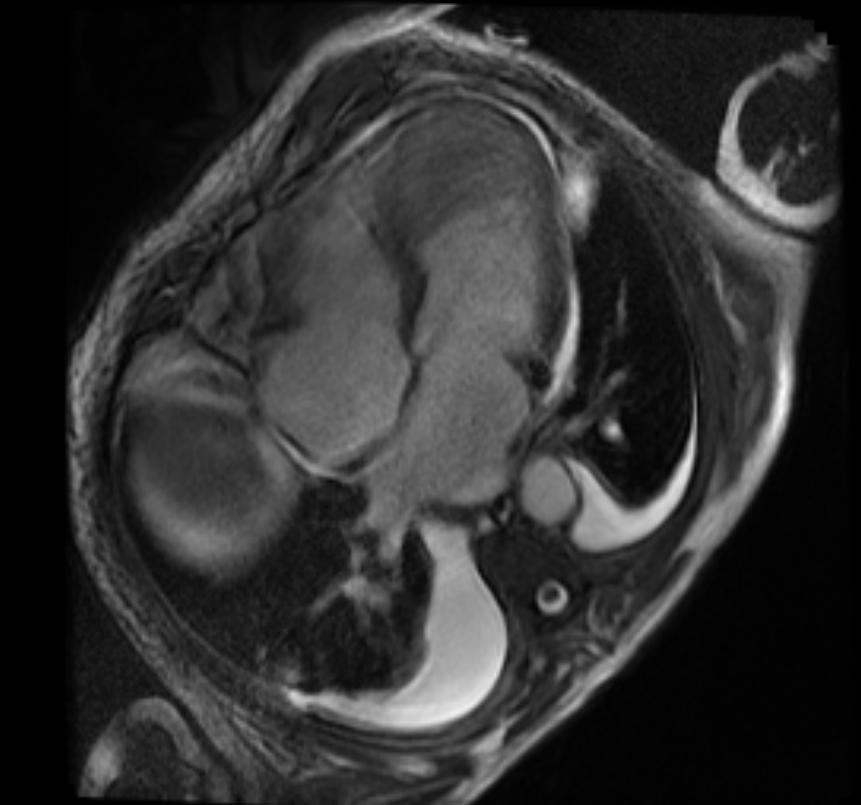
Sources of Systemic Embolism
- Left ventricular thrombus
- Left atrial thrombus
- Pelvic veins or lower extremity thrombus
- Native cardiac valves
- Prosthetic cardiac valves
- Cardiac tumors
- Aortic aneurysm
- Invasive manipulations
- Left ventricular aneurysm
-
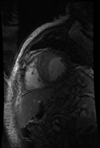 Sources of Systemic Embolism: Large left ventricular thrombus in a patient with myocardial infarction
 -
 Sources of Systemic Embolism: Large left ventricular thrombus in a patient with myocardial infarction


References
Pathophysiology
Please help WikiDoc by adding more content here. It’s easy! Click here to learn about editing.
Editor-In-Chief: C. Michael Gibson, M.S., M.D. [1]
Pathophysiology
The formation of a thrombus is usually caused by the top three causes, known as (Virchow‘s triad): (Classically, thrombosis is caused by abnormalities in one or more of the following)
- The composition of the blood (hypercoagulability)
- Quality of the vessel wall (endothelial cell injury)
- Nature of the blood flow (hemostasis)
To elaborate, the pathogenesis includes:
- An injury to the vessel’s wall (such as by trauma, infection, or turbulent flow at bifurcations);
- By the slowing or stagnation of blood flow past the point of injury (which may occur after long periods of sedentary behavior (for example, sitting on a long airplane flight);
- By a blood state of hypercoagulability (caused for example, by genetic deficiencies or autoimmune disorders).
High altitude has also been known to induce thrombosis [1] [2]. Occasionally, abnormalities in coagulation are to blame. Intravascular coagulation follows, forming a structureless mass of red blood cells, leukocytes, and fibrin.
Gross Pathology
Thromboembolism
A 67-year-old male was hospitalized because of extensive atherosclerotic cardiovascular disease. Following surgery, during which diseased portions of the femoral arteries were bypassed, he developed massive pulmonary embolism and expired. At autopsy, thrombi were found in the femoral and iliac veins, as well as in the larger pulmonary arteries.


Thromboembolism: Testes

Thromboembolism: Bowel Infarction

Coronary Thrombosis

Artificial Heart Valve Thrombosis


Microscopic Pathology
Thromboembolism


Coronary Thrombosis


Sources of Systemic Embolism
- Left ventricular thrombus
- Left atrial thrombus
- Pelvic veins or lower extremity thrombus
- Native cardiac valves
- Prosthetic cardiac valves
- Cardiac tumors
- Aortic aneurysm
- Invasive manipulations
- Left ventricular aneurysm
References
- ↑ Kuipers S, Cannegieter SC, Middeldorp S, Robyn L, Büller HR, et al. The Absolute Risk of Venous Thrombosis after Air Travel: A Cohort Study of 8,755 Employees of International Organisations PLoS Medicine Vol. 4, No. 9, e290 doi:10.1371/journal.PMID 0040290
- ↑ http://www.mounteverest.net/news.php?news=16349 Mount Everest experience
Causes
Life Threatening Causes
Life-threatening causes include conditions which may result in death or permanent disability within 24 hours if left untreated.
Common Causes
Causes by Organ System
| Cardiovascular | No underlying causes |
| Chemical/Poisoning | No underlying causes |
| Dental | No underlying causes |
| Dermatologic | No underlying causes |
| Drug Side Effect |
Axitinib, Cytomegalovirus immune globulin Dexamethasone, Doxorubicin Hydrochloride, Erythropoietin, Ethynodiol diacetate and ethinyl estradiol, Follitropin beta, Norgestimate and Ethinyl estradiol, Norgestrel and Ethinyl estradiol, Lenvatinib, Prednisolone, Sorafenib |
| Ear Nose Throat | No underlying causes |
| Endocrine | No underlying causes |
| Environmental | No underlying causes |
| Gastroenterologic | No underlying causes |
| Genetic | No underlying causes |
| Hematologic | No underlying causes |
| Iatrogenic | No underlying causes |
| Infectious Disease | No underlying causes |
| Musculoskeletal/Orthopedic | No underlying causes |
| Neurologic | No underlying causes |
| Nutritional/Metabolic | No underlying causes |
| Obstetric/Gynecologic | No underlying causes |
| Oncologic | No underlying causes |
| Ophthalmologic | No underlying causes |
| Overdose/Toxicity | No underlying causes |
| Psychiatric | No underlying causes |
| Pulmonary | No underlying causes |
| Renal/Electrolyte | No underlying causes |
| Rheumatology/Immunology/Allergy | No underlying causes |
| Sexual | No underlying causes |
| Trauma | No underlying causes |
| Urologic | No underlying causes |
| Miscellaneous | No underlying causes |
Causes in Alphabetical Order
|
|
|
|
References
Differentiating Thromboembolism from other Diseases
Please help WikiDoc by adding content here. It’s easy! Click here to learn about editing.
References
Epidemiology and Demographics
Please help WikiDoc by adding more content here. It’s easy! Click here to learn about editing.
Editor-In-Chief: C. Michael Gibson, M.S., M.D. [1]
Epidemiology and Demographics
Incidence
In the United States:
- 300,000–600,000 people have deep venous thrombosis (DVT) or pulmonary embolism (PE).
- 200,000–400,000 people have deep venous thrombosis.
- Nearly one-third of people who have had deep venous thrombosis have post-thrombotic syndrome, a chronic disabling condition characterized by swelling, pain, discoloration, and scaling in the affected limb.
- 100,000–200,000 people have a pulmonary embolism.
- Nearly one-third of people (30,000–60,000) who have a pulmonary embolism die.
- 5-8% of people have thrombophilia (inherited blood clotting disorders).
References
Risk Factors
Editor-In-Chief: C. Michael Gibson, M.S., M.D. [1]
Risk Factors
Almost anyone can have thromboembolic event. However, certain factors can increase the risk of developing this condition. The risk increases even more for someone who has more than one risk factor at the same time.
- Sedentary life style
- Elderly
- Atrial fibrillation
- Sepsis
- Prolonged bed rest and / or immobility
- Pregnancy
- Hyperemesis
- Dehydration
- Cesarean delivery
- Congestive heart failure
- Nephrotic syndrome
- Severe varicose veins
- Family history of thromboembolism
- Thrombophilia
- Factor V Leiden mutation
- Prothrombin G20210A mutation
- Decreased antithrombin level
- Decreased Protein C level
- Decreased Protein S level
- Antiphospholipid syndrome
- Long air travel
- Oral contraceptives
- Obesity
- Smoking
- Minor injuries[1]
Following is a List of Factors that Increase the Risk of Developing Deep Vein Thrombosis
- Injury to the vein, often caused by:
- Fractures,
- Severe muscle injury,
- Major surgery (particularly involving the abdomen, pelvis, hip, or legs).
- Slow blood flow, often caused by:
- Confinement to bed (e.g., due to a medical condition or after surgery);
- Limited movement (e.g., a cast on a leg to help heal an injured bone;
- Sitting for a long time, especially with crossed legs; or
- Paralysis.
- Increased estrogen, often caused by:
- Birth control pills;
- Hormone replacement therapy, sometimes used after menopause; or
- Pregnancy, for up to 6 weeks after giving birth.
- Certain chronic medical illnesses, such as:
- Trauma
- Multiple trauma
- CNS/spinal cord injury
- Burns
- Lower extremity fractures
- Other risk factors include:
- Previous DVT
- Family history of DVT
- Age (risk increases as age increases)
- Obesity
- Smoking
- High blood pressure
- A catheter located in a central vein
- Inherited clotting disorders. An inherited clotting disorder might be suspected when a person has repeated DVTs that cannot be linked to any specific cause (such as recent surgery) or develops DVT in a vein at an unusual location, such as a vein in the liver, kidney, or brain.
References
- ↑ Karlijn J. van Stralen, MSc; Frits R. Rosendaal, MD, PhD; Carine J. M. Doggen, PhD (January 14, 2008). “Minor Injuries as a Risk Factor for Venous Thrombosis”. Arch Intern Med. 168 No. 1: 21–26. doi:10.1001/archinternmed.2007.5. PMID 18195191.
Screening
Please help WikiDoc by adding content here. It’s easy! Click here to learn about editing.
References
Natural History, Complications and Prognosis
Please help WikiDoc by adding more content here. It’s easy! Click here to learn about editing.
Editor-In-Chief: C. Michael Gibson, M.S., M.D. [1]
Complications
- Postthrombotic syndrome
- Chronic thromboembolic pulmonary hypertension
References
Diagnosis
Diagnosis
Diagnostic Criteria | History and Symptoms | Physical Examination | Laboratory Findings | EKG | Chest X ray | CT | MRI | Ultrasound | Other Imaging Findings | Other Diagnostic Studies
Treatment
Treatment
Medical Therapy | Surgery | Primary Prevention | Secondary Prevention | Cost-Effectiveness of Therapy | Future or Investigational Therapies
Related Chapters
Related Chapters
- Atrial fibrillation
- Cerebrovascular accident
- Deep venous thrombosis
- Infective endocarditis
- Pulmonary embolism
- Thrombosis
- Embolism
Template:Skin and subcutaneous tissue symptoms and signs Template:Nervous and musculoskeletal system symptoms and signs Template:Urinary system symptoms and signs Template:Cognition, perception, emotional state and behaviour symptoms and signs Template:Speech and voice symptoms and signs Template:General symptoms and signs
Looking for the patient version?
© 2026 MyEClinic – IFTM Institut für Telematik in der Medizin GmbH