
Mantle cell lymphoma
For patient information click here
Editor-In-Chief: C. Michael Gibson, M.S., M.D. [1]; Associate Editor(s)-in-Chief: Ali Akram, M.B.B.S.[2] Sowminya Arikapudi, M.B,B.S. [3]
Synonyms and keywords: Mantle cell B-cell lymphoma, Mantle zone lymphoma, MCL
Overview
Editor-In-Chief: C. Michael Gibson, M.S., M.D. [1]; Associate Editor(s)-in-Chief: Ali Akram, M.B.B.S.[2] Sowminya Arikapudi, M.B,B.S. [3]
Overview
Mantle cell lymphoma is a subtype of B-cell lymphoma characterized by the presence of CD5 positive antigen-naive, pregerminal center, B-cells within the mantle zone that surrounds normal germinal center follicles. Mantle cell lymphoma accounts for 4–8% of all adult non-Hodgkin’s lymphomas (NHL). It has an incidence of approximately 1-2/100000 and tends to occur more in males, Caucasian race, with a median age of about 60 years.The translocation t(11;14)(q13;q32) is considered the precipitating oncogenic event that induces cell cycle deregulation in mantle cell lymphoma due to overexpression of cyclin D1. In addition to the pathogonomic translocation, MCL progression is controlled by secondary genetic abberations and dysregulated signaling pathways involved in DNA damage repair, proliferation, and apoptosis. According to the revised 2016 World Health Organization classification of lymphoid neoplasms, mantle cell lymphoma (MCL) can be broadly classified into classical MCL and leukemic nonnodal MCL. In-situ mantle cell neoplasia (ISMCN) is considered a separate entity, often as a precursor lesion, to the mantle cell lymphoma. The causes of mantle cell lymphoma have not been clearly identified. There are no established risk factors for mantle cell lymphoma. However, recently weak associations have been observed in the development of mantle cell lymphoma with the exposure to European strains of Borellia burgdoferi, family history of hematologic malignancy and Genetic polymorphisms in the pro-inflammatory cytokine IL-10. Mantle cell lymphoma must be differentiated from other diseases that present similarly with B symptoms (fever, night sweats and unexplained weight loss), lymphocytosis, lymphadenopathy, hepatosplenomegaly, and bone marrow involvement. Mantle cell lymphoma must be differentiated from other diseases such as diffuse large B cell lymphoma, mucosa-associated lymphatic tissue lymphoma (MALT), follicular lymphoma, small lymphocytic lymphoma/chronic lymphocytic leukemia, lymphoplasmacytoid lymphoma/Immunocytoma, marginal zone lymphoma, lymphoblastic lymphoma, burkitt lymphoma and reactive hyperplasia. Screening for mantle cell lymphoma is not recommended. The prognosis of mantle cell lymphoma has historically been very poor. However, recently improvements have been made and the median survival has increased from 3-4 years to 5-7 years. It is very important to stratify the patients according to their biological risk to better direct the therapeutic approaches. Tissue biopsy (nodal or extranodal) is the gold standard test for the diagnosis of mantle cell lymphoma. The most common symptoms of mantle cell lymphoma include fever, weight loss, night sweats, fatigue, skin rash, abdominal pain, bone pain, loss of appetite, and painless swelling in the neck, axilla, groin, thorax and abdomen. Common physical examination findings of mantle cell lymphoma include fever, rash, splenomegaly, hepatomegaly, peripheral lymphadenopathy, and central lymphadenopathy. Laboratory tests for mantle cell lymphoma include complete blood count (CBC), comprehensive metabolic panel, LDH levels, Hepatitis B testing if treatment with rituximab is planned, uric acid levels, beta-2-microglobulin, pregnancy testing in woman of child-bearing age, flow cytometry, immunohistochemistry, genetic testing and FISH. Ultrasound may be helpful in the diagnosis of mantle cell lymphoma. Findings on an ultrasound helpful in the diagnosis of mantle cell lymphoma include lymphadenopathy, splenomegaly and hepatomegaly. There are no echocardiography findings associated with mantle cell lymphoma. However, an echocardiography may be helpful in following patients who might be at risk of anthracycline-induced cardiotoxicity due to chemotherapy. CT scan may be helpful in the diagnosis of mantle cell lymphoma. CT scans of the chest, abdominal and pelvic are done to check for lymphadenopathy and is helpful in the staging the disease. MRI may be helpful in the diagnosis of mantle cell lymphoma involving the CNS. PET scan (positron emission tomography) integrated with CT scan may be helpful in the staging and treatment response assessment of mantle cell lymphoma. Other diagnostic studies for the diagnosis of mantle cell lymphoma include bone marrow aspiration, lumbar puncture, colonoscopy, upper endoscopy, laparoscopy, and laparotomy.The mainstay of treatment for mantle cell lymphoma is chemotherapy. However, immunotherapy, radioimmunotherapy, targeted therapy using newer biologic agents and stem cell transplantation are also used along with chemotherapy to treat the disease. Mantle cell lymphoma shows a heterogeneous clinical behavior, with some patients having indolent disease whereas a vast majority show aggressive presentation. Most of the patients eventually relapse and have disease progression after treatment. Hence, mantle cell lymphoma is still considered an incurable disease and there is no consensus among oncologists about its optimal treatment. It is therefore recommended that mantle cell lymphoma patients are seen by physicians having extensive experience in dealing with mantle cell lymphoma and they are also encouraged to participate in clinical trials to get the latest treatments. Recent advances in the understanding of the pathogenesis of mantle cell lymphoma have led to the development of targeted therapies which have shown potential promise as effective therapeutic approaches in the future.
Historical Perspective
In 1982, Weisenburger first proposed the concept of ‘mantle-zone lymphoma’ and in 1992, Banks first coined the term mantle cell lymphoma (MCL). In 1994, MCL was included into the Revised European-American Classification of Lymphoid Neoplasms (REAL) classification, and later also in the World Health Organization (WHO) Classification of Tumors of Haematopoietic and Lymphoid Tissues
Classification
According to the revised 2016 World Health Organization classification of lymphoid neoplasms, mantle cell lymphoma (MCL) can be broadly classified into two types:
- Classical MCL.
- Leukemic nonnodal MCL.
In-situ mantle cell neoplasia (ISMCN) is considered a separate entity, often as a precursor lesion, to the mantle cell lymphoma.
Pathophysiology
The translocation t(11;14)(q13;q32) is considered the precipitating oncogenic event that induces cell cycle deregulation in mantle cell lymphoma due to overexpression of cyclin D1. In addition to the pathogonomic translocation, MCL progression is controlled by secondary genetic abberations and dysregulated signaling pathways involved in DNA damage repair, proliferation, and apoptosis.
Causes
The causes of mantle cell lymphoma have not been clearly identified.
Differentiating Mantle Cell Lymphoma from Other Diseases
Mantle cell lymphoma must be differentiated from other diseases that present similarly with B symptoms (fever, night sweats and unexplained weight loss), lymphocytosis, lymphadenopathy, hepatosplenomegaly, and bone marrow involvement. Mantle cell lymphoma must be differentiated from other diseases such as diffuse large B cell lymphoma, mucosa-associated lymphatic tissue lymphoma (MALT), follicular lymphoma, small lymphocytic lymphoma/chronic lymphocytic leukemia, lymphoplasmacytoid lymphoma/Immunocytoma, marginal zone lymphoma, lymphoblastic lymphoma, burkitt lymphoma and reactive hyperplasia.
Epidemiology and Demographics
Mantle cell lymphoma accounts for 4–8% of all adult non-Hodgkin’s lymphomas (NHL). It has an incidence of approximately 1-2/100000 and tends to occur more in males, Caucasian race, with a median age of about 60 years.
Risk Factors
There are no established risk factors for mantle cell lymphoma. However, recently weak associations have been observed in the development of mantle cell lymphoma with the exposure to European strains of Borellia burgdoferi., family history of hematologic malignancy and Genetic polymorphisms in the pro-inflammatory cytokine IL-10.
Screening
Screening for mantle cell lymphoma is not recommended.
Natural History, Complications, and Prognosis
The prognosis of mantle cell lymphoma has historically been very poor. However, recently improvements have been made and the median survival has increased from 3-4 years to 5-7 years. It is very important to stratify the patients according to their biological risk to better direct the therapeutic approaches.
Diagnosis
Diagnostic Study of Choice
Tissue biopsy (nodal or extranodal) is the gold standard test for the diagnosis of mantle cell lymphoma.
History and Symptoms
The most common symptoms of mantle cell lymphoma include fever, weight loss, night sweats, fatigue, skin rash, abdominal pain, bone pain, loss of appetite, and painless swelling in the neck, axilla, groin, thorax and abdomen.
Physical Examination
Common physical examination findings of mantle cell lymphoma include fever, rash, splenomegaly, hepatomegaly, peripheral lymphadenopathy, and central lymphadenopathy.
Laboratory Findings
Laboratory tests for mantle cell lymphoma include complete blood count (CBC), comprehensive metabolic panel, LDH levels, Hepatitis B testing if treatment with rituximab is planned, uric acid levels, beta-2-microglobulin, pregnancy testing in woman of child-bearing age, flow cytometry, immunohistochemistry, genetic testing and FISH.
Electrocardiogram
There are no ECG findings associated with mantle cell lymphoma.
X-ray
Presence of nodules and pleural effusion on chest x ray may be suggestive of mantle cell lymphoma.
Echocardiography and Ultrasound
Ultrasound may be helpful in the diagnosis of mantle cell lymphoma. Findings on an ultrasound helpful in the diagnosis of mantle cell lymphoma include lymphadenopathy, splenomegaly and hepatomegaly. There are no echocardiography findings associated with mantle cell lymphoma. However, an echocardiography may be helpful in following patients who might be at risk of anthracycline-induced cardiotoxicity due to chemotherapy.
CT scan
CT scan may be helpful in the diagnosis of mantle cell lymphoma. CT scans of the chest, abdominal and pelvic are done to check for lymphadenopathy and is helpful in the staging the disease.
MRI
MRI may be helpful in the diagnosis of mantle cell lymphoma involving the CNS.
Other Imaging Findings
PET scan (positron emission tomography) integrated with CT scan may be helpful in the staging and treatment response assessment of mantle cell lymphoma.
Other Diagnostic Studies
Other diagnostic studies for the diagnosis of mantle cell lymphoma include bone marrow aspiration, lumbar puncture, colonoscopy, upper endoscopy, laparoscopy, and laparotomy.
Treatment
Medical Therapy
The mainstay of treatment for mantle cell lymphoma is chemotherapy. However, immunotherapy, radioimmunotherapy, targeted therapy using newer biologic agents and stem cell transplantation are also used along with chemotherapy to treat the disease. Mantle cell lymphoma shows a heterogeneous clinical behavior, with some patients having indolent disease whereas a vast majority show aggressive presentation. Most of the patients eventually relapse and have disease progression after treatment. Hence, mantle cell lymphoma is still considered an incurable disease and there is no consensus among oncologists about its optimal treatment. It is therefore recommended that mantle cell lymphoma patients are seen by physicians having extensive experience in dealing with mantle cell lymphoma and they are also encouraged to participate in clinical trials to get the latest treatments.
Interventions
The mainstay of treatment for mantle cell lymphoma is medical therapy.
Surgery
The mainstay of treatment for mantle cell lymphoma is medical therapy. However, surgical therapy has proven curative in a case of MCL with a single protruding lesion presenting as intussusception.
Primary Prevention
There are no established measures for the primary prevention of mantle cell lymphoma.
Secondary Prevention
There are no established measures for the secondary prevention of mantle cell lymphoma.
Future or investigational therapies:
Recent advances in the understanding of the pathogenesis of mantle cell lymphoma have led to the development of targeted therapies which have shown potential promise as effective therapeutic approaches in the future. In addition to the ongoing assessment of new monoclonal antibody-based therapies, the continued development of targeted molecular signaling inhibitors based on the underlying biology of MCL is an approach that will potentially yield fruitful results in this disease. Some of the current therapies under clinical investigation are the BCL-2 inhibitor venetoclax (ABT-199), the phosphatydilinosytol 3-kinase δ (PI3K δ) inhibitor idelalisib, chimeric antigen receptor T-cell (CAR-T) therapy in relapsed MCL, androgen receptor (AR) blockers like enzalutamide as means of decreasing MCL cell proliferation and a few next-generation proteasome inhibitors (carfilzomib, oprozomib, ixazomib) in bortezomib-resistant MCL patients.
References
Historical Perspective
Editor-In-Chief: C. Michael Gibson, M.S., M.D. [1]; Associate Editor(s)-in-Chief: Ali Akram, M.B.B.S.[2]
Overview
In 1982, Weisenburger first proposed the concept of mantle-zone lymphoma and in 1992, Banks first coined the term mantle cell lymphoma (MCL). In 1994, MCL was included into the Revised European-American Classification of Lymphoid Neoplasms (REAL) classification, and later also in the World Health Organization (WHO) Classification of Tumors of Haematopoietic and Lymphoid Tissues
Historical Perspective
Discovery
- In 1964, Lennert first described the disease as ‘centrocytic’ lymphoma.
- In 1982, Weisenburger first proposed the concept of ‘mantle-zone lymphoma’.[1]
- In 1985, Pileri et al. reported on 18 cases of small B-cell lymphomas which displayed a mantle-fashion growth around reactive germinal centers.[2]
- In 1992, Banks first coined the term mantle cell lymphoma (MCL).[3]
- In 1994, MCL was included into the Revised European-American Classification of Lymphoid Neoplasms (REAL) classification, and later also in the World Health Organization (WHO) Classification of Tumors of Haematopoietic and Lymphoid Tissues.
References
- ↑ D. D. Weisenburger, H. Kim & H. Rappaport (1982). “Mantle-zone lymphoma: a follicular variant of intermediate lymphocytic lymphoma”. Cancer. 49 (7): 1429–1438. PMID 6895860. Unknown parameter
|month=ignored (help) - ↑ Pileri S, Rivano MT, Gobbi M, Taruscio D, Lennert K (1985). “Neoplastic and reactive follicles within B-cell malignant lymphomas. A morphological and immunological study of 30 cases”. Hematol Oncol. 3 (4): 243–60. PMID 3936767.
- ↑ Banks PM, Chan J, Cleary ML, Delsol G, De Wolf-Peeters C, Gatter K; et al. (1992). “Mantle cell lymphoma. A proposal for unification of morphologic, immunologic, and molecular data”. Am J Surg Pathol. 16 (7): 637–40. PMID 1530105.
Classification
Editor-In-Chief: C. Michael Gibson, M.S., M.D. [1]; Associate Editor(s)-in-Chief: Ali Akram, M.B.B.S.[2]
Overview
According to the revised 2016 World Health Organization classification of lymphoid neoplasms, mantle cell lymphoma (MCL) can be broadly classified into two types include classical MCL and leukemic nonnodal MCL. In-situ mantle cell neoplasia (ISMCN) is considered a separate entity, often as a precursor lesion, to the mantle cell lymphoma.
Classification
Classical MCL
- Classical MCL typically develops from immunoglobulin heavy-chain variable region gene(IGHV)-unmutated or minimally mutated B cells that usually express SOX11 (SRY-Box 11 gene).[1][2][3]
- Classical MCL usually involves the lymph nodes and other extranodal sites.
- However, these cells can acquire further molecular and cytogenetic abnormalities leading to the more aggressive:[4][5]
- Blastoid MCL
- Pleomorphic MCL
Leukemic nonnodal MCL
- Leukemic nonnodal MCL typically develops from immunoglobulin heavy-chain variable region gene(IGHV)-mutated B cells that do not express SOX11 (SRY-Box 11 gene).[1][2]
- Leukemic nonnodal MCL involve the peripheral blood (PB), bone marrow and the spleen.
- Although these are commonly clinically indolent in nature further mutations ,especially of TP53, can lead to more aggressive disease.
In-situ mantle cell neoplasia (ISMCN)
- It is characterized by the presence of cyclin D1 positive cells mostly in the inner mantle zones of follicles in lymphoid tissues that do not otherwise suggest the diagnosis of a MCL.[6]
- The in situ lesion stage may be a common step in both SOX11-negative and positive subtypes of MCL as some in situ lesions express SOX11, whereas others are SOX11 negative.[7]
- ISMCN is often found incidentally, occasionally in association with other types of lymphomas.
References
- ↑ 1.0 1.1 Swerdlow SH, Campo E, Pileri SA, Harris NL, Stein H, Siebert R; et al. (2016). “The 2016 revision of the World Health Organization classification of lymphoid neoplasms”. Blood. 127 (20): 2375–90. doi:10.1182/blood-2016-01-643569. PMC 4874220. PMID 26980727.
- ↑ 2.0 2.1 Jares P, Colomer D, Campo E (2012). “Molecular pathogenesis of mantle cell lymphoma”. J Clin Invest. 122 (10): 3416–23. doi:10.1172/JCI61272. PMC 3461905. PMID 23023712.
- ↑ Jaffe ES, Harris NL, Stein H, Isaacson PG (December 2008). “Classification of lymphoid neoplasms: the microscope as a tool for disease discovery”. Blood. 112 (12): 4384–99. doi:10.1182/blood-2008-07-077982. PMC 2954680. PMID 19029456.
- ↑ Fisher RI (1996). “Mantle-cell lymphoma: classification and therapeutic implications”. Ann. Oncol. 7 Suppl 6: S35–9. PMID 9010577.
- ↑ Pileri SA, Falini B (November 2009). “Mantle cell lymphoma”. Haematologica. 94 (11): 1488–92. doi:10.3324/haematol.2009.013359. PMC 2770958. PMID 19880776.
- ↑ Carvajal-Cuenca A, Sua LF, Silva NM, Pittaluga S, Royo C, Song JY; et al. (2012). “In situ mantle cell lymphoma: clinical implications of an incidental finding with indolent clinical behavior”. Haematologica. 97 (2): 270–8. doi:10.3324/haematol.2011.052621. PMC 3269489. PMID 22058203.
- ↑ Christian B, Zhao W, Hamadani M, Sotomayor EM, Navarro W, Devine SM; et al. (2010). “Mantle cell lymphoma 12 years after allogeneic bone marrow transplantation occurring simultaneously in recipient and donor”. J Clin Oncol. 28 (31): e629–32. doi:10.1200/JCO.2010.29.8992. PMID 20733121.
Pathophysiology
Editor-In-Chief: C. Michael Gibson, M.S., M.D. [1]; Associate Editor(s)-in-Chief: Ali Akram, M.B.B.S.[2] Sowminya Arikapudi, M.B,B.S. [3]
Overview
The translocation t(11;14)(q13;q32) is considered the precipitating oncogenic event that induces cell cycle deregulation in mantle cell lymphoma due to overexpression of cyclin D1. In addition to the pathogonomic translocation, MCL progression is controlled by secondary genetic abberations and dysregulated signaling pathways involved in DNA damage repair, proliferation, and apoptosis.
Pathophysiology
Pathogenesis
- The translocation t(11;14)(q13;q32) is considered the precipitating oncogenic event that induces cell cycle deregulation due to overexpression of cyclin D1.
- This translocation juxtaposes the CCDN1 gene encoding cyclin D1 to the immunoglobulin heavy chain (IgH) leading to its overexpression. However, less commonly, mutations in CCDN2 and CCDN3 have also been identified in cases of mantle cell lymphoma lacking the t(11;14) translocation.[1]
- In addition to the pathogonomic translocation, MCL progression is controlled by secondary genetic abberations and dysregulated signaling pathways involved in DNA damage repair, proliferation, and apoptosis.[2]
- These secondary genetic alterations and dysregulated signaling pathways are as follows:
- SOX11, a SOX family transcription factor, has recently been identified as an important molecular feature of MCL regardless of cyclin D1 status.[13]
Genetics
Genes involved in the pathogenesis of mantle cell lymphoma include:
Microscopic Pathology
- The typical (classical) morphology of mantle cell lymphoma is seen in about 90% of cases:[14]
- Cells are slightly larger than lymphocytes.
- The nucleus is indented or bean-shaped.
- Prominent nucleoli are missing.
- The chromatin structure is less condensed and not as bright as in blasts like centroblasts.
- The cytoplasmic rim is narrow and not basophilic.
- The remaining 10% are the morphological variants:[15]
- Small cell type: They resemble CLL cytologically but the prolymphocytes and paraimmunoblasts are usually missing. This variant usually shows a very low proliferative activity.
- Pleomorphic and blastic type(together known as the blastoid variant): The pleomorphic type are similar to diffuse large B-cell lymphoma (DLBCL). The blastic type resemble lymphoblastic lymphoma or leukemia and have monomorphic roundish blasts. The blastoid variant usually have numerous mitotic figures and are very highly proliferative.
- Scattered non-granumloma forming epithelioid macrophages, hyalinized vessels and a few reactive T cells or mast cells are seen along with the lymphoma cells in MCL. Follicular dendritic cells (FDC) are a hallmark of mantle cell lymphoma and may also be involved in its pathogenesis.[16]
- Three types of growth pattern are observed in mantle cell lymphoma:[17]
- Mantle zone: Nodular with germinal centers
- Nodular: >50% nodular growth
- Diffuse: <50% nodular growth
References
- ↑ Itziar Salaverria, Cristina Royo, Alejandra Carvajal-Cuenca, Guillem Clot, Alba Navarro, Alejandra Valera, Joo Y. Song, Renata Woroniecka, Grzegorz Rymkiewicz, Wolfram Klapper, Elena M. Hartmann, Pierre Sujobert, Iwona Wlodarska, Judith A. Ferry, Philippe Gaulard, German Ott, Andreas Rosenwald, Armando Lopez-Guillermo, Leticia Quintanilla-Martinez, Nancy L. Harris, Elaine S. Jaffe, Reiner Siebert, Elias Campo & Silvia Bea (2013). “CCND2 rearrangements are the most frequent genetic events in cyclin D1(-) mantle cell lymphoma”. Blood. 121 (8): 1394–1402. doi:10.1182/blood-2012-08-452284. PMID 23255553. Unknown parameter
|month=ignored (help) - ↑ Patricia Perez-Galan, Martin Dreyling & Adrian Wiestner (2011). “Mantle cell lymphoma: biology, pathogenesis, and the molecular basis of treatment in the genomic era”. Blood. 117 (1): 26–38. doi:10.1182/blood-2010-04-189977. PMID 20940415. Unknown parameter
|month=ignored (help) - ↑ Pedro Jares, Dolors Colomer & Elias Campo (2012). “Molecular pathogenesis of mantle cell lymphoma”. The Journal of clinical investigation. 122 (10): 3416–3423. doi:10.1172/JCI61272. PMID 23023712. Unknown parameter
|month=ignored (help) - ↑ Silvia Bea, Rafael Valdes-Mas, Alba Navarro, Itziar Salaverria, David Martin-Garcia, Pedro Jares, Eva Gine, Magda Pinyol, Cristina Royo, Ferran Nadeu, Laura Conde, Manel Juan, Guillem Clot, Pedro Vizan, Luciano Di Croce, Diana A. Puente, Monica Lopez-Guerra, Alexandra Moros, Gael Roue, Marta Aymerich, Neus Villamor, Lluis Colomo, Antonio Martinez, Alexandra Valera, Jose I. Martin-Subero, Virginia Amador, Luis Hernandez, Maria Rozman, Anna Enjuanes, Pilar Forcada, Ana Muntanola, Elena M. Hartmann, Maria J. Calasanz, Andreas Rosenwald, German Ott, Jesus M. Hernandez-Rivas, Wolfram Klapper, Reiner Siebert, Adrian Wiestner, Wyndham H. Wilson, Dolors Colomer, Armando Lopez-Guillermo, Carlos Lopez-Otin, Xose S. Puente & Elias Campo (2013). “Landscape of somatic mutations and clonal evolution in mantle cell lymphoma”. Proceedings of the National Academy of Sciences of the United States of America. 110 (45): 18250–18255. doi:10.1073/pnas.1314608110. PMID 24145436. Unknown parameter
|month=ignored (help) - ↑ J. M. Adams & S. Cory (2007). “The Bcl-2 apoptotic switch in cancer development and therapy”. Oncogene. 26 (9): 1324–1337. doi:10.1038/sj.onc.1210220. PMID 17322918. Unknown parameter
|month=ignored (help) - ↑ Y. Li, M. N. Bouchlaka, J. Wolff, K. M. Grindle, L. Lu, S. Qian, X. Zhong, N. Pflum, P. Jobin, B. S. Kahl, J. C. Eickhoff, S. M. Wuerzberger-Davis, S. Miyamoto, C. J. Thomas, D. T. Yang, C. M. Capitini & L. Rui (2016). “FBXO10 deficiency and BTK activation upregulate BCL2 expression in mantle cell lymphoma”. Oncogene. 35 (48): 6223–6234. doi:10.1038/onc.2016.155. PMID 27157620. Unknown parameter
|month=ignored (help) - ↑ Joseph D. Khoury, L. Jeffrey Medeiros, George Z. Rassidakis, Timothy J. McDonnell, Lynne V. Abruzzo & Raymond Lai (2003). “Expression of Mcl-1 in mantle cell lymphoma is associated with high-grade morphology, a high proliferative state, and p53 overexpression”. The Journal of pathology. 199 (1): 90–97. doi:10.1002/path.1254. PMID 12474231. Unknown parameter
|month=ignored (help) - ↑ Jenny Zhang, Dereje Jima, Andrea B. Moffitt, Qingquan Liu, Magdalena Czader, Eric D. Hsi, Yuri Fedoriw, Cherie H. Dunphy, Kristy L. Richards, Javed I. Gill, Zhen Sun, Cassandra Love, Paula Scotland, Eric Lock, Shawn Levy, David S. Hsu, David Dunson & Sandeep S. Dave (2014). “The genomic landscape of mantle cell lymphoma is related to the epigenetically determined chromatin state of normal B cells”. Blood. 123 (19): 2988–2996. doi:10.1182/blood-2013-07-517177. PMID 24682267. Unknown parameter
|month=ignored (help) - ↑ Lan V. Pham, Archito T. Tamayo, Linda C. Yoshimura, Piao Lo & Richard J. Ford (2003). “Inhibition of constitutive NF-kappa B activation in mantle cell lymphoma B cells leads to induction of cell cycle arrest and apoptosis”. Journal of immunology (Baltimore, Md. : 1950). 171 (1): 88–95. PMID 12816986. Unknown parameter
|month=ignored (help) - ↑ Edgar Gil Rizzatti, Roberto Passetto Falcao, Rodrigo Alexandre Panepucci, Rodrigo Proto-Siqueira, Wilma Terezinha Anselmo-Lima, Oswaldo Keith Okamoto & Marco Antonio Zago (2005). “Gene expression profiling of mantle cell lymphoma cells reveals aberrant expression of genes from the PI3K-AKT, WNT and TGFbeta signalling pathways”. British journal of haematology. 130 (4): 516–526. doi:10.1111/j.1365-2141.2005.05630.x. PMID 16098065. Unknown parameter
|month=ignored (help) - ↑ Robert Kridel, Barbara Meissner, Sanja Rogic, Merrill Boyle, Adele Telenius, Bruce Woolcock, Jay Gunawardana, Christopher Jenkins, Chris Cochrane, Susana Ben-Neriah, King Tan, Ryan D. Morin, Stephen Opat, Laurie H. Sehn, Joseph M. Connors, Marco A. Marra, Andrew P. Weng, Christian Steidl & Randy D. Gascoyne (2012). “Whole transcriptome sequencing reveals recurrent NOTCH1 mutations in mantle cell lymphoma”. Blood. 119 (9): 1963–1971. doi:10.1182/blood-2011-11-391474. PMID 22210878. Unknown parameter
|month=ignored (help) - ↑ Jamie N. Anastas & Randall T. Moon (2013). “WNT signalling pathways as therapeutic targets in cancer”. Nature reviews. Cancer. 13 (1): 11–26. doi:10.1038/nrc3419. PMID 23258168. Unknown parameter
|month=ignored (help) - ↑ Ana Mozos, Cristina Royo, Elena Hartmann, Daphne De Jong, Cristina Baro, Alexandra Valera, Kai Fu, Dennis D. Weisenburger, Jan Delabie, Shih-Sung Chuang, Elaine S. Jaffe, Carmen Ruiz-Marcellan, Sandeep Dave, Lisa Rimsza, Rita Braziel, Randy D. Gascoyne, Francisco Sole, Armando Lopez-Guillermo, Dolors Colomer, Louis M. Staudt, Andreas Rosenwald, German Ott, Pedro Jares & Elias Campo (2009). “SOX11 expression is highly specific for mantle cell lymphoma and identifies the cyclin D1-negative subtype”. Haematologica. 94 (11): 1555–1562. doi:10.3324/haematol.2009.010264. PMID 19880778. Unknown parameter
|month=ignored (help) - ↑ Markus Tiemann, Carsten Schrader, Wolfram Klapper, Martin H. Dreyling, Elias Campo, Andrew Norton, Francoise Berger, Philip Kluin, German Ott, Stephano Pileri, Ennio Pedrinis, Alfred C. Feller, Hartmut Merz, Dirk Janssen, Martin L. Hansmann, Han Krieken, Peter Moller, Harald Stein, Michael Unterhalt, Wolfgang Hiddemann & Reza Parwaresch (2005). “Histopathology, cell proliferation indices and clinical outcome in 304 patients with mantle cell lymphoma (MCL): a clinicopathological study from the European MCL Network”. British journal of haematology. 131 (1): 29–38. doi:10.1111/j.1365-2141.2005.05716.x. PMID 16173960. Unknown parameter
|month=ignored (help) - ↑ Markus Tiemann, Carsten Schrader, Wolfram Klapper, Martin H. Dreyling, Elias Campo, Andrew Norton, Francoise Berger, Philip Kluin, German Ott, Stephano Pileri, Ennio Pedrinis, Alfred C. Feller, Hartmut Merz, Dirk Janssen, Martin L. Hansmann, Han Krieken, Peter Moller, Harald Stein, Michael Unterhalt, Wolfgang Hiddemann & Reza Parwaresch (2005). “Histopathology, cell proliferation indices and clinical outcome in 304 patients with mantle cell lymphoma (MCL): a clinicopathological study from the European MCL Network”. British journal of haematology. 131 (1): 29–38. doi:10.1111/j.1365-2141.2005.05716.x. PMID 16173960. Unknown parameter
|month=ignored (help) - ↑ Carsten Schrader, Peter Meusers, Gunter Brittinger, Dirk Janssen, Afshin Teymoortash, Jens U. Siebmann, Reza Parwaresch & Markus Tiemann (2006). “Growth pattern and distribution of follicular dendritic cells in mantle cell lymphoma: a clinicopathological study of 96 patients”. Virchows Archiv : an international journal of pathology. 448 (2): 151–159. doi:10.1007/s00428-005-0049-5. PMID 16133361. Unknown parameter
|month=ignored (help) - ↑ L. H. Argatoff, J. M. Connors, R. J. Klasa, D. E. Horsman & R. D. Gascoyne (1997). “Mantle cell lymphoma: a clinicopathologic study of 80 cases”. Blood. 89 (6): 2067–2078. PMID 9058729. Unknown parameter
|month=ignored (help)
-
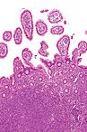 Intermediate magnification micrograph of mantle cell lymphoma of the terminal ileum. Endoscopic biopsy. H&E stain. Histomorphologic features: Monomorphic small lymphoid cells less than twice the size of a resting lymphocyte. Abundant mitoses. Sclerosed blood vessels. Scattered epithelioid histiocytes.
 -
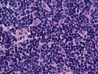 Mantle cell lymphoma. Notice the irregular nuclear contours of the medium-sized lymphoma cells and the presence of a pink histiocyte. By immunohistochemistry the lymphoma cells expressed CD20, CD5 and Cyclin D1 (high power view, H&E).
 -
 Lymph node with mantle cell lymphoma (low power view, H&E).
 -
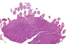 Micrograph of terminal ileum with mantle cell lymphoma (bottom of image). H&E stain.
 -
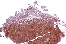 Micrograph of terminal ileum with mantle cell lymphoma (bottom of image – brown colour). Cyclin D1 immunostain.


References
Causes
Editor-In-Chief: C. Michael Gibson, M.S., M.D. [1]; Associate Editor(s)-in-Chief: Ali Akram, M.B.B.S.[2] Sowminya Arikapudi, M.B,B.S. [3]
Overview
The causes of mantle cell lymphoma have not been clearly identified.
Causes
- Mantle cell lymphoma is thought to be caused when the somatic cells acquire non-inherited genetic mutations.
- The typical mutation occurring in mantle cell lymphoma due to the “reciprocal translocation” between chromosome 11 and chromosome 14 t(11;14) causes the over expression of cyclin D1 which results in uncontrolled growth of B cells leading to lymphoma development.
- However, the initiating factors of these genetic alterations are generally not identifiable. People with no particular risk factors for lymphoma development are commonly affected by these alterations.
References
Differentiating Mantle cell lymphoma from other Diseases
Editor-In-Chief: C. Michael Gibson, M.S., M.D. [1]; Associate Editor(s)-in-Chief: Ali Akram, M.B.B.S.[2] Sowminya Arikapudi, M.B,B.S. [3]
Overview
Mantle cell lymphoma must be differentiated from other diseases that present similarly with B symptoms (fever, night sweats and unexplained weight loss), lymphocytosis, lymphadenopathy, hepatosplenomegaly, and bone marrow involvement.
Differential Diagnosis
Mantle cell lymphoma should be differentiated from other conditions presenting with fever, fatigue, lymphadenopathy, abdominal pain, rash and soft tissue swelling. The differentials include the following:[1][2][3][4][5][6][7][8][9][10][11][12][13][14][15][16][17][18][19][20][21][22][23][24][25]
| Category of Disease | Diseases | Signs and symptoms | Laboratory findings | |||||||||||||||||||
|---|---|---|---|---|---|---|---|---|---|---|---|---|---|---|---|---|---|---|---|---|---|---|
| Fever | Fatigue | Arthralgia | Myalgia | Soft tissue swelling/serositis | Skin rash | Weight loss | Dyspnea | Sore throat | Lymphadenopathy | Complete blood count (CBC) | Liver function tests (LFTs) |
Inflammatory markers |
Autoantibodies |
Diagnostic tests | ||||||||
| Erythrocyte sedimentation rate (ESR) | C- reactive protein (CRP) | Anti-nuclear antibodies (ANA) | Rheumatoid factor (RF) | Anti- glomerular basement membrane (anti-GBM) | Anti-dsDNA | Anti-Jo1/ Anti Mi2 | ANCA | |||||||||||||||
Infections |
HIV | + | + | + | + | +/- | – | + | +/- | + /- | + | ↑ | ↑ | – | – | – | – | – | – | |||
| Herpesviridae | + | + | + | + | + |
|
– | – | +/- | + | – | ↑ | ↑ | – | – | – | – | – | – | |||
| Measles | + | + | + | + | – |
|
– | – | + | + | – | ↑ | ↑ | – | – | – | – | – | – | |||
| Viral hepatitis | + | + | – | +/- | – | – | +/- | – | – | +/- | ↑ | ↑ | – | – | – | – | – | – | ||||
| Parvovirus B19 | + | + | + | +/- | – |
|
– | – | – | + |
|
↑ | ↑ | – | – | – | – | – | – | |||
| Infective endocarditis | + | + | + | +/- | – | +/- | + | – | + | – | ↑ | ↑ | – | – | – | – | – | – | Blood cultures, ultrasonography | |||
| Borreliosis, Brucellosis, Yersiniosis | + | + | + | + | – |
|
– | – | – | + | ↑ | ↑ | – | – | – | – | – | – | Serology, PCR | |||
| Syphilis and Jarisch-Herxheimer reaction | + | + | + | + | – |
|
– | – | + | + | ↑ | ↑ | – | – | – | – | – | – | Serology, PCR | |||
| Toxoplasmosis | + | + | – | + | – |
|
– | – | + | + |
|
– | – | – | – | – | – | Serology, PCR | ||||
Neoplasia |
Malignant lymphoma | + | + | – | +/- | +/- | + | + | – | + |
|
↑ | ↑ | – | – | – | – | – | – | CT, PET/CT, Bone marrow examination, lymph node biopsy | ||
| Multicentric Castleman disease | + | + | – | – | + | – | + | + | – | + | – | ↑ | ↑ | – | – | – | – | – | – | Lymph node biopsy | ||
| Angioimmunoblastic T cell lymphoma | + | + | – | – | – |
|
+ | – | – | + | ↑ | ↑ | – | – | – | – | – | – | Lymph node biopsy | |||
Drug hypersensitivity |
Drug reaction with eosinophilia and systemic symptoms | + | + | + | + | +/- |
|
– | + | – | – | – | ↑ | ↑ | – | – | – | – | – | – | Eosinophil count, skin biopsy | |
| Autoimmune conditions | Systemic lupus erythematosus | + | + | + | +/- | + |
|
+ | + | – | +/- | ↑ | ↑ | + | + | – | + | – | – | Antinuclear autoantibodies | ||
| Inflammatory myositis | + | + | – | + (weakness > pain) | – | – | – | – | +/- | – | ↑ | ↑ | +/- | +/- | – | – | + | – | Idem, muscle biopsy | |||
| Rheumatoid arthritis | + | + | + | – | + | – | + | – | + | – | ↑ | ↑ | +/- | +/- | – | – | – | – | Anti-citrullinated peptids autoantibodies, rheumatoid factor | |||
| Systemic vasculitides | + | + | + | – | + |
|
– | +/- | – | +/- | – | ↑ | ↑ | – | – | +/- | – | – | + | ANCA, tissue biopsy, arteriography | ||
| Familial Mediterranean fever | + | + | + | + | + |
|
+ | + (due to pain) | – | +/- |
|
– | ↑ | ↑ | – | – | – | – | – | – | Familial history, MEFV gene analysis | |
| Mevalonate kinase deficiency | + | + | + | + | – |
|
+ | – | + | + |
|
– | ↑ | ↑ | – | – | – | – | – | – | Urinary mevalonic acid, mevalonate kinase analysis | |
| Reactive arthritis | + | + | + | – | – |
|
– | + (Aortic insufficiency) | – | + | – | ↑ | ↑ | – | – | – | – | – | – | HLA B27, magnetic resonance imaging | ||
Miscellaneous |
Sarcoidosis | + | + | + | – | + |
|
+ | + | – | + | ↑ | ↑ | – | – | – | – | – | – |
| ||
| Neutrophilic dermatosis | ||||||||||||||||||||||
| Kikuchi–Fujimoto disease | ||||||||||||||||||||||
Mantle cell lymphoma must be differentiated from other diseases such as:[26][27][28]
Differentials Based on Cell Markers
Based on the expression of cell surface markers, the table below differentiates mantle cell lymphoma from other diseases that cause similar clinical presentations:[29]
| Differential Diagnosis | Surface Immunoglobulin | CD5 | CD22/FMC7 | CD23 | CD79b | CD103 |
|---|---|---|---|---|---|---|
|
Chronic lymphocytic leukemia |
Weakly positive |
Positive |
Negative |
Positive |
Negative |
Positive/Negative |
|
Strongly positive |
Negative |
Positive |
Negative |
Positive |
Negative | |
|
Strongly positive |
Negative |
Positive |
Negative |
Positive/Negative |
Positive | |
|
Positive |
Positive |
Strongly positive |
Negative |
Strongly positive |
Negative | |
|
Strongly positive |
Negative |
Positive |
Negative |
Strongly positive |
Negative |
Other Differentials
- Diffuse large B cell lymphoma
- Mucosa-Associated Lymphatic Tissue lymphoma (MALT)
- Follicular lymphoma
- Small lymphocytic lymphoma / Chronic lymphocytic leukemia
- Lymphoplasmacytoid lymphoma / Immunocytoma
- Marginal zone lymphoma
- Lymphoblastic lymphoma
- Burkitt lymphoma
- Reactive hyperplasia
References
- ↑ Ejilemele AA, Nwauche CA, Ejele OA (December 2007). “Pattern of abnormal liver enzymes in HIV patients presenting at a Nigerian Tertiary Hospital”. Niger Postgrad Med J. 14 (4): 306–9. PMID 18163139.
- ↑ Gøransson LG, Omdal R, Husby G (March 1992). “[Adult-onset Still’s disease. Diagnosis, differential diagnosis and treatment]”. Tidsskr. Nor. Laegeforen. (in Norwegian). 112 (9): 1155–5. PMID 1579936.
- ↑ Hatakka A, Klein J, He R, Piper J, Tam E, Walkty A (September 2011). “Acute hepatitis as a manifestation of parvovirus B19 infection”. J. Clin. Microbiol. 49 (9): 3422–4. doi:10.1128/JCM.00575-11. PMC 3165617. PMID 21734024.
- ↑ Yaguchi D, Marui N, Matsuo M (2015). “Three Adult Cases of HPV-B19 Infection with Concomitant Leukopenia and Low Platelet Counts”. Clin Med Insights Case Rep. 8: 19–22. doi:10.4137/CCRep.S18085. PMC 4345940. PMID 25780346.
- ↑ Díaz F, Collazos J (March 2000). “Hepatic dysfunction due to parvovirus B19 infection”. J. Infect. Chemother. 6 (1): 63–4. doi:10.1007/s101560000023. PMID 11810534.
- ↑ “watermark.silverchair.com” (PDF).
- ↑ Shetty RK, Vivek G, Naha K, Bekkam S (January 2013). “Right-sided infective endocarditis presenting with purpuric skin rash and cardiac failure in a patient without fever”. BMJ Case Rep. 2013. doi:10.1136/bcr-2012-007841. PMC 3603787. PMID 23355575.
- ↑ Aucott JN, Crowder LA, Yedlin V, Kortte KB (2012). “Bull’s-Eye and Nontarget Skin Lesions of Lyme Disease: An Internet Survey of Identification of Erythema Migrans”. Dermatol Res Pract. 2012: 451727. doi:10.1155/2012/451727. PMC 3485866. PMID 23133445.
- ↑ Karaali Z, Baysal B, Poturoglu S, Kendir M (May 2011). “Cutaneous manifestations in brucellosis”. Indian J Dermatol. 56 (3): 339–40. doi:10.4103/0019-5154.82505. PMC 3132922. PMID 21772606.
- ↑ La Spada E, Micalizzi A, La Spada M, Quartarano P, Nugara G, Soresi M, Affronti M, Montalto G (September 2008). “[Abnormal liver function in brucellosis]”. Infez Med (in Italian). 16 (3): 148–53. PMID 18843212.
- ↑ French P (January 2007). “Syphilis”. BMJ. 334 (7585): 143–7. doi:10.1136/bmj.39085.518148.BE. PMC 1779891. PMID 17235095.
- ↑ “Syphilis: Review with Emphasis on Clinical, Epidemiologic, and Some Biologic Features”.
- ↑ Baveja S, Garg S, Rajdeo A (March 2014). “Syphilitic hepatitis: an uncommon manifestation of a common disease”. Indian J Dermatol. 59 (2): 209. doi:10.4103/0019-5154.127711. PMC 3969699. PMID 24700957.
- ↑ Mawhorter SD, Effron D, Blinkhorn R, Spagnuolo PJ (May 1992). “Cutaneous manifestations of toxoplasmosis”. Clin. Infect. Dis. 14 (5): 1084–8. PMID 1600010.
- ↑ Flegr J, Prandota J, Sovičková M, Israili ZH (2014). “Toxoplasmosis–a global threat. Correlation of latent toxoplasmosis with specific disease burden in a set of 88 countries”. PLoS ONE. 9 (3): e90203. doi:10.1371/journal.pone.0090203. PMC 3963851. PMID 24662942.
- ↑ Furtado JM, Smith JR, Belfort R, Gattey D, Winthrop KL (July 2011). “Toxoplasmosis: a global threat”. J Glob Infect Dis. 3 (3): 281–4. doi:10.4103/0974-777X.83536. PMC 3162817. PMID 21887062.
- ↑ Ripert C (March 2000). “[Reactive hypereosinophilia in parasitic diseases]”. Rev Prat (in French). 50 (6): 602–7. PMID 10808314.
- ↑ Alvarado-Esquivel C, Torres-Berumen JL, Estrada-Martínez S, Liesenfeld O, Mercado-Suarez MF (May 2011). “Toxoplasma gondii infection and liver disease: a case-control study in a northern Mexican population”. Parasit Vectors. 4: 75. doi:10.1186/1756-3305-4-75. PMC 3105944. PMID 21569516.
- ↑ Han T, Stutzman L (July 1967). “Mode of spread in patients with localized malignant lymphoma”. Arch. Intern. Med. 120 (1): 1–7. PMID 5339237.
- ↑ Saeed-Abdul-Rahman I, Al-Amri AM (September 2012). “Castleman disease”. Korean J Hematol. 47 (3): 163–77. doi:10.5045/kjh.2012.47.3.163. PMC 3464333. PMID 23071471.
- ↑ Saeed-Abdul-Rahman I, Al-Amri AM (September 2012). “Castleman disease”. Korean J Hematol. 47 (3): 163–77. doi:10.5045/kjh.2012.47.3.163. PMC 3464333. PMID 23071471.
- ↑ Papadavid E, Panayiotides I, Dalamaga M, Katoulis A, Economopoulos T, Stavrianeas N (2010). “Cutaneous involvement in angioimmunoblastic T-cell lymphoma”. Indian J Dermatol. 55 (3): 279–80. doi:10.4103/0019-5154.70704. PMC 2965920. PMID 21063526.
- ↑ Brockow K, Przybilla B, Aberer W, Bircher AJ, Brehler R, Dickel H, Fuchs T, Jakob T, Lange L, Pfützner W, Mockenhaupt M, Ott H, Pfaar O, Ring J, Sachs B, Sitter H, Trautmann A, Treudler R, Wedi B, Worm M, Wurpts G, Zuberbier T, Merk HF (2015). “Guideline for the diagnosis of drug hypersensitivity reactions: S2K-Guideline of the German Society for Allergology and Clinical Immunology (DGAKI) and the German Dermatological Society (DDG) in collaboration with the Association of German Allergologists (AeDA), the German Society for Pediatric Allergology and Environmental Medicine (GPA), the German Contact Dermatitis Research Group (DKG), the Swiss Society for Allergy and Immunology (SGAI), the Austrian Society for Allergology and Immunology (ÖGAI), the German Academy of Allergology and Environmental Medicine (DAAU), the German Center for Documentation of Severe Skin Reactions and the German Federal Institute for Drugs and Medical Products (BfArM)”. Allergo J Int. 24 (3): 94–105. doi:10.1007/s40629-015-0052-6. PMC 4479479. PMID 26120552.
- ↑ Medlej-Hashim M, Loiselet J, Lefranc G, Mégarbané A (2004). “[Familial Mediterranean Fever (FMF): from diagnosis to treatment]”. Sante (in French). 14 (4): 261–6. PMID 15745878.
- ↑ Zhang S (May 2016). “Natural history of mevalonate kinase deficiency: a literature review”. Pediatr Rheumatol Online J. 14 (1): 30. doi:10.1186/s12969-016-0091-7. PMC 4855321. PMID 27142780.
- ↑ Elias Campo, Steven H. Swerdlow, Nancy L. Harris, Stefano Pileri, Harald Stein & Elaine S. Jaffe (2011). “The 2008 WHO classification of lymphoid neoplasms and beyond: evolving concepts and practical applications”. Blood. 117 (19): 5019–5032. doi:10.1182/blood-2011-01-293050. PMID 21300984. Unknown parameter
|month=ignored (help) - ↑ Alejandra Carvajal-Cuenca, Luz F. Sua, Nhora M. Silva, Stefania Pittaluga, Cristina Royo, Joo Y. Song, Rachel L. Sargent, Blanca Espinet, Fina Climent, Samuel A. Jacobs, Jan Delabie, Kikkeri N. Naresh, Adam Bagg, Pierre Brousset, Roger A. Warnke, Sergi Serrano, Nancy Lee Harris, Steven H. Swerdlow, Elaine S. Jaffe & Elias Campo (2012). “In situ mantle cell lymphoma: clinical implications of an incidental finding with indolent clinical behavior”. Haematologica. 97 (2): 270–278. doi:10.3324/haematol.2011.052621. PMID 22058203. Unknown parameter
|month=ignored (help) - ↑ Gabor Barna, Lilla Reiniger, Peter Tatrai, Laszlo Kopper & Andras Matolcsy (2008). “The cut-off levels of CD23 expression in the differential diagnosis of MCL and CLL”. Hematological oncology. 26 (3): 167–170. doi:10.1002/hon.855. PMID 18381689. Unknown parameter
|month=ignored (help) - ↑ Hoffbrand V, Moss P. Essential Haematology. John Wiley & Sons; 2011
Epidemiology and Demographics
Editor-In-Chief: C. Michael Gibson, M.S., M.D. [1]; Associate Editor(s)-in-Chief: Ali Akram, M.B.B.S.[2]
Overview
Mantle cell lymphoma accounts for 4–8% of all adult non-Hodgkin lymphoma (NHL). It has an incidence of approximately 1-2 per 100,000 individuals worldwide. The male to female ratio is approximately 4 to 1 and the median age is about 60 years.
Epidemiology and Demographics
Incidence
- The annual incidence of mantle cell lymphoma has increased during recent decades to 1–2 per 100,000 individuals worldwide.[1]
Age
Race
- Mantle cell lymphoma tends to affect Caucasians more than African-Americans.[3]
Gender
- Males are more commonly affected by mantle cell lymphoma than females. The male to female ratio is approximately 4 to 1.[4]
References
- ↑ M. Dreyling, C. Geisler, O. Hermine, H. C. Kluin-Nelemans, S. Le Gouill, S. Rule, O. Shpilberg, J. Walewski & M. Ladetto (2014). “Newly diagnosed and relapsed mantle cell lymphoma: ESMO Clinical Practice Guidelines for diagnosis, treatment and follow-up”. Annals of oncology : official journal of the European Society for Medical Oncology. 25 Suppl 3: iii83–iii92. doi:10.1093/annonc/mdu264. PMID 25210087. Unknown parameter
|month=ignored (help) - ↑ Arati A. Inamdar, Andre Goy, Nehad M. Ayoub, Christen Attia, Lucia Oton, Varun Taruvai, Mark Costales, Yu-Ting Lin, Andrew Pecora & K. Stephen Suh (2016). “Mantle cell lymphoma in the era of precision medicine-diagnosis, biomarkers and therapeutic agents”. Oncotarget. 7 (30): 48692–48731. doi:10.18632/oncotarget.8961. PMID 27119356. Unknown parameter
|month=ignored (help) - ↑ Yuhong Zhou, Haijun Wang, Wenjing Fang, Jorge E. Romaguer, Yanxia Zhang, Kay B. Delasalle, Larry Kwak, Qing Yi, Xianglin L. Du & Michael Wang (2008). “Incidence trends of mantle cell lymphoma in the United States between 1992 and 2004”. Cancer. 113 (4): 791–798. doi:10.1002/cncr.23608. PMID 18615506. Unknown parameter
|month=ignored (help) - ↑ Arati A. Inamdar, Andre Goy, Nehad M. Ayoub, Christen Attia, Lucia Oton, Varun Taruvai, Mark Costales, Yu-Ting Lin, Andrew Pecora & K. Stephen Suh (2016). “Mantle cell lymphoma in the era of precision medicine-diagnosis, biomarkers and therapeutic agents”. Oncotarget. 7 (30): 48692–48731. doi:10.18632/oncotarget.8961. PMID 27119356. Unknown parameter
|month=ignored (help)
Risk Factors
Editor-In-Chief: C. Michael Gibson, M.S., M.D. [1]; Associate Editor(s)-in-Chief: Ali Akram, M.B.B.S.[2] Sowminya Arikapudi, M.B,B.S. [3]
Overview
There are no established risk factors for mantle cell lymphoma. However, recently weak associations have been observed in the development of mantle cell lymphoma such as the exposure to European strains of Borrelia burgdorferi, family history of hematologic malignancy, and genetic polymorphisms in the pro-inflammatory cytokine IL-10.
Risk Factors
- There are no established risk factors for mantle cell lymphoma. Although, the pathogenesis of mantle cell lymphoma at the cellular level is well understood now, there is not enough information on the risk factors that precipitate the cellular events.
- However, recently weak associations have been observed in the development of mantle cell lymphoma include following:[1][2][3]
- Exposure to European strains of Borrelia burgdorferi
- Family history of hematologic malignancy
- Genetic polymorphisms in the pro-inflammatory cytokine IL-10
References
- ↑ Schöllkopf C, Melbye M, Munksgaard L, Smedby KE, Rostgaard K, Glimelius B; et al. (2008). “Borrelia infection and risk of non-Hodgkin lymphoma”. Blood. 111 (12): 5524–9. doi:10.1182/blood-2007-08-109611. PMC 2972577. PMID 18424667.
- ↑ Wang SS, Slager SL, Brennan P, Holly EA, De Sanjose S, Bernstein L; et al. (2007). “Family history of hematopoietic malignancies and risk of non-Hodgkin lymphoma (NHL): a pooled analysis of 10 211 cases and 11 905 controls from the International Lymphoma Epidemiology Consortium (InterLymph)”. Blood. 109 (8): 3479–88. doi:10.1182/blood-2006-06-031948. PMC 1852242. PMID 17185468.
- ↑ Skibola CF, Bracci PM, Nieters A, Brooks-Wilson A, de Sanjosé S, Hughes AM; et al. (2010). “Tumor necrosis factor (TNF) and lymphotoxin-alpha (LTA) polymorphisms and risk of non-Hodgkin lymphoma in the InterLymph Consortium”. Am J Epidemiol. 171 (3): 267–76. doi:10.1093/aje/kwp383. PMC 2842204. PMID 20047977.
Screening
Editor-In-Chief: C. Michael Gibson, M.S., M.D. [1]; Associate Editor(s)-in-Chief: Ali Akram, M.B.B.S.[2] Sowminya Arikapudi, M.B,B.S. [3]
Overview
There is insufficient evidence to recommend routine screening for Mantle cell lymphoma.
Screening
There is insufficient evidence to recommend routine screening for Mantle cell lymphoma.
References
Natural History, Complications and Prognosis
Editor-In-Chief: C. Michael Gibson, M.S., M.D. [1]; Associate Editor(s)-in-Chief:
Ali Akram, M.B.B.S.[2] Sowminya Arikapudi, M.B,B.S. [3]
Overview
The prognosis of mantle cell lymphoma has historically been very poor. However, recently improvements have been made and the median survival has increased from 3-4 years to 5-7 years.[1] It is very important to stratify the patients according to their biological risk to better direct the therapeutic approaches.
Natural History
- Mantle cell lymphoma is generally considered to be an aggressive disease with patients having early relapses and poor long-term survival rates.
- However, rare forms of the disease which show an indolent behavior, have better outcomes. [2]
Prognosis
- The evolution of disease in mantle cell lymphoma is highly heterogeneous. Therefore, it is very important to stratify the patients according to their biological risk to better direct the therapeutic approaches.
- The Mantle Cell Lymphoma International Prognostic Index (MIPI) is now widely used as a prognostic model in MCL patients. It uses the following four parameters to assess the prognosis (Age, ECOG performance, LDH levels, WBC Count):
| Points | Age (years) | ECOG | LDH (upper limit) | WBC Count (x 109/L) |
|---|---|---|---|---|
| 0 | <50 | 0/1 | <0.67 | <6700 |
| 1 | 50-59 | – | 0.67-0.99 | 6700-9999 |
| 2 | 60-69 | 1/2 | 1-1.49 | 10000-14999 |
| 3 | >70 | – | >1.50 | >15000 |
| Risk | Points | Survival at five years |
|---|---|---|
| Low | 0-3 | 60% alive at 5 years |
| Medium | 4-5 | 51 months |
| High | >5 | 29 months |
- The tumor proliferation marker, Ki-67, is also an important prognostic marker for mantle cell lymphoma. Values of >30% are associated with a poor progression free survival.[3]
- Presence of blastoid or pleomorphic morphologic characteristics have also shown to have poor outcomes.[4]
- Other markers which have shown to adversely affect prognosis are as follows:[5][6][7][8][9][10][11][12]
- High expression of eukaryotic initiation factor 4E
- Myc overexpression
- Increased expression of SOX11
- Low TCL1 expression
- TP53 deletion
- Lack of hypermutated heavy chain immunoglobulin variable regions
- Increased absolute levels of the following in the serum:
References
- ↑ P. Martin, A. Chadburn, P. Christos, R. Furman, J. Ruan, M. A. Joyce, E. Fusco, P. Glynn, R. Elstrom, R. Niesvizky, E. J. Feldman, T. B. Shore, M. W. Schuster, S. Ely, D. M. Knowles, S. Chen-Kiang, M. Coleman & J. P. Leonard (2008). “Intensive treatment strategies may not provide superior outcomes in mantle cell lymphoma: overall survival exceeding 7 years with standard therapies”. Annals of oncology : official journal of the European Society for Medical Oncology. 19 (7): 1327–1330. doi:10.1093/annonc/mdn045. PMID 18349031. Unknown parameter
|month=ignored (help) - ↑ Pedro Jares & Elias Campo (2008). “Advances in the understanding of mantle cell lymphoma”. British journal of haematology. 142 (2): 149–165. doi:10.1111/j.1365-2141.2008.07124.x. PMID 18410453. Unknown parameter
|month=ignored (help) - ↑ Eva Hoster, Andreas Rosenwald, Francoise Berger, Heinz-Wolfram Bernd, Sylvia Hartmann, Christoph Loddenkemper, Thomas F. E. Barth, Nicole Brousse, Stefano Pileri, Grzegorz Rymkiewicz, Roman Kodet, Stephan Stilgenbauer, Roswitha Forstpointner, Catherine Thieblemont, Michael Hallek, Bertrand Coiffier, Ursula Vehling-Kaiser, Reda Bouabdallah, Lothar Kanz, Michael Pfreundschuh, Christian Schmidt, Vincent Ribrag, Wolfgang Hiddemann, Michael Unterhalt, Johanna C. Kluin-Nelemans, Olivier Hermine, Martin H. Dreyling & Wolfram Klapper (2016). “Prognostic Value of Ki-67 Index, Cytology, and Growth Pattern in Mantle-Cell Lymphoma: Results From Randomized Trials of the European Mantle Cell Lymphoma Network”. Journal of clinical oncology : official journal of the American Society of Clinical Oncology. 34 (12): 1386–1394. doi:10.1200/JCO.2015.63.8387. PMID 26926679. Unknown parameter
|month=ignored (help) - ↑ German Ott, Andreas Rosenwald & Elias Campo (2013). “Understanding MYC-driven aggressive B-cell lymphomas: pathogenesis and classification”. Hematology. American Society of Hematology. Education Program. 2013: 575–583. doi:10.1182/asheducation-2013.1.575. PMID 24319234.
- ↑ Veronica Fernandez, Olga Salamero, Blanca Espinet, Francesc Sole, Cristina Royo, Alba Navarro, Francisca Camacho, Silvia Bea, Elena Hartmann, Virginia Amador, Luis Hernandez, Claudio Agostinelli, Rachel L. Sargent, Maria Rozman, Marta Aymerich, Dolors Colomer, Neus Villamor, Steven H. Swerdlow, Stefano A. Pileri, Francesc Bosch, Miguel A. Piris, Emili Montserrat, German Ott, Andreas Rosenwald, Armando Lopez-Guillermo, Pedro Jares, Sergi Serrano & Elias Campo (2010). “Genomic and gene expression profiling defines indolent forms of mantle cell lymphoma”. Cancer research. 70 (4): 1408–1418. doi:10.1158/0008-5472.CAN-09-3419. PMID 20124476. Unknown parameter
|month=ignored (help) - ↑ Mohamad B. Sonbol, Matthew J. Maurer, Mary J. Stenson, Cristine Allmer, Betsy R. LaPlant, George J. Weiner, William R. Macon, James R. Cerhan, Thomas E. Witzig & Mamta Gupta (2014). “Elevated soluble IL-2Ralpha, IL-8, and MIP-1beta levels are associated with inferior outcome and are independent of MIPI score in patients with mantle cell lymphoma”. American journal of hematology. 89 (12): E223–E227. doi:10.1002/ajh.23838. PMID 25164110. Unknown parameter
|month=ignored (help) - ↑ Julia Slotta-Huspenina, Ina Koch, Laurence de Leval, Gisela Keller, Margit Klier, Karin Bink, Marcus Kremer, Mark Raffeld, Falko Fend & Leticia Quintanilla-Martinez (2012). “The impact of cyclin D1 mRNA isoforms, morphology and p53 in mantle cell lymphoma: p53 alterations and blastoid morphology are strong predictors of a high proliferation index”. Haematologica. 97 (9): 1422–1430. doi:10.3324/haematol.2011.055715. PMID 22315488. Unknown parameter
|month=ignored (help) - ↑ M. Meggendorfer, W. Kern, C. Haferlach, T. Haferlach & S. Schnittger (2013). “SOX11 overexpression is a specific marker for mantle cell lymphoma and correlates with t(11;14) translocation, CCND1 expression and an adverse prognosis”. Leukemia. 27 (12): 2388–2391. doi:10.1038/leu.2013.141. PMID 23648671. Unknown parameter
|month=ignored (help) - ↑ Thomas E. Witzig, Matthew J. Maurer, Thomas M. Habermann, Brian K. Link, Ivana N. M. Micallef, Grzegorz S. Nowakowski, Stephen M. Ansell, Joseph P. Colgan, David J. Inwards, Luis F. Porrata, Svetomir N. Markovic, Patrick B. Johnston, Yi Lin, Carrie Thompson, Mamta Gupta, Jerry A. Katzmann & James R. Cerhan (2014). “Elevated monoclonal and polyclonal serum immunoglobulin free light chain as prognostic factors in B- and T-cell non-Hodgkin lymphoma”. American journal of hematology. 89 (12): 1116–1120. doi:10.1002/ajh.23839. PMID 25228125. Unknown parameter
|month=ignored (help) - ↑ Changhoon Yoo, Dok Hyun Yoon, Shin Kim, Jooryung Huh, Chan-Sik Park, Chan-Jeong Park, Sang-Wook Lee & Cheolwon Suh (2016). “Serum beta-2 microglobulin as a prognostic biomarker in patients with mantle cell lymphoma”. Hematological oncology. 34 (1): 22–27. doi:10.1002/hon.2188. PMID 25689467. Unknown parameter
|month=ignored (help) - ↑ Ji-Young Choe, Ji Yun Yun, Hee Young Na, Jooryung Huh, Su-Jin Shin, Hyun-Jung Kim, Jin Ho Paik, Young A. Kim, Soo Jeong Nam, Yoon Kyung Jeon, Gyeongsin Park & Ji Eun Kim (2016). “MYC overexpression correlates with MYC amplification or translocation, and is associated with poor prognosis in mantle cell lymphoma”. Histopathology. 68 (3): 442–449. doi:10.1111/his.12760. PMID 26100211. Unknown parameter
|month=ignored (help) - ↑ Su-Jin Shin, Jin Roh, Hee Jeong Cha, Yoo Duk Choi, Jin-Man Kim, Soo Kee Min, Ji Eun Kim, Dae-Woon Eom, Hojung Lee, Hyun-Jung Kim, Dok Hyun Yoon, Cheolwon Suh & Jooryung Huh (2015). “TCL1 expression predicts overall survival in patients with mantle cell lymphoma”. European journal of haematology. 95 (6): 583–594. doi:10.1111/ejh.12539. PMID 25688912. Unknown parameter
|month=ignored (help)
Diagnosis
Diagnosis
Diagnostic study of choice | History and Symptoms | Physical Examination | Laboratory Findings | Electrocardiogram | X-Ray Findings | Echocardiography and Ultrasound | CT-Scan Findings | MRI Findings | Other Imaging Findings | Other Diagnostic Studies
Treatment
Treatment
Medical Therapy | Surgery | Cost-Effectiveness of Therapy | Future or Investigational Therapies
Looking for the patient version?
© 2026 MyEClinic – IFTM Institut für Telematik in der Medizin GmbH