

Health Dictionary
Reliable, easy-to-read patient information on common medical conditions — written in plain language, free of jargon, and grounded in current evidence. Browse the most frequent diagnoses, understand what they mean for everyday life, and learn which steps and questions help you have a focused conversation with your doctor.
Stomach cancer
Introduction
For patient information click here
Synonyms and keywords: Gastric cancer, Gastric carcinoma, Carcinoma of stomach
Overview
Overview
Stomach cancer (gastric cancer) is a malignant neoplasm arising from the stomach, of which more than 90% are adenocarcinomas; less common types include gastric lymphoma, gastrointestinal stromal tumor, and neuroendocrine tumors. Gastric cancer is the fifth most commonly diagnosed cancer and the fifth leading cause of cancer death worldwide, with an estimated 968,350 new cases and 659,853 deaths in 2022. Age-standardized incidence and mortality have declined for decades as Helicobacter pylori prevalence has fallen, but incidence remains highest in East Asia, Eastern Europe, and parts of Central and South America. In the United States, the median age at diagnosis is 68 years, men are affected nearly twice as often as women, and early-onset disease (diagnosis before age 50) is rising. Billroth performed the first successful gastrectomy for gastric cancer in 1881.
Gastric adenocarcinoma develops through two biologically distinct pathways. The intestinal type arises through the Correa cascade (chronic gastritis → atrophic gastritis → intestinal metaplasia → dysplasia → carcinoma), which is overwhelmingly initiated by chronic H. pylori infection. The diffuse type usually arises de novo and is centered on loss of E-cadherin; its hereditary form, hereditary diffuse gastric cancer, is caused by germline CDH1 pathogenic variants. H. pylori, an IARC Group 1 carcinogen, is associated with about 90% of non-cardia cancers. Other risk factors include tobacco smoking, heavy alcohol use, high-salt and salt-preserved foods, processed meat, obesity, prior partial gastrectomy, autoimmune gastritis/pernicious anemia, and family history; cardia and gastroesophageal junction cancers are more closely linked to obesity and gastroesophageal reflux disease. About 10% of cases show familial clustering, but only 1–3% are attributable to known germline variants. The Cancer Genome Atlas (TCGA) defines four molecular subtypes: Epstein-Barr virus–positive, microsatellite instability-high, genomically stable, and chromosomal instability. Gastric cancer is classified by anatomic site, TNM stage, histology (Laurén and WHO), macroscopic pattern (Borrmann and Paris), and molecular subtype, but only stage and predictive biomarkers currently change management.
Early gastric cancer is often asymptomatic. Advanced disease presents with abdominal pain, weight loss, early satiety, bloating, dysphagia, hematemesis, or melena. Signs of advanced or metastatic disease include Virchow’s node, Sister Mary Joseph nodule, Blumer’s shelf, the Leser-Trelat sign, and Trousseau’s syndrome. Gastric cancer must be differentiated from gastric lymphoma, gastric metastasis, gastritis, benign gastric ulcer, and Ménétrier’s disease. Endoscopic screening every 2–3 years is suggested in high-incidence regions, whereas in low-incidence regions such as the United States it is reserved for defined high-risk groups. Upper endoscopy with biopsy is the diagnostic test of choice. Staging uses the AJCC/UICC 8th edition TNM system, with separate clinical (cTNM), pathological (pTNM), and post-neoadjuvant (ypTNM) stage groups. Endoscopic ultrasonography is the most reliable method for assessing depth of invasion, contrast-enhanced CT evaluates distant spread, PET/CT helps characterize CT-detected lymphadenopathy, and staging laparoscopy detects occult peritoneal disease before neoadjuvant therapy. Stage is the strongest determinant of prognosis: five-year survival exceeds 90% for treated early gastric cancer but is poor for disseminated disease.
Endoscopic resection by endoscopic mucosal resection (EMR) or endoscopic submucosal dissection (ESD) is the treatment of choice for early gastric cancer meeting Japanese Gastric Cancer Association criteria. Gastrectomy with D2 lymphadenectomy is standard for potentially curable T2–T4 tumors, and laparoscopic and robot-assisted approaches are oncologically equivalent to open surgery. For resectable cT2 or higher or node-positive disease, perioperative chemotherapy with FLOT (fluorouracil, leucovorin, oxaliplatin, and docetaxel) is the preferred standard, and the addition of durvalumab improved event-free and overall survival in the MATTERHORN trial. For unresectable, recurrent, or metastatic disease, first-line systemic therapy is a fluoropyrimidine plus platinum backbone guided by biomarker testing: a PD-1 inhibitor (nivolumab or pembrolizumab) for HER2-negative tumors, trastuzumab (with pembrolizumab if PD-L1-positive) for HER2-positive tumors, and zolbetuximab for claudin 18.2-positive tumors. Later-line options include ramucirumab with or without paclitaxel, trastuzumab deruxtecan, and trifluridine/tipiracil. Palliative gastrectomy is not routinely recommended. Primary prevention centers on H. pylori eradication, smoking cessation, and a diet low in salt and rich in fruits and vegetables.
Historical perspective
John Jones was the first to perform a gastric resection in animals. In 1881, Billroth’s first human surgery. In 1897, Schlatter has done the first esophago-enterostomy after gastrectomy. Between 1884 to 1929, Finney’s and Rienhoff were the first to perform partial gastrectomy showing less side effects and less mortality rates.
Classification
No single scheme captures all clinically relevant information about gastric cancer. In current practice, a gastric adenocarcinoma is described along several complementary axes:[1][2]
- Anatomic site: non-cardia (true gastric) versus gastroesophageal junction (GEJ) tumors, with Siewert subtyping for junctional tumors
- Extent of disease: the AJCC/UICC 8th-edition TNM system, with separate clinical (cTNM), pathologic (pTNM), and post-neoadjuvant (ypTNM) stage groups
- Histology: the Laurén types and the WHO histologic subtypes
- Macroscopic pattern: the Borrmann classification for advanced tumors and the Paris/superficial classification for early tumors
- Molecular subtype: the TCGA and ACRG genomic classifications, plus the predictive biomarkers HER2, PD-L1, mismatch repair/microsatellite instability (MMR/MSI), and claudin 18.2 (CLDN18.2)
- Precursor lesions: gastric dysplasia graded by the WHO two-tier system and the Vienna classification
Only TNM stage and the predictive biomarkers currently change management. Histologic and molecular subtypes remain mainly descriptive and prognostic.[3][4]
Pathophysiology
Gastric adenocarcinoma develops through two biologically distinct pathways. The intestinal-type pathway is driven by inflammation and progresses stepwise through the Correa cascade. It is overwhelmingly initiated by Helicobacter pylori. The diffuse-type pathway is largely de novo and is centered on loss of E-cadherin (CDH1).[4][5][6] H. pylori is a WHO Group 1 carcinogen. Only about 1–3% of infected individuals develop cancer, which reflects the combined effect of bacterial virulence (particularly CagA), host genetics, and environmental cofactors.[4][7]
The 2014 The Cancer Genome Atlas (TCGA) classification sits on top of the Lauren histologic dichotomy. It defines four subtypes:
- Chromosomal instability (CIN)
- Microsatellite instability (MSI)
- Epstein-Barr virus–positive (EBV+)
- Genomically stable (GS)
These subtypes are independently prognostic and underpin the biomarkers that direct targeted therapy and immunotherapy: mismatch repair (MMR)/MSI, HER2, PD-L1, and EBV.[8][9][10]
About 10% of gastric cancers show familial aggregation, but only 1–3% are attributable to known germline variants. The most important of these is hereditary diffuse gastric cancer (HDGC), caused by germline CDH1 pathogenic variants. Contemporary penetrance estimates for HDGC are substantially lower than historical figures.[4][2][11]
Causes
Gastric adenocarcinoma results from an interaction between chronic infection, environmental and dietary exposures, host genetic susceptibility, and, in a small minority, high-penetrance germline variants. Chronic Helicobacter pylori infection, an IARC group 1 carcinogen, is the dominant modifiable cause. It accounts for approximately 75% of attributable risk overall and approximately 90% of non-cardia (distal) cancers.[4][12] Most intestinal-type cancers arise through the stepwise Correa cascade (chronic gastritis → atrophic gastritis → intestinal metaplasia → dysplasia → carcinoma). Diffuse-type cancers frequently arise without a recognizable precancerous lesion. When diffuse-type cancer is hereditary, it is driven by germline CDH1 (E-cadherin) inactivation.[13] About 10% of cases show familial clustering, but only about 1–3% are attributable to a known heritable germline variant.[2] The TCGA molecular classification (EBV-positive, MSI, genomically stable, chromosomal instability) has superseded older, undifferentiated oncogene lists as the framework for molecular etiology.[8]
Differential diagnosis
Stomach cancer must be differentiated from other diseases presenting with episodic abdominal pain, weight loss and loss of appetite such as gastric lymphoma, gastric metastasis, gastritis, benign gastric ulcer, Menetrier’s disease.
Epidemiology and Demographics
Gastric cancer is the fifth most commonly diagnosed cancer and the fifth leading cause of cancer death worldwide. GLOBOCAN estimated 968,350 new cases and 659,853 deaths in 2022.[14][9] Age-standardized incidence and mortality have declined for more than half a century, mainly because Helicobacter pylori prevalence has fallen and food refrigeration and preservation have improved. Absolute case numbers keep rising because of population growth and aging.[12] Incidence is highest in East Asia, Eastern Europe, and parts of Central and South America. In the United States, about 30,300 new cases are diagnosed each year, the median age at diagnosis is 68 years, and incidence in men is nearly twice that in women.[9] Cardia and non-cardia gastric cancer are epidemiologically distinct diseases. The incidence of early-onset disease (diagnosis before age 50) is rising in several low-incidence countries, including the United States.[4][9]
Risk Factors
Risk for gastric adenocarcinoma (>90% of stomach cancers) is dominated by chronic Helicobacter pylori infection. H. pylori is an IARC Group 1 carcinogen and is associated with roughly 90% of non-cardia (gastric body and antrum) cancers.[15][9] It drives the stepwise Correa cascade: chronic gastritis → atrophic gastritis → intestinal metaplasia → dysplasia → carcinoma.[4][12]
Other modifiable risk factors are tobacco smoking, heavy alcohol use, high dietary salt and salt-preserved foods, processed meat, and obesity. Nonmodifiable factors include older age, male sex, high-risk ancestry or immigration from high-incidence regions, prior partial gastrectomy, prior abdominal radiotherapy, autoimmune gastritis/pernicious anemia, and inherited predisposition. The most important inherited cause is a germline CDH1 pathogenic variant causing hereditary diffuse gastric cancer (HDGC).[9]
Risk-factor profiles differ by anatomic site and histology:
- Non-cardia, intestinal-type cancer is overwhelmingly H. pylori–driven and arises from precancerous mucosal lesions.
- Cardia/gastroesophageal junction (GEJ) cancer is more closely linked to obesity and gastroesophageal reflux disease (GERD).[12]
- Diffuse-type cancer can arise without a precursor inflammatory field. This includes the hereditary (CDH1) form.[16]
Screening
Gastric cancer screening aims to detect adenocarcinoma at an early, curable stage and to identify gastric premalignant conditions along the Correa cascade: atrophic gastritis (AG), gastric intestinal metaplasia (GIM), and dysplasia.[17]
How a population is screened depends on how common gastric cancer is in that region:
- High-incidence regions: endoscopic screening every 2–3 years is suggested.
- Low-incidence regions: general-population screening is not recommended.[18]
- United States: screening endoscopy is considered only for defined high-risk groups.[19]
Upper endoscopy (EGD) is the preferred screening test.[19] In the Korean national program, EGD was associated with lower gastric cancer mortality, whereas the upper GI series (UGIS) was not.[20]
The only randomized trial reported so far found a non-significant reduction in non-cardia gastric cancer mortality with endoscopic screening.[21]
Natural history, Complications and Prognosis
If left untreated, the five-year survival rates of gastric cancer range from almost no survival for patients with disseminated disease to almost 50% survival for patients with localized distal gastric cancers confined to resectable regions. Higher recurrence rates are seen in those who have piecemeal or incomplete resections. Depending on the extent of the tumor at the time of diagnosis, the prognosis may vary. However, the prognosis is generally regarded as poor. Complications of gastric cancer are ascites, gastrointestinal bleeding, distant metastasis to other organs, weight loss, recurrence of cancer, and treatment complications. The prognosis of patients with gastric cancer is related to tumor extent that includes direct tumor extension and lymph nodes involvement. The five-year survival rate for treated early gastric cancer is over 90 percent; nearly 100 percent for mucosal tumors, and 80 to 90 percent for submucosal tumors.
Staging
Gastric adenocarcinoma is staged with the tumor–node–metastasis (TNM) system maintained jointly by the American Joint Committee on Cancer (AJCC) and the Union for International Cancer Control (UICC). The current standard is the AJCC 8th edition, implemented January 1, 2018. It is the version used in the NCCN Gastric Cancer Guidelines.[22][3]
The 8th edition uses the same T, N, and M definitions in every setting. It provides three separate stage-grouping schemata for different points in care:
- clinical (cTNM), before treatment
- pathological (pTNM), after upfront resection
- post-neoadjuvant pathological (ypTNM), after preoperative therapy and resection[23][16]
Stage determines the main treatment choice: endoscopic resection, surgery alone, perioperative multimodality therapy, or palliative systemic therapy. Stage is also the strongest determinant of prognosis.[9]
History and Symptoms
Symptoms of stomach cancer include abdominal pain, bloating, weight loss, hematemesis, melena, and dysphagia. Twenty-five percent of patients have a history of gastric ulcer
Physical Examination
Patients with stomach cancer generally appear weak. Common physical examination findings include abdominal distention, palpation of an abdominal mass, and pallor. Leser-Trelat sign and presence of Virchow’s node (left supraclavicular lymphadenopathy), Sister Mary Joseph nodule (visible periumbilical nodule), Blumer’s shelf (rectal mass/shelf on rectal exam) and/or Trousseau’s syndrome (migratory phlebitis) on physical examination are highly suggestive of stomach cancer
Laboratory findings
Laboratory findings in gastric cancer include anemia of chronic disease on complete blood count, liver function tests may reveal abnormalities in liver function tests, antigens such as carcinoembryonic antigen, glycoprotein CA 125, carbohydrate antigen 19-9, cancer antigen 72-4, alpha-fetoprotein
Endoscopy and Biopsy
Biopsy may be helpful in the diagnosis of stomach cancer. It has a sensitivity of 98% to diagnose gastric cancer but may be negative in linitis plastica. It is commonly used nowadays as first line of treatment for superficial lesions.
Chest X-Ray
Chest x-ray may show spread to the lungs as a cannon-ball appearance on radiography. Advanced gastric carcinoma may be visible on an abdominal x-ray as an uneven stomach contours or small masses indenting the stomach contours
CT
Abdominal CT scan may be helpful in the diagnosis of stomach cancer. It is used to evaluate metastatic disease, especially hepatic or adnexal metastases, ascites, or distant nodal spread. Integrated PET/CT imaging can be useful to confirm malignant involvement of CT-detected lymphadenopathy. A negative PET CT is not helpful, since even large tumors with a diameter of several centimeters may not be visible on PET scan if the tumor cells have a fairly low metabolic activity.
MRI
MRI has better soft tissue sensitivity than CT.Individual layers may be better differentiated on MRI compared with CT. Hence, better T staging of stomach cancer. Water or effervescent granules are used to distend stomach to perform MRI
Echocardiography/Ultrasound
Endoscopic ultrasonography (EUS) is the most reliable diagnostic technique for evaluation of the depth of invasion of primary gastric cancers. Endoscopic ultrasonography is not the procedure of choice for detecting nodal spread.
Other imaging findings
Barium studies may be diagnostic of stomach cancer. The sensitivity of barium meals may be 14%. False-negative barium studies can occur in 50 percent of cases. There are three types of early gastric cancer which include polypoid, ulcerated, and superficial.
Other diagnostic studies
Laparoscopy has the advantage of directly visualizing the liver surface, the peritoneum, and local lymph nodes. Diagnostic laparoscopy is especially important for patients who are being considered for a trial of neoadjuvant therapy.
Medical therapy
The optimal therapy for stomach cancer depends on the stage at diagnosis and, in advanced disease, increasingly on molecular biomarkers. For patients with resectable disease, perioperative chemotherapy with the FLOT regimen (fluorouracil, leucovorin, oxaliplatin, and docetaxel) is the preferred standard for medically fit patients, having demonstrated superior overall survival compared with the older epirubicin-based ECF/ECX regimen (median overall survival 50 versus 35 months) in the phase 3 FLOT4-AIO trial.[24] The addition of perioperative immunotherapy with durvalumab to FLOT improved event-free survival (HR 0.71) and, at the final analysis, overall survival (HR 0.78; 95% CI 0.63–0.96; p=0.021) in the phase 3 MATTERHORN trial; no overall survival advantage was seen in the diffuse-type subgroup.[25][26][3] Adjuvant chemoradiotherapy and adjuvant chemotherapy with S-1 or capecitabine plus oxaliplatin remain acceptable options, particularly where perioperative therapy was not given. Medical therapy is also indicated for patients with unresectable or recurrent disease, after non-curative R2 resection, unresectable T4b disease, extensive nodal disease, hepatic metastases, peritoneal dissemination, or other M1 disease. Response to treatment should be evaluated by examinations such as CT scan, endoscopy, and contrast radiography. For unresectable, recurrent, or metastatic disease, systemic therapy is guided by biomarker testing for HER2 overexpression, programmed death-ligand 1 (PD-L1) combined positive score (CPS), claudin 18.2 (CLDN18.2), and mismatch repair/microsatellite instability (MMR/MSI) status. First-line therapy uses a fluoropyrimidine plus platinum backbone, to which a PD-1 inhibitor (nivolumab or pembrolizumab) is added in HER2-negative disease, trastuzumab is added in HER2-positive disease (with pembrolizumab for PD-L1–positive HER2-positive tumors), and zolbetuximab is added for CLDN18.2-positive tumors. For locally advanced unresectable and metastatic tumors, the goals of chemotherapy include palliation of symptoms, improvement in quality of life, and prolongation of survival; later-line options include ramucirumab with or without paclitaxel, trastuzumab deruxtecan for HER2-positive disease, and trifluridine/tipiracil.
Surgery
Surgery remains the only potentially curative treatment for stomach cancer and is central to the multidisciplinary management of gastric cancer. Endoscopic resection is the treatment of choice for early gastric cancer meeting absolute or expanded indication criteria, as defined by the Japanese Gastric Cancer Association (JGCA) 2021 (6th edition) treatment guidelines.[27] Methods for endoscopic resection include endoscopic mucosal resection (EMR) and endoscopic submucosal dissection (ESD). Side effects of endoscopy include bleeding and perforation. For T1 tumors, a gross resection margin of 2 cm should be obtained. A proximal margin of at least 3 cm is recommended for T2 or deeper tumors with an expansive growth pattern and 5 cm for those with an infiltrative growth pattern. For tumors invading the esophagus, a 5-cm margin is not necessarily required, but intraoperative frozen section examination of the resection line is preferred to confirm an R0 resection.
Lymph node dissection is defined by extent. D1 lymphadenectomy refers to dissection of only the perigastric lymph nodes (stations 1-7). D1+ lymphadenectomy adds stations 8a, 9, and (for total gastrectomy) 11p to the D1 dissection and is used for cT1 tumors not indicated for D1 alone. D2 lymphadenectomy is an extended lymph node dissection that additionally removes nodes along the hepatic, left gastric, celiac, and splenic arteries, and, when indicated, the splenic hilum; it is the standard of care for potentially curable T2-T4 tumors. D3 (super-extended) dissection adds D2 plus removal of nodes within the porta hepatis and para-aortic regions; prophylactic para-aortic dissection added to D2 did not improve survival in the randomized JCOG9501 trial and is not routinely recommended outside clinical trials.[28]
Laparoscopic and robot-assisted minimally invasive gastrectomy with D2 lymphadenectomy are now supported by long-term randomized data as oncologically equivalent, and in some outcomes superior, alternatives to open gastrectomy for both early and locally advanced gastric cancer.[29][30][31] For resectable locoregional adenocarcinoma (cT2 or higher, or node-positive), perioperative systemic chemotherapy with FLOT (fluorouracil, leucovorin, oxaliplatin, and docetaxel) — now increasingly combined with the anti-PD-L1 monoclonal antibody durvalumab — has replaced older anthracycline-based (ECF/MAGIC) regimens as the perioperative standard of care.[24][25] For metastatic disease, gastrectomy is not routinely recommended for palliation alone, based on the randomized REGATTA trial, and first-line systemic therapy — chemotherapy plus the anti-PD-1 monoclonal antibody nivolumab in appropriate candidates — is preferred over surgery-first approaches.[32][33]
Primary prevention
Effective measures for the primary prevention of stomach cancer include smoking cessation, eradication of Helicobacter pylori infection, and having a balanced diet rich in fruits and vegetables. In areas of low gastric cancer, incidence and screening for gastric cancer with upper endoscopy should be reserved for specific high-risk subgroups. Individuals at increased risk for gastric cancer include gastric adenomas, pernicious anemia, gastric intestinal metaplasia, familial adenomatous polyposis, Lynch syndrome, Peutz-Jeghers syndrome, Juvenile polyposis syndrome.
Secondary prevention
Gastric cancer secondary prevention is indicated for all patients after gastric surgeries. Physical examination, complete blood count, imaging or endoscopy are indicated to decrease chances of recurrence.
References
- ↑ Van Cutsem E, Sagaert X, Topal B, Haustermans K, Prenen H (2016). “Gastric cancer”. Lancet. 388 (10060): 2654–2664. doi:10.1016/S0140-6736(16)30354-3. PMID 27156933.
- ↑ 2.0 2.1 2.2 Smyth EC, Nilsson M, Grabsch HI, van Grieken NC, Lordick F (2020). “Gastric cancer”. Lancet. 396 (10251): 635–648. doi:10.1016/S0140-6736(20)31288-5. PMID 32861308 Check
|pmid=value (help). - ↑ 3.0 3.1 3.2 National Comprehensive Cancer Network. NCCN Clinical Practice Guidelines in Oncology: Gastric Cancer. Version 4.2026. 2026. NCCN
- ↑ 4.0 4.1 4.2 4.3 4.4 4.5 4.6 Sundar R, Nakayama I, Markar SR, Shitara K, van Laarhoven HWM, Janjigian YY, Smyth EC (2025). “Gastric cancer”. Lancet. 405 (10494): 2087–2102. doi:10.1016/S0140-6736(25)00052-2. PMID 40319897 Check
|pmid=value (help). - ↑ Gawron AJ, Shah SC, Altayar O, Davitkov P, Morgan D, Turner K, Mustafa RA (2020). “AGA Technical Review on Gastric Intestinal Metaplasia—Natural History and Clinical Outcomes”. Gastroenterology. 158 (3): 705–731.e5. doi:10.1053/j.gastro.2019.12.001. PMID 31816300.
- ↑ Bessède E, Dubus P, Mégraud F, Varon C (2015). “Helicobacter pylori infection and stem cells at the origin of gastric cancer”. Oncogene. 34 (20): 2547–55. doi:10.1038/onc.2014.187.
- ↑ Duan Y, Xu Y, Dou Y, Xu D (2025). “Helicobacter pylori and gastric cancer: mechanisms and new perspectives”. J Hematol Oncol. 18 (1): 10. doi:10.1186/s13045-024-01654-2.
- ↑ 8.0 8.1 Cancer Genome Atlas Research Network (2014). “Comprehensive molecular characterization of gastric adenocarcinoma”. Nature. 513 (7517): 202–9. doi:10.1038/nature13480. PMC 4170219. PMID 25079317.
- ↑ 9.0 9.1 9.2 9.3 9.4 9.5 9.6 Patel AK, Sethi NS, Park H (2026). “Gastric Cancer: A Review”. JAMA. 335 (5): 439–450. doi:10.1001/jama.2025.20034. PMID 41499132 Check
|pmid=value (help). - ↑ Walch HS, Borpatragohain R, Jee J, et al. (2025). “Clinical implications of The Cancer Genome Atlas molecular classification system in esophagogastric cancer”. Clin Cancer Res. 31 (10): 1912–1921. doi:10.1158/1078-0432.CCR-24-3473. PMID 40299774 Check
|pmid=value (help). - ↑ Ryan CE, Fasaye GA, Gallanis AF, et al. (2024). “Germline CDH1 variants and lifetime cancer risk”. JAMA. 332 (9): 722–729. doi:10.1001/jama.2024.10852.
- ↑ 12.0 12.1 12.2 12.3 Thrift AP, Wenker TN, El-Serag HB (May 2023). “Global burden of gastric cancer: epidemiological trends, risk factors, screening and prevention”. Nat Rev Clin Oncol. 20 (5): 338–349. doi:10.1038/s41571-023-00747-0. PMID 36959359 Check
|pmid=value (help). - ↑ Decourtye-Espiard L, Guilford P (April 2023). “Hereditary diffuse gastric cancer”. Gastroenterology. 164 (5): 719–735. doi:10.1053/j.gastro.2023.01.038. PMID 36740198 Check
|pmid=value (help). - ↑ Bray F, Laversanne M, Sung H, Ferlay J, Siegel RL, Soerjomataram I, Jemal A (2024). “Global cancer statistics 2022: GLOBOCAN estimates of incidence and mortality worldwide for 36 cancers in 185 countries”. CA Cancer J Clin. 74 (3): 229–263. doi:10.3322/caac.21834. PMID 38572751 Check
|pmid=value (help). - ↑ IARC Working Group on the Evaluation of Carcinogenic Risks to Humans (1994). Schistosomes, liver flukes and Helicobacter pylori. IARC Monographs on the Evaluation of Carcinogenic Risks to Humans. 61. Lyon: International Agency for Research on Cancer. PMID 7715068.
- ↑ 16.0 16.1 Hirata Y, Noorani A, Song S, Wang L, Ajani JA (2023). “Early stage gastric adenocarcinoma: clinical and molecular landscapes”. Nat Rev Clin Oncol. 20 (7): 453–469. doi:10.1038/s41571-023-00767-w. PMC 11869628 Check
|pmc=value (help). PMID 37264184 Check|pmid=value (help). - ↑ Morgan DR, Corral JE, Li D, Montgomery EA, Riquelme A, Kim JJ, Sauer B, Shah SC (2025). “ACG Clinical Guideline: Diagnosis and Management of Gastric Premalignant Conditions”. Am J Gastroenterol. 120 (4): 709–737. doi:10.14309/ajg.0000000000003350. PMID 40072510 Check
|pmid=value (help). - ↑ Dinis-Ribeiro M, Libânio D, Uchima H, Spaander MCW, Bornschein J, Matysiak-Budnik T, Tziatzios G, Santos-Antunes J, Areia M, Chapelle N, Esposito G, Fernandez-Esparrach G, Kunovsky L, Garrido M, Tacheci I, Link A, Marcos P, Marcos-Pinto R, Moreira L, Pereira AC, Pimentel-Nunes P, Romanczyk M, Fontes F, Hassan C, Bisschops R, Feakins R, Schulz C, Triantafyllou K, Carneiro F, Kuipers EJ (2025). “Management of epithelial precancerous conditions and early neoplasia of the stomach (MAPS III): European Society of Gastrointestinal Endoscopy (ESGE), European Helicobacter and Microbiota Study Group (EHMSG) and European Society of Pathology (ESP) Guideline update 2025”. Endoscopy. 57 (5): 504–554. doi:10.1055/a-2529-5025. PMID 40112834 Check
|pmid=value (help). - ↑ 19.0 19.1 Shah SC, Wang AY, Wallace MB, Hwang JH (2025). “AGA Clinical Practice Update on Screening and Surveillance in Individuals at Increased Risk for Gastric Cancer in the United States: Expert Review”. Gastroenterology. 168 (2): 405–416.e1. doi:10.1053/j.gastro.2024.11.001. PMID 39718517 Check
|pmid=value (help). - ↑ Jun JK, Choi KS, Lee HY, Suh M, Park B, Song SH, Jung KW, Lee CW, Choi IJ, Park EC, Lee D (2017). “Effectiveness of the Korean National Cancer Screening Program in Reducing Gastric Cancer Mortality”. Gastroenterology. 152 (6): 1319–1328.e7. doi:10.1053/j.gastro.2017.01.029. PMID 28147224.
- ↑ Liu M, Yang W, Guo C, Liu Z, Li F, Liu A, Yang H, Shen L, Wu Q, Duan L, Wang H, Tian H, Shi C, Pan Y, Liu Y, Liu F, Cai H, He Z, Ke Y (2026). “Effect of endoscopic screening for non-cardia gastric cancer: a 12-year report of a population-based randomized trial”. BMC Med. 24: 529. doi:10.1186/s12916-026-05043-z. PMID 42410417 Check
|pmid=value (help). - ↑ Amin MB, Edge SB, Greene FL, et al. (eds.) (2017). AJCC Cancer Staging Manual (8th ed.). New York: Springer.
- ↑ In H, Ravetch E, Langdon-Embry M, Palis B, Ajani JA, Hofstetter WL, Kelsen DP, Sano T (2018). “The newly proposed clinical and post-neoadjuvant treatment staging classifications for gastric adenocarcinoma for the American Joint Committee on Cancer (AJCC) staging”. Gastric Cancer. 21 (1): 1–9. doi:10.1007/s10120-017-0765-y. PMID 28948368.
- ↑ 24.0 24.1 Al-Batran SE, Homann N, Pauligk C, Goetze TO, Meiler J, Kasper S, Kopp HG, Mayer F, Haag GM, Luley K, Lindig U, Schmiegel W, Pohl M, Stoehlmacher J, Folprecht G, Probst S, Prasnikar N, Fischbach W, Mahlberg R, Trojan J, Koenigsmann M, Martens UM, Thuss-Patience P, Egger M, Block A, Heinemann V, Illerhaus G, Moehler M, Schenk M, Kullmann F, Behringer DM, Heike M, Pink D, Teschendorf C, Löhr C, Bernhard H, Schuch G, Rethwisch V, von Weikersthal LF, Hartmann JT, Kneba M, Daum S, Schulmann K, Weniger J, Belle S, Gaiser T, Oduncu FS, Güntner M, Hozaeel W, Reichart A, Jäger E, Kraus T, Mönig S, Bechstein WO, Schuler M, Schmalenberg H, Hofheinz RD (May 2019). “Perioperative chemotherapy with fluorouracil plus leucovorin, oxaliplatin, and docetaxel versus fluorouracil or capecitabine plus cisplatin and epirubicin for locally advanced, resectable gastric or gastro-oesophageal junction adenocarcinoma (FLOT4): a randomised, phase 2/3 trial”. Lancet. 393 (10184): 1948–1957. doi:10.1016/S0140-6736(18)32557-1. PMID 30982686.
- ↑ 25.0 25.1 Janjigian YY, Al-Batran SE, Wainberg ZA, Kim YW, Elimova E, Barros-Sotolongo A, Boku N, Chao J, Ho G, Lorenzen S, Aprile G, Chau I, Sun W, Kim JG, Rueschoff J, Chen LT, Bhagia P, Zhao J, Watson P, Molife LR, Shitara K (2025). “Perioperative Durvalumab in Gastric and Gastroesophageal Junction Cancer”. N Engl J Med. 393 (3): 217–230. doi:10.1056/NEJMoa2503701. PMID 40454643 Check
|pmid=value (help). Vancouver style error: initials (help) - ↑ Janjigian YY, Al-Batran SE, Wainberg ZA, et al. (September 2026). “Perioperative durvalumab plus fluorouracil, leucovorin, oxaliplatin, and docetaxel for resectable gastric and gastro-oesophageal junction adenocarcinoma (MATTERHORN): final results of overall survival and event-free survival by pathological response in a global, randomised, double-blind, placebo-controlled, multicentre, phase 3 trial”. Lancet. 408 (10560): 1114–1128. doi:10.1016/S0140-6736(26)01254-7. PMID 42716074 Check
|pmid=value (help). - ↑ Japanese Gastric Cancer Association (2023). “Japanese Gastric Cancer Treatment Guidelines 2021 (6th edition)”. Gastric Cancer. 26 (1): 1–25. doi:10.1007/s10120-022-01331-8. PMC 9813208 Check
|pmc=value (help). PMID 36342574 Check|pmid=value (help). - ↑ Sasako M, Sano T, Yamamoto S, Kurokawa Y, Nashimoto A, Kurita A; et al. (2008). “D2 lymphadenectomy alone or with para-aortic nodal dissection for gastric cancer”. N Engl J Med. 359 (5): 453–62. doi:10.1056/NEJMoa0707035. PMID 18669424.
- ↑ Son SY, Hur H, Hyung WJ, Park YK, Lee HJ, An JY, Kim W, Kim HI, Kim HH, Ryu SW, Kim MC, Kong SH, Cho GS, Kim JJ, Park DJ, Ryu KW, Kim YW, Kim JW, Lee JH, Yang HK, Han SU (2022). “Laparoscopic vs Open Distal Gastrectomy for Locally Advanced Gastric Cancer: 5-Year Outcomes of the KLASS-02 Randomized Clinical Trial”. JAMA Surg. 157 (10): 879–886. doi:10.1001/jamasurg.2022.2749. PMC 9301593 Check
|pmc=value (help). PMID 35857305 Check|pmid=value (help). - ↑ Huang C, Liu H, Hu Y, Sun Y, Su X, Cao H, Hu J, Wang K, Suo J, Tao K, He X, Wei H, Ying M, Hu W, Du X, Yu J, Zheng C, Liu F, Li Z, Zhao G, Zhang J, Chen P, Li G (2022). “Laparoscopic vs Open Distal Gastrectomy for Locally Advanced Gastric Cancer: Five-Year Outcomes From the CLASS-01 Randomized Clinical Trial”. JAMA Surg. 157 (1): 9–17. doi:10.1001/jamasurg.2021.5104. PMC 8529527 Check
|pmc=value (help). PMID 34668963 Check|pmid=value (help). - ↑ Hu Y, Hyung WJ, Huang H, Huang C, Yang HK, Sun Y, Park YK, Su X, Lee HJ, Cao H, An JY, Hu J, Kim W, Wang K, Kim HI, Suo J, Kim HH, Tao K, Ryu SW, He X, Hur H, Wei H, Kim MC, Kong SH, Ying M, Cho GS, Hu W, Kim JJ, Du X, Park DJ, Yu J, Ryu KW, Liu H, Kim YW, Li Z, Kim JW, Ji J, Lee JH, Li G, Han SU (2025). “Long-term outcomes of laparoscopic distal gastrectomy for locally advanced gastric cancer: An individual patient data meta-analysis of KLASS-02 and CLASS-01 randomized controlled trials”. Chin J Cancer Res. 37 (3): 365–376. doi:10.21147/j.issn.1000-9604.2025.03.06. PMC 12240251 Check
|pmc=value (help). PMID 40642493 Check|pmid=value (help). - ↑ Fujitani K, Yang HK, Mizusawa J, Kim YW, Terashima M, Han SU, Iwasaki Y, Hyung WJ, Takagane A, Park DJ, Yoshikawa T, Hahn S, Nakamura K, Park CH, Kurokawa Y, Bang YJ, Park BJ, Sasako M, Tsujinaka T (2016). “Gastrectomy plus chemotherapy versus chemotherapy alone for advanced gastric cancer with a single non-curable factor (REGATTA): a phase 3, randomised controlled trial”. Lancet Oncol. 17 (3): 309–318. doi:10.1016/S1470-2045(15)00553-7. PMID 26822397.
- ↑ Janjigian YY, Shitara K, Moehler M, Garrido M, Salman P, Shen L, Wyrwicz L, Yamaguchi K, Skoczylas T, Campos Bragagnoli A, Liu T, Schenker M, Yanez P, Tehfe M, Kowalyszyn R, Karamouzis MV, Bruges R, Zander T, Pazo-Cid R, Hitre E, Feeney K, Cleary JM, Poulart V, Cullen D, Lei M, Xiao H, Kondo K, Li M, Ajani JA (2021). “First-line nivolumab plus chemotherapy versus chemotherapy alone for advanced gastric, gastro-oesophageal junction, and oesophageal adenocarcinoma (CheckMate 649): a randomised, open-label, phase 3 trial”. Lancet. 398 (10294): 27–40. doi:10.1016/S0140-6736(21)00797-2. PMID 34102137 Check
|pmid=value (help).
Editor-In-Chief:
C. Michael Gibson, M.S., M.D. [1]; Associate Editor(s)-in-Chief: Fahad Hasan, M.D.[2] Omer Kamal, M.D.[3], Parminder Dhingra, M.D. [4], Mohammed Abdelwahed M.D[5]
Historical Perspective
Overview
John Jones was the first to perform a gastric resection in animals. In 1881, Billroth’s first human surgery. In 1897, Schlatter has done the first esophago-enterostomy after gastrectomy. Between 1884 to 1929, Finney’s and Rienhoff were the first to perform partial gastrectomy showing less side effects and less mortality rates.
Gastric cancer historical perspective
- Around 1800, Dr. John Jones was the first to perform a gastric resection in animals [1]
- In 1881, Billroth’s performed the first human gastric surgery [2]
- In 1885, Billroth developed a second resection method. He used anterior gastro-enterostomy that was called Billroth II operation[3]
- In 1897, Schlatter has done the first esophago-enterostomy after gastrectomy[4]
- In 1898, a successful gastrectomy was performed on a patient without any complications by Mcdonald.[5]
- Gastric cancer was the most common cancer in 1990 In 1990[6]
- Between 1884 to 1929, Finney’s and Rienhoff were the first to perform partial gastrectomy showing fewer side effects and fewer mortality rates[7]
- In 1940s, Coller, Kay, and McIntyre published a study of all regional lymph nodes[8]
- Between November 1950 and January 1953, Sunderland et al. studied lymph node metastasis associated with gastric cancer[9]
References
- ↑ Jones RC (April 2004). “History of the Department of Surgery at Baylor University Medical Center”. Proc (Bayl Univ Med Cent). 17 (2): 130–67. PMC 1200650. PMID 16200098.
- ↑ Pach R, Orzel-Nowak A, Scully T (2008). “Ludwik Rydygier–contributor to modern surgery”. Gastric Cancer. 11 (4): 187–91. doi:10.1007/s10120-008-0482-7. PMID 19132478.
- ↑ Aldini NN (1995). “The first Billroth II gastric resection as reported by an Italian medical journal”. Gesnerus. 52 (3–4): 290–302. PMID 8851060.
- ↑ Santoro R, Ettorre GM, Santoro E (October 2014). “Subtotal gastrectomy for gastric cancer”. World J. Gastroenterol. 20 (38): 13667–80. doi:10.3748/wjg.v20.i38.13667. PMC 4194551. PMID 25320505.
- ↑ ROBINSON JO (December 1960). “The history of gastric surgery”. Postgrad Med J. 36: 706–13. PMC 2482051. PMID 13742310.
- ↑ Parkin DM, Pisani P, Ferlay J (March 1999). “Estimates of the worldwide incidence of 25 major cancers in 1990”. Int. J. Cancer. 80 (6): 827–41. PMID 10074914.
- ↑ McConville SR, Crookes PF (January 2007). “The history of gastric surgery: the contribution of the Belfast School”. Ulster Med J. 76 (1): 31–6. PMC 1940305. PMID 17288303.
- ↑ Ueno H, Mochizuki H, Hashiguchi Y, Hase K (August 2001). “Prognostic determinants of patients with lateral nodal involvement by rectal cancer”. Ann. Surg. 234 (2): 190–7. PMC 1422005. PMID 11505064.
- ↑ Smith JW, Shiu MH, Kelsey L, Brennan MF (December 1991). “Morbidity of radical lymphadenectomy in the curative resection of gastric carcinoma”. Arch Surg. 126 (12): 1469–73. PMID 1842175.
Editor-In-Chief:
C. Michael Gibson, M.S., M.D. [1]; Associate Editor(s)-in-Chief: Omer Kamal, M.D.[2] Mohammed Abdelwahed M.D[3]
Classification
Overview
No single scheme captures all clinically relevant information about gastric cancer. In current practice, a gastric adenocarcinoma is described along several complementary axes:[1][2]
- Anatomic site: non-cardia (true gastric) versus gastroesophageal junction (GEJ) tumors, with Siewert subtyping for junctional tumors
- Extent of disease: the AJCC/UICC 8th-edition TNM system, with separate clinical (cTNM), pathologic (pTNM), and post-neoadjuvant (ypTNM) stage groups
- Histology: the Laurén types and the WHO histologic subtypes
- Macroscopic pattern: the Borrmann classification for advanced tumors and the Paris/superficial classification for early tumors
- Molecular subtype: the TCGA and ACRG genomic classifications, plus the predictive biomarkers HER2, PD-L1, mismatch repair/microsatellite instability (MMR/MSI), and claudin 18.2 (CLDN18.2)
- Precursor lesions: gastric dysplasia graded by the WHO two-tier system and the Vienna classification
Only TNM stage and the predictive biomarkers currently change management. Histologic and molecular subtypes remain mainly descriptive and prognostic.[3][4]
Classification
Anatomic classification and Siewert subtyping
Non-cardia and cardia/GEJ cancers differ in epidemiology, etiology, and treatment. Location should therefore be assigned first.[1] Adenocarcinomas involving the GEJ are subtyped by the Siewert classification, which uses the position of the tumor epicenter relative to the anatomic junction.[5][3]
| Siewert type | Epicenter location | Description | Staging and treatment pathway (AJCC 8th / NCCN) |
|---|---|---|---|
| Type I | 1–5 cm above the EGJ | Distal esophageal adenocarcinoma, often Barrett-associated | Esophageal |
| Type II | 1 cm above to 2 cm below the EGJ | True cardia carcinoma | Esophageal/EGJ |
| Type III | 2–5 cm below the EGJ | Subcardial carcinoma infiltrating the junction from below | Gastric |
- Under the AJCC 8th edition, a tumor that involves the EGJ and has its epicenter no more than 2 cm into the proximal stomach is staged as esophageal. A tumor with its epicenter more than 2 cm distal to the EGJ is staged as gastric, even if the EGJ is involved.[6][7]
- Genomically, Siewert type I and II tumors resemble lower esophageal adenocarcinoma: they are predominantly chromosomal-instability (CIN) tumors with Barrett metaplasia and p53 pathway dysregulation. Siewert type III tumors share features with gastric cancer, which supports staging them as gastric.[7]
TNM staging (AJCC/UICC 8th edition)
The AJCC/UICC 8th edition, implemented in 2018, is the main classification used for prognosis and treatment selection. It defines separate stage groups for clinical (cTNM), pathologic (pTNM), and post-neoadjuvant (ypTNM) settings. Using the stage group that matches the clinical setting avoids misclassification.[8][6][3]
| Primary tumor (T) | |
|---|---|
| Tis | Carcinoma in situ: intraepithelial tumor or high-grade dysplasia without invasion of the lamina propria |
| T1a | Invades the lamina propria or muscularis mucosae |
| T1b | Invades the submucosa |
| T2 | Invades the muscularis propria |
| T3 | Penetrates the subserosal connective tissue without invading the visceral peritoneum or adjacent structures |
| T4a | Invades the serosa (visceral peritoneum) |
| T4b | Invades adjacent structures or organs |
| Regional lymph nodes (N) | |
| N0 | No regional lymph node metastasis |
| N1 | 1–2 regional nodes |
| N2 | 3–6 regional nodes |
| N3a | 7–15 regional nodes |
| N3b | ≥16 regional nodes |
| Distant metastasis (M) | |
| M0 | No distant metastasis |
| M1 | Distant metastasis, including positive peritoneal cytology without visible implants |
Key changes from the 7th edition:[6][9]
- N3 was formally split into N3a and N3b for stage grouping. As a result, T1N3b, T2N3b, and T3N3b were upstaged, and several T4 combinations were downstaged.
- Separate clinical and post-neoadjuvant (ypTNM) stage groupings were introduced.
- Siewert type III tumors were reassigned from esophageal to gastric staging.
- In Korean and SEER validation cohorts, the 8th edition predicted survival more accurately than the 7th edition and produced more homogeneous stage IIB–IIIC subgroups.[9]
Histologic classification
Laurén classification
The Laurén classification (1965) remains the scheme most widely used for subgroup analysis in clinical trials, although its independent predictive value is limited.[10][1][2]
| Type | Morphology | Clinical and biologic associations |
|---|---|---|
| Intestinal | Gland-forming, well to moderately differentiated | Arises on chronic gastritis and intestinal metaplasia, usually associated with Helicobacter pylori; older patients; often distal/antral; environmental etiology |
| Diffuse | Poorly cohesive single cells or signet-ring cells without gland formation | Younger patients; worse prognosis; associated with CDH1 (E-cadherin) alterations and hereditary diffuse gastric cancer; prone to peritoneal spread; signet-ring morphology often chemoresistant |
| Mixed / indeterminate | Mixed components, or features not classifiable | About 20% of cases; mixed tumors behave aggressively, with worse overall survival |
WHO classification
Morphology remains the basis of diagnosis, and the predominant histologic pattern defines the subtype. The WHO 6th edition (2026) moves toward an integrated histomolecular framework. It incorporates genotype–phenotype correlation and requires predictive-biomarker assessment.[11][12]
| WHO subtype | Key features | Approximate Laurén correlate |
|---|---|---|
| Tubular adenocarcinoma | Most common subtype (about 45% in Europe, up to 64% in Japan); includes the newly recognized crawling-type variant | Intestinal |
| Papillary adenocarcinoma | Rare; associated with liver metastasis | Intestinal |
| Poorly cohesive carcinoma (PCC) | Subclassified in the 6th edition into signet-ring cell type and non-signet-ring cell type; includes the concept of pure signet-ring cell carcinoma | Diffuse |
| Mucinous adenocarcinoma | Extracellular mucin-predominant | Intestinal |
| Mixed adenocarcinoma | Glandular and poorly cohesive components; formally recognized since the 5th edition | Mixed |
| Rare variants | Carcinoma with lymphoid stroma (medullary; often EBV– or MSI-associated), AFP-producing/hepatoid adenocarcinoma, micropapillary adenocarcinoma, adenocarcinoma of fundic-gland type, sarcomatoid carcinoma; gastroblastoma and other GLI1-altered neoplasms newly delineated | — |
- A European consensus defines signet-ring cell carcinoma within the PCC category as a tumor made up of more than 90% signet-ring cells. The consensus also recommends classifying tumors according to the current WHO edition.[13]
- WHO terminology has changed over successive editions. Signet-ring cell carcinoma was folded into poorly cohesive carcinoma in the 4th edition, mixed adenocarcinoma was recognized in the 5th edition, and PCC was subclassified in the 6th edition.[11]
Macroscopic classification
Borrmann classification (advanced gastric cancer)
The Borrmann classification (1926) describes the gross growth pattern of advanced gastric cancer and is associated with molecular features, disease extent, and survival.[14][15]
| Borrmann type | Gross appearance | Clinical associations |
|---|---|---|
| Type I | Polypoid, well-demarcated mass | Earlier stage; intestinal histology; best prognosis |
| Type II | Ulcerated, with elevated, sharply defined margins | — |
| Type III | Ulcerated, with infiltrative, poorly defined margins | Most common type in some series; relative frequency of types II and III varies by cohort |
| Type IV | Diffusely infiltrative (linitis plastica) | Diffuse histology; younger patients; peritoneal dissemination; worst prognosis |
Paris / superficial classification (early gastric cancer)
Early gastric cancer is described with the superficial (type 0) classification rather than the Borrmann system.[16][14][2]
- 0-I: protruded
- 0-II: superficial, subdivided into 0-IIa (elevated), 0-IIb (flat), and 0-IIc (depressed)
- 0-III: excavated
Molecular classification
TCGA classification (2014)
| Subtype | Approximate frequency | Defining and associated features |
|---|---|---|
| EBV-positive | ~9% | Proximal stomach; extreme DNA hypermethylation (CIMP); recurrent PIK3CA, ARID1A, and BCOR mutations; PD-L1/PD-L2 overexpression; immunotherapy-sensitive |
| Microsatellite unstable (MSI) | ~22% | Older patients; high tumor mutational burden; MLH1 promoter silencing; highly immunogenic; best prognosis |
| Genomically stable (GS) | ~20% | Diffuse histology; CDH1 and RHOA mutations; CLDN18–ARHGAP fusions |
| Chromosomal instability (CIN) | ~50% | Intestinal histology; GEJ/cardia predominance; TP53 mutation; receptor tyrosine kinase amplifications (including ERBB2/HER2, VEGFA, FGFR2) |
ACRG classification (2015)
The ACRG classification is expression-based. It was developed and validated in East Asian cohorts and stratifies prognosis more strongly than TCGA.[18]
| ACRG subtype | Features | Prognosis |
|---|---|---|
| MSI | Hypermutated | Best |
| MSS/TP53-active | Intact p53 activity | Intermediate |
| MSS/TP53-inactive | Loss of p53 activity | Intermediate |
| MSS/EMT | Mesenchymal phenotype; CDH1 loss; diffuse histology; youngest patients; peritoneal metastasis | Worst |
- TCGA and ACRG overlap with Laurén/WHO histology: GS corresponds broadly to diffuse histology, and CIN and MSI to intestinal histology. However, the two genomic systems do not map cleanly onto each other. In addition, TCGA subtypes stratify survival only weakly in Asian (ACRG) cohorts.[18][2]
- Neither system is used for routine treatment selection. Prognostic and predictive validity is robust mainly for the MSI and EBV subtypes.[4]
Clinically actionable biomarkers
These biomarkers are the molecular classifiers that currently change management.[3][19]
| Biomarker | Testing and positivity definition | Clinical relevance |
|---|---|---|
| HER2 (ERBB2) | IHC first, with reflex ISH for IHC 2+ (equivocal). Positive = IHC 3+, or IHC 2+ with ISH-positive result (HER2:CEP17 ratio ≥2.0; NCCN also accepts average HER2 copy number ≥6)[20] | Overexpressed or amplified in ~15–25%, more often in intestinal-type and proximal/GEJ tumors; directs HER2-targeted therapy (trastuzumab; zanidatamab). Marked intratumoral heterogeneity: obtain multiple biopsies (≥5) and consider testing primary and metastatic sites.[2][21] |
| PD-L1 (CPS) | Combined positive score | CPS ≥1 determines addition of an immune checkpoint inhibitor (pembrolizumab, nivolumab, tislelizumab) to first-line chemotherapy |
| MMR / MSI | IHC for MMR proteins or MSI testing; universal in newly diagnosed patients | dMMR/MSI-H is prognostically favorable and strongly predictive of checkpoint-inhibitor benefit |
| CLDN18.2 | IHC positive = moderate-to-strong membranous staining in ≥75% of tumor cells | Directs zolbetuximab in HER2-negative advanced disease[22] |
| Other | BRAF V600E, NTRK/RET fusions, tumor mutational burden; next-generation sequencing or ctDNA when tissue is limited or at progression | Tumor-agnostic targeted therapy options |
- NCCN recommends universal MMR/MSI and PD-L1 testing at diagnosis, and HER2 and CLDN18.2 testing in documented or suspected advanced or metastatic adenocarcinoma.[3]
Precursor lesions and gastric dysplasia
Gastric dysplasia (non-invasive epithelial neoplasia) is the recognized precursor of intestinal-type adenocarcinoma. It is graded with the WHO two-tier system:[23][12]
- Low-grade dysplasia (LGD)
- High-grade dysplasia (HGD)
- “Indefinite for dysplasia” is used when inflammation or regenerative change obscures interpretation.
This two-tier system replaced the older three-tier (mild/moderate/severe) grading. Earlier schemes such as the Padova international classification and the Japanese Group I–V biopsy categories have been largely superseded by the Vienna and WHO systems.[24][23]
Vienna classification of gastrointestinal epithelial neoplasia (revised)
| Category | Diagnosis |
|---|---|
| 1 | Negative for neoplasia/dysplasia |
| 2 | Indefinite for neoplasia/dysplasia |
| 3 | Mucosal low-grade neoplasia (low-grade adenoma/dysplasia) |
| 4 | Mucosal high-grade neoplasia: 4.1 high-grade adenoma/dysplasia; 4.2 non-invasive carcinoma (carcinoma in situ); 4.3 suspicious for invasive carcinoma; 4.4 intramucosal carcinoma |
| 5 | Submucosal invasion by carcinoma |
- The Vienna scheme was created to reconcile a long-standing East–West discrepancy. Japanese pathologists diagnose “adenocarcinoma” on cytoarchitectural atypia alone, whereas Western/WHO pathologists reserve “carcinoma” for invasive lesions.[23][26]
WHO 6th edition (2026) refinements
- Gastric dysplasia, intestinal-type adenomas, and gastric-type adenomas are consolidated into a single “gastric dysplasia” section, mirroring the MAPS III management framework.[27][12]
- Surface epithelium–derived lesions are divided into:[27]
- Intestinal-type
- Foveolar-type, including indolent flat-elevated and “raspberry-type” lesions in H. pylori-naïve stomachs
- Serrated dysplasia/adenoma
- Subsurface lesions are classified separately: pyloric gland adenoma, oxyntic gland adenoma, and pit/crypt dysplasia.[27]
- Serrated dysplasia is rare but high-risk and warrants complete resection.[27]
- Interobserver variability is substantial, especially for indefinite and low-grade lesions. Dysplasia should be confirmed by an experienced GI pathologist, and active H. pylori should be eradicated before definitive grading.[28]
Correlation across classification systems
| Phenotype | Laurén / WHO | Macroscopic | Molecular | Typical features |
|---|---|---|---|---|
| Diffuse/infiltrative | Diffuse / poorly cohesive (signet-ring) | Borrmann IV (linitis plastica) | TCGA GS; ACRG MSS/EMT | Younger patients, CDH1 loss, peritoneal spread, worst prognosis |
| Intestinal/proximal | Intestinal / tubular, papillary | Borrmann I–III | TCGA CIN | GEJ/cardia predominance, TP53 mutation, enriched HER2 amplification |
| Immunogenic | Intestinal; carcinoma with lymphoid stroma | — | TCGA MSI or EBV; ACRG MSI | Favorable prognosis (MSI); checkpoint-inhibitor sensitivity |
Areas of uncertainty
- The prognostic value of histologic subtype varies across studies, partly because classification is applied inconsistently. This is particularly true for signet-ring cell tumors.[2]
- Genomic classifications derive largely from resected primary tumors. Data on metastases and premalignant lesions are limited.[4]
- Whether Siewert type II tumors should be managed with an esophageal or a gastric approach remains debated, and molecular profiling may eventually supplement purely anatomic GEJ classification.[7]
- East–West diagnostic criteria for dysplasia and early cancer still diverge despite the Vienna consensus. Management of low-grade non-invasive neoplasia (surveillance versus endoscopic resection) is not uniformly defined.[26]
- HER2 heterogeneity limits the reproducibility of HER2 classification in gastric cancer compared with breast cancer.[21]
References
- ↑ 1.0 1.1 1.2 Van Cutsem E, Sagaert X, Topal B, Haustermans K, Prenen H (2016). “Gastric cancer”. Lancet. 388 (10060): 2654–2664. doi:10.1016/S0140-6736(16)30354-3. PMID 27156933.
- ↑ 2.0 2.1 2.2 2.3 2.4 2.5 2.6 Smyth EC, Nilsson M, Grabsch HI, van Grieken NC, Lordick F (2020). “Gastric cancer”. Lancet. 396 (10251): 635–648. doi:10.1016/S0140-6736(20)31288-5. PMID 32861308 Check
|pmid=value (help). - ↑ 3.0 3.1 3.2 3.3 3.4 National Comprehensive Cancer Network. NCCN Clinical Practice Guidelines in Oncology: Gastric Cancer. Version 4.2026. 2026. NCCN
- ↑ 4.0 4.1 4.2 Sundar R, Nakayama I, Markar SR, Shitara K, van Laarhoven HWM; et al. (2025). “Gastric cancer”. Lancet. doi:10.1016/S0140-6736(25)00052-2.
- ↑ Siewert JR, Stein HJ (1998). “Classification of adenocarcinoma of the oesophagogastric junction”. Br J Surg. 85 (11): 1457–1459. doi:10.1046/j.1365-2168.1998.00940.x. PMID 9823902.
- ↑ 6.0 6.1 6.2 Kumagai K, Sano T (2021). “Revised points and disputed matters in the eighth edition of the TNM staging system for gastric cancer”. Jpn J Clin Oncol. 51 (7): 1024–1027. doi:10.1093/jjco/hyab069. PMID 33987661 Check
|pmid=value (help). - ↑ 7.0 7.1 7.2 Nakauchi M, Walch HS, Nussenzweig S, Carr R, Vos E, Berger MF; et al. (2024). “Genomic landscape of adenocarcinomas across the gastroesophageal junction: moving on from the Siewert classification”. Ann Surg. PMID 38841851 Check
|pmid=value (help). - ↑ Amin MB, Greene FL, Edge SB, Compton CC, Gershenwald JE, Brookland RK; et al. (2017). “The Eighth Edition AJCC Cancer Staging Manual: continuing to build a bridge from a population-based to a more “personalized” approach to cancer staging”. CA Cancer J Clin. 67 (2): 93–99. doi:10.3322/caac.21388. PMID 28094848.
- ↑ 9.0 9.1 Son T, Sun J, Choi S, Cho M, Kwon IG, Kim HI; et al. (2019). “Multi-institutional validation of the 8th AJCC TNM staging system for gastric cancer: analysis of survival data from high-volume Eastern centers and the SEER database”. J Surg Oncol. 120 (4): 676–684. PMID 31338834.
- ↑ Laurén P (1965). “The two histological main types of gastric carcinoma: diffuse and so-called intestinal-type carcinoma. An attempt at a histo-clinical classification”. Acta Pathol Microbiol Scand. 64: 31–49. PMID 14320675.
- ↑ 11.0 11.1 11.2 Kushima R. Gastric carcinoma classification in the WHO 6th edition (2026): updated framework and emerging entities. Virchows Arch. 2026.
- ↑ 12.0 12.1 12.2 Arends MJ, Esposito I, Gill AJ, Hruban RH, Khoury JD, Kojima M; et al. (2026). “Changes in the 6th edition of the World Health Organization classification of tumours of the digestive system”. Histopathology. doi:10.1111/his.70116.
- ↑ Mariette C, Carneiro F, Grabsch HI; et al. (2019). “Consensus on the pathological definition and classification of poorly cohesive gastric carcinoma”. Gastric Cancer. 22 (1): 1–9. doi:10.1007/s10120-018-0868-0. PMID 30167905.
- ↑ 14.0 14.1 Japanese Gastric Cancer Association (2011). “Japanese classification of gastric carcinoma: 3rd English edition”. Gastric Cancer. 14 (2): 101–112. doi:10.1007/s10120-011-0041-5. PMID 21573743.
- ↑ Issin G, Demir F, Cagatay DV, Bakkaloglu I. Macroscopic growth patterns and their molecular implications in gastric carcinoma: toward risk stratification via Borrmann classification. BMC Gastroenterol. 2026;26(1).
- ↑ Participants in the Paris Workshop (2003). “The Paris endoscopic classification of superficial neoplastic lesions: esophagus, stomach, and colon”. Gastrointest Endosc. 58 (6 Suppl): S3–43. doi:10.1016/s0016-5107(03)02159-x. PMID 14652541.
- ↑ 17.0 17.1 Cancer Genome Atlas Research Network (2014). “Comprehensive molecular characterization of gastric adenocarcinoma”. Nature. 513 (7517): 202–209. doi:10.1038/nature13480. PMID 25079317.
- ↑ 18.0 18.1 18.2 Cristescu R, Lee J, Nebozhyn M, Kim KM, Ting JC, Wong SS; et al. (2015). “Molecular analysis of gastric cancer identifies subtypes associated with distinct clinical outcomes”. Nat Med. 21 (5): 449–456. doi:10.1038/nm.3850. PMID 25894828.
- ↑ Patel AK, Sethi NS, Park H (2026). “Gastric cancer: a review”. JAMA. doi:10.1001/jama.2025.20034.
- ↑ Bartley AN, Washington MK, Colasacco C, Ventura CB, Ismaila N, Benson AB; et al. (2017). “HER2 testing and clinical decision making in gastroesophageal adenocarcinoma: guideline from the College of American Pathologists, American Society for Clinical Pathology, and the American Society of Clinical Oncology”. J Clin Oncol. 35 (4): 446–464. doi:10.1200/JCO.2016.69.4836. PMID 28129523.
- ↑ 21.0 21.1 Robbins CJ, Bates KM, Rimm DL. HER2 testing: evolution and update for a companion diagnostic assay. Nat Rev Clin Oncol. 2025.
- ↑ Shitara K, Lordick F, Bang YJ, Enzinger P, Ilson D, Shah MA; et al. (2023). “Zolbetuximab plus mFOLFOX6 in patients with CLDN18.2-positive, HER2-negative, untreated, locally advanced unresectable or metastatic gastric or gastro-oesophageal junction adenocarcinoma (SPOTLIGHT): a multicentre, randomised, double-blind, phase 3 trial”. Lancet. 401 (10389): 1655–1668. doi:10.1016/S0140-6736(23)00620-7. PMID 37068504 Check
|pmid=value (help). - ↑ 23.0 23.1 23.2 23.3 Schlemper RJ, Riddell RH, Kato Y, Borchard F, Cooper HS, Dawsey SM; et al. (2000). “The Vienna classification of gastrointestinal epithelial neoplasia”. Gut. 47 (2): 251–255. doi:10.1136/gut.47.2.251. PMID 10896917.
- ↑ Rugge M, Correa P, Dixon MF, Hattori T, Leandro G, Lewin K; et al. (2000). “Gastric dysplasia: the Padova international classification”. Am J Surg Pathol. 24 (2): 167–76. PMID 10680883.
- ↑ Dixon MF (2002). “Gastrointestinal epithelial neoplasia: Vienna revisited”. Gut. 51 (1): 130–131. doi:10.1136/gut.51.1.130. PMID 12077106.
- ↑ 26.0 26.1 Kato M (2015). “Diagnosis and therapies for gastric non-invasive neoplasia”. World J Gastroenterol. PMID 26640329.
- ↑ 27.0 27.1 27.2 27.3 Ushiku T (2026). “Gastric dysplasia: an integrated classification of surface epithelium-derived precursor lesions”. Virchows Arch. PMID 42841970 Check
|pmid=value (help). - ↑ Shah SC, Wang AY, Wallace MB, Hwang JH. AGA Clinical Practice Update on screening and surveillance in individuals at increased risk for gastric cancer in the United States: expert review. Gastroenterology. 2025.
Editor-In-Chief: C. Michael Gibson, M.S., M.D. [1];Associate Editor(s)-in-Chief: Fahad Hasan, M.D.[2] Omer Kamal, M.D.[3], Parminder Dhingra, M.D. [4], Mohammed Abdelwahed M.D[5]
Pathophysiology
Overview
Gastric adenocarcinoma develops through two biologically distinct pathways. The intestinal-type pathway is driven by inflammation and progresses stepwise through the Correa cascade. It is overwhelmingly initiated by Helicobacter pylori. The diffuse-type pathway is largely de novo and is centered on loss of E-cadherin (CDH1).[1][2][3] H. pylori is a WHO Group 1 carcinogen. Only about 1–3% of infected individuals develop cancer, which reflects the combined effect of bacterial virulence (particularly CagA), host genetics, and environmental cofactors.[1][4]
The 2014 The Cancer Genome Atlas (TCGA) classification sits on top of the Lauren histologic dichotomy. It defines four subtypes:
- Chromosomal instability (CIN)
- Microsatellite instability (MSI)
- Epstein-Barr virus–positive (EBV+)
- Genomically stable (GS)
These subtypes are independently prognostic and underpin the biomarkers that direct targeted therapy and immunotherapy: mismatch repair (MMR)/MSI, HER2, PD-L1, and EBV.[5][6][7]
About 10% of gastric cancers show familial aggregation, but only 1–3% are attributable to known germline variants. The most important of these is hereditary diffuse gastric cancer (HDGC), caused by germline CDH1 pathogenic variants. Contemporary penetrance estimates for HDGC are substantially lower than historical figures.[1][8][9]
Pathophysiology
General mechanism
- Gastric adenocarcinoma arises when chronic injury, infection, and accumulated genomic and epigenomic damage shift gastric epithelial cells toward:[1][10]
- Reduced apoptosis
- Increased proliferation
- Aberrant differentiation
- Helicobacter pylori is the dominant cause of gastric adenocarcinoma.
- Population-attributable risk is approximately 75% overall and approximately 89–90% for non-cardia cancer.[1][4][2]
- In a prospective Japanese cohort, gastric cancer developed in infected individuals but not in uninfected individuals.[11]
- A causal-inference analysis of 6.8 million Koreans estimated that H. pylori infection raised gastric cancer risk approximately 6-fold (RR 6.40, 95% CI 6.05–6.77).[12]
- Cofactors that amplify risk among infected individuals include:[13]
- More virulent strains
- Pro-inflammatory host genotypes
- High-salt or smoked diets
- Low iron levels
- Smoking
- Hypochlorhydria from parietal cell loss permits colonization by pro-inflammatory, genotoxic non-H. pylori gastric microbiota.[13][14]
Intestinal-type pathway (Correa cascade)
The Correa cascade describes the stepwise histologic progression of intestinal-type gastric carcinogenesis:[15][2]
Template:Family tree/summary= Correa cascade of intestinal-type gastric carcinogenesis| Normal gastric mucosa | |||||||||
| Chronic non-atrophic gastritis (H. pylori infection) | |||||||||
| Chronic atrophic gastritis (parietal cell loss, hypochlorhydria) | |||||||||
| Intestinal metaplasia | |||||||||
| Dysplasia (low → high grade) | |||||||||
| Invasive intestinal-type adenocarcinoma | |||||||||
- Progression risk:
- Annual progression from intestinal metaplasia to cancer is low, approximately 0.13–0.3% per year.
- Risk is highest with incomplete (type III) metaplasia and with extensive corpus involvement (antrum plus corpus).[2][16][17]
- CagA and the type IV secretion system:
- The cag pathogenicity island encodes a type IV secretion system that injects CagA into epithelial cells.
- CagA is phosphorylated at EPIYA motifs and binds SHP-2.
- It then aberrantly activates ERK–MAPK, Wnt/β-catenin, STAT3, and Hippo/YAP signaling.
- The result is increased proliferation, inhibition of apoptosis, invasion, and stem-cell expansion.[4][18][19]
- CagA-positive strains confer higher cancer risk than CagA-negative strains.[4][13]
- Inflammation and genotoxicity:
- VacA (notably s1/m1 genotypes), lipopolysaccharide, peptidoglycan, and neutrophil-activating protein amplify NF-κB-driven inflammation.
- This inflammation generates reactive oxygen and nitrogen species that cause DNA double-strand breaks and genomic instability.[4][20][21]
- Current evidence does not support nitric oxide alone as the principal mediator.
- Epigenetic field cancerization:
- H. pylori-induced inflammation upregulates DNA methyltransferases and activation-induced cytidine deaminase.
- This causes promoter hypermethylation and silencing of tumor-suppressor genes, including CDH1, CDKN2A/p16, and MLH1.
- Somatic TP53 mutations also accumulate.
- These field changes can persist after the organism is eradicated.[10][22]
- Driver mutations: TP53, KRAS, and APC/Wnt-pathway alterations accumulate as dysplasia emerges.[10][23]
- Refinement of the cascade: Single-cell and lineage-tracing studies add several cellular-level layers beneath the tissue-level sequence:[23][24][25]
- Spasmolytic polypeptide-expressing metaplasia (SPEM)
- Pyloric metaplasia
- Stem-cell plasticity
- Non-sequential and parallel trajectories
- Effect of eradication: Eradication of H. pylori reduces cancer incidence but has limited, if any, effect on established intestinal metaplasia.[17][26]
Diffuse-type pathway
- Initiating event: Loss of E-cadherin (CDH1) initiates diffuse-type cancer.
- Tumors are poorly cohesive.
- Intracytoplasmic mucin displaces the nucleus, producing signet-ring cells.
- Diffuse mural infiltration produces linitis plastica.[1][2][3]
- No defined precursor: Unlike intestinal-type disease, there is no well-defined precursor cascade. Tumors arise at a younger age and carry a worse prognosis.[1][27]
- Mechanism:
- E-cadherin is lost through somatic mutation, loss of heterozygosity, and promoter hypermethylation.
- Loss destabilizes adherens junctions and disrupts cell polarity and tension sensing.
- This enables epithelial–mesenchymal transition and single-cell invasion.[28][25]
- In murine models, E-cadherin loss in CD44+ gastric cells alone is sufficient to initiate multifocal signet-ring carcinoma. TP53 loss mainly accelerates progression.[28]
- Molecular correlate: Diffuse-type disease maps closely onto the TCGA genomically stable subtype, which is enriched for CDH1 and RHOA mutations and CLDN18–ARHGAP fusions.[5][29]
- Open question: Whether diffuse-type cancer is purely de novo, or shares early injury and field effects with the intestinal pathway, remains unresolved.[25][3]
Epstein–Barr virus–associated gastric cancer
- Burden: EBV is present in approximately 8–10% of gastric cancers worldwide, which is the largest absolute burden of any EBV-associated malignancy.[30]
- Clonality: Tumor cells carry a monoclonal EBV genome, which implies transformation from a single infected cell.[30][31]
- Mechanism:
- Latent viral products (EBNA1, LMP2A, EBERs, and BART miRNAs) drive DNA methyltransferase upregulation.
- This produces an extreme CpG-island methylator phenotype (CIMP) that silences tumor-suppressor genes.
- PIK3CA mutations and PD-L1/PD-L2 (CD274/PDCD1LG2) amplification are characteristic.
- TP53 is usually wild-type.[5][31][32][14]
- Phenotype: EBV+ tumors are typically:[31][32][33]
- Proximal in location
- More common in men
- Poorly differentiated with dense lymphoid stroma
- Associated with lower nodal metastasis
- Highly sensitive to immunotherapy
- Prognosis: Most data favor a better prognosis, but at least one analysis found no significant survival effect.[33][34]
Molecular Classification
Molecular subtype reflects tumor biology better than histology or anatomic location.[5][6][35] In a Memorial Sloan Kettering cohort of 1,438 patients, TCGA subtype was independently associated with cancer-specific survival after adjustment for stage and grade. Subtype was also more influential than tumor location in predicting treatment response.[7]
| Subtype | Approximate frequency | Key molecular features | Clinical and therapeutic relevance |
|---|---|---|---|
| Chromosomal instability (CIN) | ~50% | Aneuploidy, TP53 mutation, receptor tyrosine kinase–RAS amplifications (ERBB2/HER2, EGFR, FGFR2, KRAS), VEGFA | Most common subtype; enriched at the gastroesophageal junction and cardia and in Lauren intestinal-type tumors; HER2 amplification directs trastuzumab-based therapy, and VEGFR2 biology underlies ramucirumab[5][35][36] |
| Microsatellite instability (MSI) | ~20–22% | Mismatch repair deficiency (often MLH1 promoter hypermethylation); high tumor mutational burden | Older patients, female predominance, antral location; favorable prognosis; high response to PD-1/PD-L1 immune checkpoint inhibitors[5][6][37] |
| EBV-positive | ~9–10% | Extreme CpG-island hypermethylation; PIK3CA, ARID1A, and BCOR mutations; PD-L1/PD-L2 and JAK2 amplification; dense lymphoid stroma | Proximal stomach, younger men; marked immune infiltration; immunotherapy-responsive[5][31][14] |
| Genomically stable (GS) | ~20% | CDH1 and RHOA mutations, CLDN18–ARHGAP fusions, epithelial–mesenchymal transition features | Diffuse histology, younger patients; tendency toward peritoneal metastasis; poorer prognosis[5][29][38] |
- Anatomic distribution: GS and EBV+ tumors favor the proximal stomach, CIN concentrates at the gastroesophageal junction and cardia, and MSI favors the antrum.[6][36]
- ACRG classification: The Asian Cancer Research Group (ACRG) scheme defines four subtypes and overlaps substantially with TCGA:[8][39][38]
- MSI
- MSS/EMT (mesenchymal-like)
- MSS/TP53-active
- MSS/TP53-inactive
- The distinctive ACRG contribution is the MSS/EMT subtype. It is characterized by CDH1 loss, CLDN18–ARHGAP fusions, strong peritoneal-metastasis propensity, and the worst prognosis.
- Limitations in routine practice: TCGA and ACRG subtypes are not yet routine treatment determinants.
Genetics and Hereditary Syndromes
Hereditary gastric cancer syndromes
| Syndrome | Gene(s) | Key features |
|---|---|---|
| Hereditary diffuse gastric cancer (HDGC) | CDH1 (germline pathogenic variants in ~20–40% of clinically defined families); CTNNA1 (~2%) | Autosomal dominant; diffuse-type gastric cancer and lobular breast cancer; see below[28][8] |
| Gastric adenocarcinoma and proximal polyposis of the stomach (GAPPS) | APC promoter 1B point variants | Autosomal dominant fundic gland polyposis restricted to the proximal stomach[8] |
| Familial intestinal gastric cancer (FIGC) | Not defined | Intestinal-type cancers aggregating in families without gastric polyposis[1] |
| Lynch syndrome | MLH1, MSH2, MSH6, PMS2 | Elevated gastric cancer risk with MMR-deficient tumors[8] |
| Familial adenomatous polyposis | APC | Elevated gastric cancer risk[8] |
| Other syndromes | TP53 (Li-Fraumeni syndrome), STK11 (Peutz-Jeghers syndrome), BMPR1A/SMAD4 (juvenile polyposis syndrome), PTEN (Cowden syndrome), hereditary breast and ovarian cancer genes | Elevated gastric cancer risk[8] |
Hereditary diffuse gastric cancer
- Second hit: Neoplastic transformation requires somatic inactivation of the second CDH1 allele.[28]
- Occult intramucosal foci:
- Penetrance has been revised downward:
- Historical estimates from high-risk kindreds were approximately 70% in men and 56% in women.[41]
- NCCN-endorsed cumulative risks through age 80 are approximately 37–42% in men and 25–33% in women.[40]
- In a North American cohort of families with CDH1 variants, lifetime cumulative risk of advanced gastric cancer was 10.3% in male carriers and 6.5% in female carriers.[9]
- Risk is substantially higher with a strong family history; the estimate was 38% with 3 or more affected first-degree relatives.[9]
- Breast cancer risk: Female carriers have an approximately 37–55% lifetime risk of lobular breast cancer, which requires separate surveillance.[9][42]
- In a genotype-first European multicenter study, CDH1 pathogenic and likely pathogenic variants were positively associated with diffuse gastric cancer and lobular breast cancer.
- No such association was found for missense variants of uncertain significance.[43]
- Surveillance limitations and surgery:
- Surveillance endoscopy has limited sensitivity for multifocal superficial signet-ring foci and usually cannot distinguish pT1a from pT1b disease.
- Carriers are therefore offered risk-reducing total gastrectomy, typically between ages 20 and 30.
- Surveillance is reserved for carriers who defer surgery.[27][40][44][45]
- Decision-making: No genotype–phenotype predictor exists, and risk varies widely between families. Surgery-versus-surveillance decisions are therefore increasingly individualized.[9][40]
Gross and Microscopic Pathology
- Lauren classification:[1][3][22]
- Intestinal type: Gland-forming tumors arising on a background of atrophy and intestinal metaplasia. They are associated with H. pylori and occur predominantly in older men.
- Diffuse type: Poorly cohesive, often signet-ring cells infiltrating singly. Extensive mural infiltration produces a rigid, thickened stomach wall (linitis plastica).
- Combined use: The Lauren classification combined with molecular subtyping, rather than exhaustive histologic typing schemes, is what currently informs trials and management.[1][39]
-
 Diffuse-type gastric adenocarcinoma producing linitis plastica (diffuse mural thickening). Case courtesy of Dr Andrew Ryan, Radiopaedia.org, rID: 16159
 -
 Poorly differentiated gastric adenocarcinoma (H&E, 400×). Public Domain, https://commons.wikimedia.org/w/index.php?curid=182915
 -
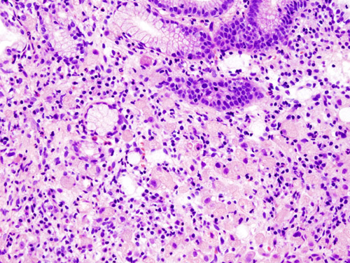 Signet-ring cell carcinoma: intracytoplasmic mucin displaces the nucleus to the cell periphery. CC BY-SA 3.0, https://commons.wikimedia.org/w/index.php?curid=502927


Clinical Correlations
- Test and treat H. pylori: Testing and eradication are indicated in patients with gastric neoplasia, premalignant lesions, or a family history of gastric cancer.
- High-risk premalignant mucosa: The following features identify mucosa at highest risk:[2][17]
- Extensive (antrum plus corpus) atrophy or intestinal metaplasia
- Incomplete intestinal metaplasia
- Persistent H. pylori infection
- Age older than 50
- Family history of gastric cancer
- Biomarker testing: Testing of gastric adenocarcinoma for MMR/MSI, HER2, PD-L1, and EBV follows directly from subtype biology.[6][8][7]
- MSI-high and EBV+ tumors predict checkpoint-inhibitor benefit.
- HER2 amplification (CIN subtype) directs trastuzumab-based therapy.
- Suspected HDGC: Patients should be referred to expert centers for CDH1/CTNNA1 testing and genetic counseling.[40][27]
Areas of Uncertainty
- Role of intestinal metaplasia: Whether metaplasia is an independent “point of no return” or a bystander is debated, as is which subset progresses. Chromosomal-instability features within metaplasia may identify progressors.[16][17][23]
- Natural history in CDH1 carriers: The progression rate from pT1a foci to advanced cancer, and the optimal timing of gastrectomy versus surveillance, remain uncertain.[9][40]
- Prognostic weight of EBV: The independent prognostic weight of EBV positivity is unsettled.[33][34]
- Origin of diffuse-type cancer: Whether it is purely de novo or shares an early field effect with the intestinal pathway is not established.[25]
References
- ↑ 1.00 1.01 1.02 1.03 1.04 1.05 1.06 1.07 1.08 1.09 1.10 1.11 Sundar R, Nakayama I, Markar SR, et al. (2025). “Gastric cancer”. Lancet. 405 (10494): 2087–2102. doi:10.1016/S0140-6736(25)00052-2.
- ↑ 2.0 2.1 2.2 2.3 2.4 2.5 Gawron AJ, Shah SC, Altayar O, Davitkov P, Morgan D, Turner K, Mustafa RA (2020). “AGA Technical Review on Gastric Intestinal Metaplasia—Natural History and Clinical Outcomes”. Gastroenterology. 158 (3): 705–731.e5. doi:10.1053/j.gastro.2019.12.001. PMID 31816300.
- ↑ 3.0 3.1 3.2 3.3 Bessède E, Dubus P, Mégraud F, Varon C (2015). “Helicobacter pylori infection and stem cells at the origin of gastric cancer”. Oncogene. 34 (20): 2547–55. doi:10.1038/onc.2014.187.
- ↑ 4.0 4.1 4.2 4.3 4.4 Duan Y, Xu Y, Dou Y, Xu D (2025). “Helicobacter pylori and gastric cancer: mechanisms and new perspectives”. J Hematol Oncol. 18 (1): 10. doi:10.1186/s13045-024-01654-2.
- ↑ 5.0 5.1 5.2 5.3 5.4 5.5 5.6 5.7 Cancer Genome Atlas Research Network (2014). “Comprehensive molecular characterization of gastric adenocarcinoma”. Nature. 513 (7517): 202–9. doi:10.1038/nature13480. PMC 4170219. PMID 25079317.
- ↑ 6.0 6.1 6.2 6.3 6.4 Patel AK, Sethi NS, Park H (2026). “Gastric cancer: a review”. JAMA. 335 (5): 439–450. doi:10.1001/jama.2025.20034.
- ↑ 7.0 7.1 7.2 Walch HS, Borpatragohain R, Jee J, et al. (2025). “Clinical implications of The Cancer Genome Atlas molecular classification system in esophagogastric cancer”. Clin Cancer Res. 31 (10): 1912–1921. doi:10.1158/1078-0432.CCR-24-3473. PMID 40299774 Check
|pmid=value (help). - ↑ 8.0 8.1 8.2 8.3 8.4 8.5 8.6 8.7 8.8 Smyth EC, Nilsson M, Grabsch HI, van Grieken NC, Lordick F (2020). “Gastric cancer”. Lancet. 396 (10251): 635–648. doi:10.1016/S0140-6736(20)31288-5.
- ↑ 9.0 9.1 9.2 9.3 9.4 9.5 Ryan CE, Fasaye GA, Gallanis AF, et al. (2024). “Germline CDH1 variants and lifetime cancer risk”. JAMA. 332 (9): 722–729. doi:10.1001/jama.2024.10852.
- ↑ 10.0 10.1 10.2 Baba H, Ishimoto T, Baba Y, Hayashi H, Iwatsuki M (2026). “Advances in gastric cancer research: insights into carcinogenesis, the tumor microenvironment, metastasis, and factors influencing prognosis”. Ann Gastroenterol Surg. 10 (4): 984–994. doi:10.1002/ags3.70195.
- ↑ Uemura N, Okamoto S, Yamamoto S, et al. (2001). “Helicobacter pylori infection and the development of gastric cancer”. N Engl J Med. 345 (11): 784–9. doi:10.1056/NEJMoa001999. PMID 11556297.
- ↑ Oh HJ, Kim CH, Park HW, Park B (2025). “Quantifying the effects of the Correa pathway from Helicobacter pylori infection to gastric cancer: causal inference found in 6.8 million Koreans”. BMC Cancer. 26 (1): 174. doi:10.1186/s12885-025-15507-9.
- ↑ 13.0 13.1 13.2 Malfertheiner P, Camargo MC, El-Omar E, et al. (2023). “Helicobacter pylori infection”. Nat Rev Dis Primers. 9 (1): 19. doi:10.1038/s41572-023-00431-8.
- ↑ 14.0 14.1 14.2 Wizenty J, Sigal M (2025). “Helicobacter pylori, microbiota and gastric cancer — principles of microorganism-driven carcinogenesis”. Nat Rev Gastroenterol Hepatol. 22 (5): 296–313. doi:10.1038/s41575-025-01042-2.
- ↑ Correa P (1988). “A human model of gastric carcinogenesis”. Cancer Res. 48 (13): 3554–60. PMID 3288329.
- ↑ 16.0 16.1 Yeoh KG, Tan P (2022). “Mapping the genomic diaspora of gastric cancer”. Nat Rev Cancer. 22 (2): 71–84. doi:10.1038/s41568-021-00412-7.
- ↑ 17.0 17.1 17.2 17.3 17.4 Sugano K, Moss SF, Kuipers EJ (2023). “Gastric intestinal metaplasia: real culprit or innocent bystander as a precancerous condition for gastric cancer?”. Gastroenterology. 165 (6): 1352–1366.e1. doi:10.1053/j.gastro.2023.08.028.
- ↑ Hatakeyama M (2004). “Oncogenic mechanisms of the Helicobacter pylori CagA protein”. Nat Rev Cancer. 4 (9): 688–94. doi:10.1038/nrc1433. PMID 15343275.
- ↑ Wan C, Wang P, Xu Y, et al. (2025). “Mechanism and role of H. pylori CagA-induced NLRP3 inflammasome in gastric cancer immune cell infiltration”. Sci Rep. 15 (1): 14335. doi:10.1038/s41598-025-98301-8.
- ↑ El Tekle G, Garrett WS (2023). “Bacteria in cancer initiation, promotion and progression”. Nat Rev Cancer. 23 (9): 600–618. doi:10.1038/s41568-023-00594-2.
- ↑ Müller A, He J (2023). “A double whammy on gastric cancer risk”. N Engl J Med. 388 (13): 1225–1229. doi:10.1056/NEJMe2215503.
- ↑ 22.0 22.1 Ajani JA, Lee J, Sano T, et al. (2017). “Gastric adenocarcinoma”. Nat Rev Dis Primers. 3: 17036. doi:10.1038/nrdp.2017.36.
- ↑ 23.0 23.1 23.2 Zhang T, Wang Z, Zhang B, Tang X (2026). “Refining the Correa cascade: gastric stem cell plasticity, niche remodelling, and parallel pathways to neoplasia”. Pharmacol Res. 230: 108315. doi:10.1016/j.phrs.2026.108315.
- ↑ Cornish SK, Choi E, Mills JC, Goldenring JR (2026). “The origins and progression of pyloric metaplasia following gastric mucosal injury”. Physiol Rev. 106 (4): 2441–2471. doi:10.1152/physrev.00042.2025.
- ↑ 25.0 25.1 25.2 25.3 Chen X, Wang S, Li J (2026). “Reframing early gastric carcinogenesis through lineage, niche, and evolution”. Cancer Metastasis Rev. 45 (3): 67. doi:10.1007/s10555-026-10376-5.
- ↑ Kapteijn N, Marijnissen FE, Pluimers J, et al. (2026). “Dietary factors associated with the progression of gastric intestinal metaplasia: a multicenter, prospective cohort study in a Western population”. Clin Transl Gastroenterol. 17 (5): e01006. doi:10.14309/ctg.0000000000001006. Vancouver style error: initials (help)
- ↑ 27.0 27.1 27.2 Vos EL, Salo-Mullen EE, Tang LH, et al. (2020). “Indications for total gastrectomy in CDH1 mutation carriers and outcomes of risk-reducing minimally invasive and open gastrectomies”. JAMA Surg. 155 (11): 1050–1057. doi:10.1001/jamasurg.2020.3356.
- ↑ 28.0 28.1 28.2 28.3 28.4 Decourtye-Espiard L, Guilford P (2023). “Hereditary diffuse gastric cancer”. Gastroenterology. 164 (5): 719–735. doi:10.1053/j.gastro.2023.01.038.
- ↑ 29.0 29.1 29.2 Liabeuf D, Oshima M, Stange DE, Sigal M (2022). “Stem cells, Helicobacter pylori, and mutational landscape: utility of preclinical models to understand carcinogenesis and to direct management of gastric cancer”. Gastroenterology. 162 (4): 1067–1087. doi:10.1053/j.gastro.2021.12.252.
- ↑ 30.0 30.1 Hirabayashi M, Georges D, Clifford GM, de Martel C (2023). “Estimating the global burden of Epstein-Barr virus-associated gastric cancer: a systematic review and meta-analysis”. Clin Gastroenterol Hepatol. 21 (4): 922–930.e21. doi:10.1016/j.cgh.2022.07.042. PMID 35963539 Check
|pmid=value (help). - ↑ 31.0 31.1 31.2 31.3 Damania B, Kenney SC, Raab-Traub N (2022). “Epstein-Barr virus: biology and clinical disease”. Cell. 185 (20): 3652–3670. doi:10.1016/j.cell.2022.08.026.
- ↑ 32.0 32.1 Yang J, Liu Z, Zeng B, Hu G, Gan R (2020). “Epstein-Barr virus-associated gastric cancer: a distinct subtype”. Cancer Lett. 495: 191–199. doi:10.1016/j.canlet.2020.09.019.
- ↑ 33.0 33.1 33.2 Zhao Y, Zhang J, Cheng A, et al. (2020). “Gastric cancer: genome damaged by bugs”. Oncogene. 39 (17): 3427–3442. doi:10.1038/s41388-020-1241-4. Vancouver style error: initials (help)
- ↑ 34.0 34.1 Ozawa H, Wang Y, Withers HG, et al. (2025). “MUC1-C auto-regulatory complex with EBNA1 is responsible for latent Epstein-Barr virus-associated gastric cancer progression”. Oncogene. 44 (38): 3609–3624. doi:10.1038/s41388-025-03519-5.
- ↑ 35.0 35.1 Felismino TC, Coimbra F (2023). “Lessons in gastric adenocarcinoma from TCGA”. Nat Rev Cancer. 23 (10): 655. doi:10.1038/s41568-023-00606-1. Vancouver style error: initials (help)
- ↑ 36.0 36.1 Nakamura Y, Kawazoe A, Lordick F, Janjigian YY, Shitara K (2021). “Biomarker-targeted therapies for advanced-stage gastric and gastro-oesophageal junction cancers: an emerging paradigm”. Nat Rev Clin Oncol. 18 (8): 473–487. doi:10.1038/s41571-021-00492-2.
- ↑ Van Cutsem E, Sagaert X, Topal B, Haustermans K, Prenen H (2016). “Gastric cancer”. Lancet. 388 (10060): 2654–2664. doi:10.1016/S0140-6736(16)30354-3.
- ↑ 38.0 38.1 Johnston FM, Beckman M (2019). “Updates on management of gastric cancer”. Curr Oncol Rep. 21 (8): 67. doi:10.1007/s11912-019-0820-4.
- ↑ 39.0 39.1 Chen J, Zhang X, Zhao Y (2026). “Advances in the molecular subtyping of gastric cancer”. World J Surg Oncol. 24 (1): 314. doi:10.1186/s12957-026-04437-w.
- ↑ 40.0 40.1 40.2 40.3 40.4 40.5 National Comprehensive Cancer Network. NCCN Clinical Practice Guidelines in Oncology: Genetic/Familial High-Risk Assessment: Colorectal, Endometrial, Esophageal, and Gastric. Version 1.2026 (updated June 16, 2026). https://www.nccn.org
- ↑ van der Post RS, Vogelaar IP, Manders P, et al. (2015). “Accuracy of hereditary diffuse gastric cancer testing criteria and outcomes in patients with a germline mutation in CDH1”. Gastroenterology. 149 (4): 897–906.e19. doi:10.1053/j.gastro.2015.06.003. PMID 26072394.
- ↑ Corso G (2022). “Pleiotropic cancer manifestations of germline CDH1 mutations: risks and management”. J Surg Oncol. 125 (8): 1326–1331. doi:10.1002/jso.26847.
- ↑ Garcia-Pelaez J, Barbosa-Matos R, Lobo S, et al. (2023). “Genotype-first approach to identify associations between CDH1 germline variants and cancer phenotypes: a multicentre study by the European Reference Network on Genetic Tumour Risk Syndromes”. Lancet Oncol. 24 (1): 91–106. doi:10.1016/S1470-2045(22)00643-X. PMID 36436516 Check
|pmid=value (help). - ↑ Laszkowska M, Silver ER, Schrope B, et al. (2020). “Optimal timing of total gastrectomy to prevent diffuse gastric cancer in individuals with pathogenic variants in CDH1”. Clin Gastroenterol Hepatol. 18 (4): 822–829.e4. doi:10.1016/j.cgh.2019.06.009.
- ↑ Hirata Y, Noorani A, Song S, Wang L, Ajani JA (2023). “Early stage gastric adenocarcinoma: clinical and molecular landscapes”. Nat Rev Clin Oncol. 20 (7): 453–469. doi:10.1038/s41571-023-00767-w.
Editor-In-Chief: C. Michael Gibson, M.S., M.D. [1]; Associate Editor(s)-in-Chief: Fahad Hasan, M.D.[2] Omer Kamal, M.D.[3], Parminder Dhingra, M.D. [4], Mohammed Abdelwahed M.D[5]
Causes
Overview
Gastric adenocarcinoma results from an interaction between chronic infection, environmental and dietary exposures, host genetic susceptibility, and, in a small minority, high-penetrance germline variants. Chronic Helicobacter pylori infection, an IARC group 1 carcinogen, is the dominant modifiable cause. It accounts for approximately 75% of attributable risk overall and approximately 90% of non-cardia (distal) cancers.[1][2] Most intestinal-type cancers arise through the stepwise Correa cascade (chronic gastritis → atrophic gastritis → intestinal metaplasia → dysplasia → carcinoma). Diffuse-type cancers frequently arise without a recognizable precancerous lesion. When diffuse-type cancer is hereditary, it is driven by germline CDH1 (E-cadherin) inactivation.[3] About 10% of cases show familial clustering, but only about 1–3% are attributable to a known heritable germline variant.[4] The TCGA molecular classification (EBV-positive, MSI, genomically stable, chromosomal instability) has superseded older, undifferentiated oncogene lists as the framework for molecular etiology.[5]
Causes
Infectious Causes
Helicobacter pylori
- H. pylori is the single most important and most modifiable cause of gastric adenocarcinoma.
- Attributable risk: approximately 75% of global gastric cancer and approximately 90% of non-cardia cancers. In Japan, fewer than 1% of gastric adenocarcinomas are considered unrelated to H. pylori.[1][7]
- Anatomic specificity: the association is strong for non-cardia cancer (pooled OR approximately 2.97) and essentially null for cardia cancer (OR approximately 0.99).[8]
- Test method affects measured risk: serology reflects lifetime infection and yields higher odds ratios than histology. Histology underestimates infection when atrophic gastritis has reduced the bacterial load.[2]
- Low absolute risk per infected person: about 44% of adults worldwide are infected, but only about 1–3% of infected individuals develop gastric cancer. Strain virulence, host genetics, and co-exposures modulate this risk.[1][9]
- Mechanism (Correa cascade): chronic infection causes the following changes, which drive progression from normal mucosa to carcinoma:[10][11]
- Virulence factors: CagA (delivered through the type IV secretion system) and VacA, particularly the s1/m1 genotype, confer substantially higher risk than less virulent strains.[11] CagA also activates the NLRP3 inflammasome, which shapes immune-cell infiltration in gastric cancer.[12]
- Histologic subtype: H. pylori is associated with both intestinal-type and diffuse-type cancer. Chronic inflammation is not required for diffuse-type carcinogenesis, which implies a distinct mechanism.[1]
- Causal evidence: a cluster-randomized trial in Linqu County, China, enrolled 180,284 participants with 11.8 years of follow-up. Community-based eradication reduced gastric cancer incidence (intention-to-treat HR 0.86; 95% CI 0.74–0.99). The effect was larger among participants with confirmed eradication (HR 0.81; 95% CI 0.69–0.96).[13][14]
Epstein–Barr virus
- EBV is associated with approximately 5–10% of gastric cancers.[1][9]
- EBV-positive tumors have the following features:[1][5]
- Location in the proximal stomach
- Occurrence in younger patients
- Dense immune infiltration with PD-L1/PD-L2 expression
- Extensive DNA hypermethylation
- Recurrent PIK3CA, ARID1A, and BCOR mutations
Environmental and Lifestyle Causes
Environmental exposures are believed to drive most non-hereditary cases.[15]
| Exposure | Notes |
|---|---|
| Tobacco smoking | Dose- and duration-dependent; independent risk factor; contributes particularly to cardia cancer[9][16] |
| Heavy alcohol use (≥4 drinks/day) | Risk applies at heavy intake[9] |
| High-salt and salt-preserved foods (salted fish, pickled vegetables, salt-preserved meats) | Synergistic with H. pylori[9][14] |
| Obesity | Mainly cardia and GEJ adenocarcinoma[7][14] |
| GERD and Barrett’s esophagus | Drives the Western shift toward proximal tumors[7] |
| Diet low in fruits and vegetables; high in processed or red meat and ultraprocessed foods | [15] |
- Other associations: older age, male sex, lower socioeconomic status, poor hygiene, pernicious anemia, and prior gastric ulcer or partial gastrectomy.[1][17]
- Chronic atrophic gastritis and intestinal metaplasia are intermediate risk states on the causal pathway, not independent exposures.[1]
- The rising incidence of early-onset gastric cancer (age <50 years) is incompletely explained by known risk factors.[9]
Hereditary and Genetic Causes
- Hereditary gastric cancer is traditionally divided into three autosomal dominant syndromes:[4]
- Hereditary diffuse gastric cancer (HDGC)
- Familial intestinal gastric cancer
- Gastric adenocarcinoma and proximal polyposis of the stomach (GAPPS)
Hereditary diffuse gastric cancer and CDH1
- HDGC is an autosomal dominant syndrome of early-onset diffuse gastric cancer and lobular breast cancer. It is caused mainly by inactivating germline CDH1 variants.[18][19]
- Pathogenic CDH1 variants occur in approximately 1 in 5,000 to 1 in 8,000 people in unselected populations.[19]
- Mechanism: CDH1 encodes E-cadherin, a transmembrane cell–cell adhesion protein and tumor suppressor.
- Carriers inherit one inactivated allele.
- A somatic second hit (mutation, loss of heterozygosity, or promoter hypermethylation) inactivates the remaining allele.
- Loss of E-cadherin function produces poorly cohesive signet ring cell carcinoma.[19][20][21]
- Current penetrance estimates (lower than older pedigree-based figures of approximately 70% in men and 56% in women):[18][19]
- Cumulative gastric cancer risk to age 80: approximately 37–42% in men and 25–33% in women
- Lobular breast cancer risk in women: 37–55%
- Intramucosal foci: nearly all carriers harbor microscopic intramucosal (pT1a) signet ring cell foci. Cancer of any stage is found in 88–97% of risk-reducing gastrectomy specimens, but disease ≥pT1b is found in only about 2–3%. Modeled lifetime risk of advanced (≥stage 2) cancer is approximately 10% in men and 6.5% in women.[19]
- Other HDGC genes: CTNNA1 (α-E-catenin) accounts for approximately 2% of HDGC families. About 70% of clinically defined HDGC families have no identified causative gene.[3][4]
Other hereditary syndromes
| Syndrome | Gene(s) | Key feature |
|---|---|---|
| HDGC | CDH1, CTNNA1 | Diffuse/signet ring cell gastric cancer; lobular breast cancer |
| GAPPS | APC promoter 1B point variants | >100 fundic gland polyps with antral sparing; no colorectal polyposis[4][19] |
| Lynch syndrome | MLH1, MSH2, MSH6, PMS2, EPCAM | Intestinal-type, MSI-high tumors |
| Familial adenomatous polyposis | APC | Gastric polyps; elevated gastric cancer risk |
| Li-Fraumeni syndrome | TP53 | Multiple early-onset cancers |
| Peutz-Jeghers syndrome | STK11 | Hamartomatous polyps |
| Juvenile polyposis syndrome | BMPR1A, SMAD4 | Gastric juvenile polyps |
| Cowden syndrome | PTEN | Hamartomatous polyps |
| MUTYH-associated polyposis | MUTYH | Attenuated polyposis |
| Hereditary breast and ovarian cancer and related | BRCA1, BRCA2, PALB2 | Modestly increased risk[4][19] |
Molecular Etiology
The TCGA classification defines four molecular subtypes with distinct causal mechanisms.[5]
| Subtype | Approximate frequency | Defining features |
|---|---|---|
| Chromosomal instability (CIN) | ~50% | Aneuploidy; frequent TP53 mutation; receptor tyrosine kinase amplification (ERBB2, VEGFA, FGFR2); mostly intestinal type; often at the GEJ |
| Microsatellite instability (MSI) | ~22% | Mismatch-repair deficiency, commonly from MLH1 promoter hypermethylation; high tumor mutational burden |
| Genomically stable (GS) | ~20% | Diffuse-type histology; CDH1 and RHOA mutations; CLDN18–ARHGAP fusions; peritoneal spread |
| EBV-positive | ~9% | Extensive hypermethylation; PIK3CA and ARID1A mutations; PD-L1 overexpression |
- The Asian Cancer Research Group (ACRG) proposed an overlapping classification: MSI, MSS/EMT, MSS/TP53-active, and MSS/TP53-inactive. Its distinct MSS/EMT subtype is marked by CDH1 loss and peritoneal tropism.[22]
- In routine practice, formal subtype assignment is not yet standard. Only actionable markers are tested: MMR/MSI, HER2, PD-L1, and increasingly EBV and Claudin-18.2.[4][23]
- Up to about 25% of GEJ tumors fall outside the expected CIN class.[24]
Clinical Implications of Etiology
- Apply the H. pylori causal link to non-cardia cancer only. Cardia and GEJ cancers are instead linked to obesity, reflux, and Barrett’s esophagus.[8][7]
- A single negative histologic H. pylori test can be falsely negative in atrophic gastritis or intestinal metaplasia; serology captures lifetime infection.[2]
- Diffuse-type cancer often does not follow the Correa cascade.[1]
- The following findings should prompt germline CDH1 evaluation per NCCN criteria:[19]
- Diffuse gastric cancer or T1a signet ring cell carcinoma at any age
- Diffuse gastric cancer diagnosed before age 50
- Lobular breast cancer with a personal or family history of diffuse gastric cancer
- Qualifying family histories
References
- ↑ 1.0 1.1 1.2 1.3 1.4 1.5 1.6 1.7 1.8 Sundar R, Nakayama I, Markar SR, Shitara K, van Laarhoven H, Janjigian YY, Smyth EC (June 2025). “Gastric cancer”. Lancet. 405 (10494): 2087–2102. doi:10.1016/S0140-6736(25)00052-2. PMID 40319897 Check
|pmid=value (help). Vancouver style error: initials (help) - ↑ 2.0 2.1 2.2 2.3 Thrift AP, Wenker TN, El-Serag HB (May 2023). “Global burden of gastric cancer: epidemiological trends, risk factors, screening and prevention”. Nat Rev Clin Oncol. 20 (5): 338–349. doi:10.1038/s41571-023-00747-0. PMID 36959359 Check
|pmid=value (help). - ↑ 3.0 3.1 Decourtye-Espiard L, Guilford P (April 2023). “Hereditary diffuse gastric cancer”. Gastroenterology. 164 (5): 719–735. doi:10.1053/j.gastro.2023.01.038. PMID 36740198 Check
|pmid=value (help). - ↑ 4.0 4.1 4.2 4.3 4.4 4.5 Smyth EC, Nilsson M, Grabsch HI, van Grieken NC, Lordick F (August 2020). “Gastric cancer”. Lancet. 396 (10251): 635–648. doi:10.1016/S0140-6736(20)31288-5. PMID 32861308 Check
|pmid=value (help). - ↑ 5.0 5.1 5.2 Cancer Genome Atlas Research Network (September 2014). “Comprehensive molecular characterization of gastric adenocarcinoma”. Nature. 513 (7517): 202–209. doi:10.1038/nature13480. PMC 4170219. PMID 25079317.
- ↑ IARC Working Group on the Evaluation of Carcinogenic Risks to Humans (1994). “Schistosomes, liver flukes and Helicobacter pylori”. IARC Monogr Eval Carcinog Risks Hum. 61: 1–241. PMID 7715068.
- ↑ 7.0 7.1 7.2 7.3 Hirata Y, Noorani A, Song S, Wang L, Ajani JA (July 2023). “Early stage gastric adenocarcinoma: clinical and molecular landscapes”. Nat Rev Clin Oncol. 20 (7): 453–469. doi:10.1038/s41571-023-00767-w. PMC 11869628 Check
|pmc=value (help). PMID 37264184 Check|pmid=value (help). - ↑ 8.0 8.1 Nawawi KNM, El-Omar EM, Ali RAR. Screening, surveillance, and prevention of esophageal and gastric cancers. In: Gastrointestinal Oncology. 2nd ed. 2024.
- ↑ 9.0 9.1 9.2 9.3 9.4 9.5 Patel AK, Sethi NS, Park H (February 2026). “Gastric cancer: a review”. JAMA. 335 (5): 439–450. doi:10.1001/jama.2025.20034. PMID 41499132 Check
|pmid=value (help). - ↑ Liabeuf D, Oshima M, Stange DE, Sigal M (April 2022). “Stem cells, Helicobacter pylori, and mutational landscape: utility of preclinical models to understand carcinogenesis and to direct management of gastric cancer”. Gastroenterology. 162 (4): 1067–1087. doi:10.1053/j.gastro.2021.12.252. PMID 34942172 Check
|pmid=value (help). - ↑ 11.0 11.1 Tang W, Li F, Zheng H, Zhou S, Li C, Xu X, Fu J (December 2025). “Unveiling hidden players: the role of intratumoral microbiota in gastrointestinal cancer dynamics”. J Cancer Res Clin Oncol. 152 (1): 15. doi:10.1007/s00432-025-06401-9. PMC 12715092 Check
|pmc=value (help). PMID 41410942 Check|pmid=value (help). - ↑ Wan C, Wang P, Xu Y, Zhu Y, Chen H, Cao X, Gu Y (April 2025). “Mechanism and role of H. pylori CagA-induced NLRP3 inflammasome in gastric cancer immune cell infiltration”. Sci Rep. 15 (1): 14335. doi:10.1038/s41598-025-98301-8. PMC 12022178 Check
|pmc=value (help). PMID 40274924 Check|pmid=value (help). - ↑ Pan KF, Li WQ, Zhang L, Liu WD, Ma JL, Zhang Y, et al. (November 2024). “Gastric cancer prevention by community eradication of Helicobacter pylori: a cluster-randomized controlled trial”. Nat Med. 30 (11): 3250–3260. doi:10.1038/s41591-024-03153-w. PMID 39079993 Check
|pmid=value (help). - ↑ 14.0 14.1 14.2 Jayakrishnan T, Ng K (October 2025). “Early-onset gastrointestinal cancers: a review”. JAMA. 334 (15): 1373–1385. doi:10.1001/jama.2025.10218. PMID 40674064 Check
|pmid=value (help). - ↑ 15.0 15.1 Müller A, He J (March 2023). “A double whammy on gastric cancer risk”. N Engl J Med. 388 (13): 1225–1229. doi:10.1056/NEJMe2215503. PMID 36988601 Check
|pmid=value (help). - ↑ Zhao G, Liu Y, Wang H, Zhang Y, Shang L, Li L (December 2025). “Global, regional, and national burden of gastric cancer attributable to smoking and a high-sodium diet from 1990 to 2021: a global burden of disease study 2021”. BMC Gastroenterol. 26 (1): 52. doi:10.1186/s12876-025-04431-8. PMC 12828996 Check
|pmc=value (help). PMID 41340089 Check|pmid=value (help). - ↑ Walter K (June 2026). “Gastric cancer”. JAMA. 335 (24): 2176. doi:10.1001/jama.2026.3754. PMID 42166170 Check
|pmid=value (help). - ↑ 18.0 18.1 Garcia-Pelaez J, Barbosa-Matos R, Lobo S, Dias A, Garrido L, Castedo S, et al. (January 2023). “Genotype-first approach to identify associations between CDH1 germline variants and cancer phenotypes: a multicentre study by the European Reference Network on Genetic Tumour Risk Syndromes”. Lancet Oncol. 24 (1): 91–106. doi:10.1016/S1470-2045(22)00643-X. PMC 9810541 Check
|pmc=value (help). PMID 36436516 Check|pmid=value (help). - ↑ 19.0 19.1 19.2 19.3 19.4 19.5 19.6 19.7 National Comprehensive Cancer Network (2026). “NCCN Clinical Practice Guidelines in Oncology: Genetic/Familial High-Risk Assessment: Colorectal, Endometrial, Esophageal, and Gastric (HGAST-1, HGAST-A, HGAST-B)”.
- ↑ MedlinePlus Genetics, National Library of Medicine. “Hereditary diffuse gastric cancer”.
- ↑ Graziano F, Humar B, Guilford P (December 2003). “The role of the E-cadherin gene (CDH1) in diffuse gastric cancer susceptibility: from the laboratory to clinical practice”. Ann Oncol. 14 (12): 1705–13. doi:10.1093/annonc/mdg486. PMID 14630673.
- ↑ Cristescu R, Lee J, Nebozhyn M, Kim KM, Ting JC, Wong SS, et al. (May 2015). “Molecular analysis of gastric cancer identifies subtypes associated with distinct clinical outcomes”. Nat Med. 21 (5): 449–456. doi:10.1038/nm.3850. PMID 25894828.
- ↑ Röcken C (January 2023). “Predictive biomarkers in gastric cancer”. J Cancer Res Clin Oncol. 149 (1): 467–481. doi:10.1007/s00432-022-04408-0. PMC 9889517 Check
|pmc=value (help). PMID 36260159 Check|pmid=value (help). - ↑ Walch HS, Borpatragohain R, Jee J, Chatila W, Fong C, Maron SB, et al. (May 2025). “Clinical implications of The Cancer Genome Atlas molecular classification system in esophagogastric cancer”. Clin Cancer Res. 31 (10): 1912–1921. doi:10.1158/1078-0432.CCR-24-3473. PMID 40299774 Check
|pmid=value (help).
Editor-In-Chief: C. Michael Gibson, M.S., M.D. [1]; Associate Editor(s)-in-Chief: Fahad Hasan, M.D.[2] Omer Kamal, M.D.[3], Mohammed Abdelwahed M.D[4]
Differentiating Stomach Cancer from other Diseases
Overview
Stomach cancer must be differentiated from other diseases presenting with episodic abdominal pain, weight loss and loss of appetite such as gastric lymphoma, gastric metastasis, gastritis, benign gastric ulcer, Menetrier’s disease.
Differential Diagnosis
Stomach cancer must be differentiated from other diseases presenting with episodic abdominal pain, weight loss and loss of appetite. The differentials include the following:[1][2][3][4][5][6][7][8][9]
| Differential Diagnosis | ||||||||||||
| Disease | Cause | Symptoms | Diagnosis | Other findings | ||||||||
| Pain | Nausea & Vomiting | Heartburn | Belching or Bloating | Weight loss | Loss of Appetite | Stools | Endoscopy findings | |||||
| Location | Aggravating Factors | Alleviating Factors | ||||||||||
| Acute gastritis |
|
Food | Antacids | ✔ | ✔ | ✔ | – | ✔ | Black stools |
|
– | |
| Chronic gastritis |
|
Food | Antacids | ✔ | ✔ | ✔ | ✔ | ✔ | – | H. pylori gastritis
Lymphocytic gastritis
|
– | |
| Atrophic gastritis | – | – | ✔ | – | ✔ | ✔ | – | H. pylori |
| |||
| Crohn’s disease | – | – | – | – | – | ✔ | ✔ |
|
|
|||
| GERD |
|
|
|
✔
(Suspect delayed gastric emptying) |
✔ | – | – | – | – | Other symptoms:
Complications
| ||
| Peptic ulcer disease |
|
|
|
|
✔ | ✔ | – | – | – | Gastric ulcers
Duodenal ulcers
|
Other diagnostic tests | |
| Gastrinoma |
|
– | – | ✔
(suspect gastric outlet obstruction) |
✔ | – | – | – | Useful in collecting the tissue for biopsy |
Diagnostic tests
| ||
| Gastric Adenocarcinoma |
|
– | – | ✔ | ✔ | ✔ | ✔ | ✔ |
|
Esophagogastroduodenoscopy
|
Other symptoms | |
| Primary gastric lymphoma |
|
– | – | – | – | – | ✔ | – | – | Useful in collecting the tissue for biopsy | Other symptoms
| |
References
- ↑ Sugimachi K, Inokuchi K, Kuwano H, Ooiwa T (1984). “Acute gastritis clinically classified in accordance with data from both upper GI series and endoscopy”. Scand J Gastroenterol. 19 (1): 31–7. PMID 6710074.
- ↑ Sipponen P, Maaroos HI (2015). “Chronic gastritis”. Scand J Gastroenterol. 50 (6): 657–67. doi:10.3109/00365521.2015.1019918. PMC 4673514. PMID 25901896.
- ↑ Sartor RB (2006). “Mechanisms of disease: pathogenesis of Crohn’s disease and ulcerative colitis”. Nat Clin Pract Gastroenterol Hepatol. 3 (7): 390–407. doi:10.1038/ncpgasthep0528. PMID 16819502.
- ↑ Sipponen P (1989). “Atrophic gastritis as a premalignant condition”. Ann Med. 21 (4): 287–90. PMID 2789799.
- ↑ Badillo R, Francis D (2014). “Diagnosis and treatment of gastroesophageal reflux disease”. World J Gastrointest Pharmacol Ther. 5 (3): 105–12. doi:10.4292/wjgpt.v5.i3.105. PMC 4133436. PMID 25133039.
- ↑ Ramakrishnan K, Salinas RC (2007). “Peptic ulcer disease”. Am Fam Physician. 76 (7): 1005–12. PMID 17956071.
- ↑ Banasch M, Schmitz F (2007). “Diagnosis and treatment of gastrinoma in the era of proton pump inhibitors”. Wien Klin Wochenschr. 119 (19–20): 573–8. doi:10.1007/s00508-007-0884-2. PMID 17985090.
- ↑ Dicken BJ, Bigam DL, Cass C, Mackey JR, Joy AA, Hamilton SM (2005). “Gastric adenocarcinoma: review and considerations for future directions”. Ann Surg. 241 (1): 27–39. PMC 1356843. PMID 15621988.
- ↑ Ghimire P, Wu GY, Zhu L (2011). “Primary gastrointestinal lymphoma”. World J Gastroenterol. 17 (6): 697–707. doi:10.3748/wjg.v17.i6.697. PMC 3042647. PMID 21390139.
Editor-In-Chief: C. Michael Gibson, M.S., M.D. [1]Associate Editor(s)-in-Chief: Parminder Dhingra, M.D. [2]
Epidemiology and Demographics
Overview
Gastric cancer is the fifth most commonly diagnosed cancer and the fifth leading cause of cancer death worldwide. GLOBOCAN estimated 968,350 new cases and 659,853 deaths in 2022.[1][2] Age-standardized incidence and mortality have declined for more than half a century, mainly because Helicobacter pylori prevalence has fallen and food refrigeration and preservation have improved. Absolute case numbers keep rising because of population growth and aging.[3] Incidence is highest in East Asia, Eastern Europe, and parts of Central and South America. In the United States, about 30,300 new cases are diagnosed each year, the median age at diagnosis is 68 years, and incidence in men is nearly twice that in women.[2] Cardia and non-cardia gastric cancer are epidemiologically distinct diseases. The incidence of early-onset disease (diagnosis before age 50) is rising in several low-incidence countries, including the United States.[4][2]
Epidemiology and Demographics
Global Burden
- GLOBOCAN 2022 ranked gastric cancer fifth worldwide in both incidence and mortality, with 968,350 new cases and 659,853 deaths.[1][2]
- In the Global Burden of Disease (GBD) 2021 analysis, the global age-standardized incidence rate (ASIR) was 14.33 per 100,000, down from 24.76 per 100,000 in 1990. The age-standardized mortality rate (ASMR) was 11.20 per 100,000.[5]
- Over the same period, absolute incident cases rose from about 981,000 (1990) to about 1.23 million (2021).[5]
- Different modeling frameworks give different absolute counts, so figures should always be cited with their source and year rather than averaged:[1][5]
| Source (estimate year) | New cases | Deaths | Comment |
|---|---|---|---|
| GLOBOCAN 2020 | ~1.09 million | ~769,000 | Registry-based IARC estimates[3] |
| GLOBOCAN 2022 | 968,350 | 659,853 | Current IARC estimate[1] |
| GBD 2021 | ~1.23 million | — | ASIR 14.33 and ASMR 11.20 per 100,000[5] |
| GBD 2023 forecast (2050) | — | ~1.16 million | Deaths +22.5%, although ASMR is forecast to fall ~36%[6] |
Geographic Distribution
- Incidence is highest in East Asia (China, Japan, South Korea), Eastern Europe, and parts of Central and South America. It is lowest in North America and most of Africa.[3]
- In 2020, incidence was about 22.4 per 100,000 in Eastern Asia, compared with about 4.2 per 100,000 in North America and 3–4 per 100,000 in Africa.[3]
- Asia accounts for about 75% of global cases and deaths. In GBD 2021, China alone accounted for about 612,000 cases.[7][8]
- Development gradient: Incidence tracks H. pylori prevalence. Mortality is proportionally higher in medium- and low-HDI regions because of late diagnosis and limited access to treatment.[3]
- Migration: First-generation immigrants from high-incidence countries keep up to ~5-fold higher incidence and ~2-fold higher mortality than the low-incidence host population. The excess shrinks in later generations.[9] Classic studies of Japanese migrants to the United States first showed that early-life environmental exposures, rather than genetics, drive most of this risk.[10]
Secular Trends
- Age-standardized incidence and mortality have fallen worldwide. The global ASIR dropped by roughly 30–40% between 1990 and 2021.[5]
- In the United States, age-standardized incidence fell by about 1.46% per year from 1975 to 2019 (11.6 to 6.1 per 100,000).[3] The decline was about 0.94% per year between 2001 and 2015.[11]
- Rates are falling while counts are rising, because aging and population growth outweigh the falling rates. This has direct implications for service planning.[3]
United States
- Estimates for 2025 are about 30,300 new cases and 10,780 deaths.[2]
- Five-year relative survival across all stages rose from 15% in 1977 to about 36% by 2020.[2]
- Survival depends strongly on stage: about 70% for localized, 31% for regional, and 6% for distant disease. About 60% of patients present with regional or distant disease.[12] Early-stage disease amenable to endoscopic or surgical resection has a 5-year relative survival of about 75%.[2]
- Mortality fell substantially across all racial and ethnic groups from 2000 to 2019. It was lowest in the White population every year, and minoritized populations had higher mortality in almost all counties.[12]
- Gastric cancer mortality is 43% higher in U.S. counties with persistent poverty.[9]
Age
- Incidence rises sharply after age 50. Globally, the 70–74-year age group has the most incident cases and deaths.[8]
- In the United States, the median age at diagnosis is 68 years.[2] About 70% of patients are diagnosed between ages 55 and 84.[13]
- Early-onset disease (before age 50) has a distinct epidemiology and is described below.
Sex
- Gastric cancer is about 2- to 3-fold more common in men than in women, both worldwide and in the United States.[3] U.S. incidence is 8.4 per 100,000 per year in men and 4.8 per 100,000 per year in women.[2]
- Most of the male excess comes from cardia cancer, where the male-to-female incidence rate ratio is about 4.2. For non-cardia cancer the ratio is about 1.6.[3]
- Incidence in women resembles that in men but lags by roughly 10–15 years after menopause. A protective effect of estrogen has been proposed, but this remains a hypothesis.[13]
- Early-onset disease runs counter to the overall male predominance: it is relatively more common in women.[14][4]
Race and Ethnicity (United States)
- Gastric cancer is one of the few malignancies for which non-Hispanic White Americans have the lowest incidence and mortality.[12]
- Non-cardia gastric cancer incidence is 1.7- to 3.9-fold higher in Black, Hispanic, Asian/Pacific Islander, American Indian, and Alaska Native individuals than in non-Hispanic White individuals.[15]
- Korean American men and women aged 50 years or older have the highest risk: up to 12- to 14.5-fold higher non-cardia incidence than non-Hispanic White peers. Korean American men in this age group had a non-cardia incidence of about 70 per 100,000.[16][9]
- Cardia gastric cancer, by contrast, is more common in White patients.[14]
- Minoritized groups more often present with regional or distant non-cardia disease.[17]
- Race and ethnicity are surrogates for underlying drivers: H. pylori prevalence, country of origin, and social determinants of health. Aggregate categories such as “Asian” hide markedly different risk across origin groups, so risk should be assessed by country or region of birth.[9] Implications for targeted screening are covered in Stomach cancer screening.
Anatomic Subsite: Cardia vs Non-Cardia
Gastric cancer is best understood as two epidemiologically distinct diseases.[3][1]
| Feature | Non-cardia (body/antrum) | Cardia / gastroesophageal junction |
|---|---|---|
| Share of cases | ~85% of cases globally[2] | 30–40% of gastric cancers in North America and Western Europe[1] |
| Principal drivers | Chronic H. pylori infection (~90% attributable), high-salt or salt-preserved diet, smoking, low socioeconomic status[2][1] | Obesity, gastroesophageal reflux disease, Barrett’s esophagus; profile overlaps esophageal adenocarcinoma. Only ~1 in 5 cases is H. pylori-attributable globally[18][1] |
| Male:female incidence rate ratio | ~1.6 | ~4.2[3] |
| U.S. race/ethnicity pattern | Higher in Black, Hispanic, Asian/Pacific Islander, and AI/AN populations than in non-Hispanic White[15] | More common in White patients[14] |
| Secular trend | Declining for decades | U.S./UK incidence rose from the 1960s to the 1980s, then stabilized[1][18] |
Early-Onset Gastric Cancer
- Incidence of gastric cancer diagnosed before age 50 is rising, mainly in low-incidence countries. People born in 1980–1994 have roughly double the rates of those born in the 1950s.[4]
- In the United States, age-adjusted incidence in people under 50 rose from 2013 to 2022:[2]
- Women: 1.2 to 1.8 per 100,000 (average annual percent change [AAPC] 4.0%)
- Men: 1.5 to 1.6 per 100,000 (AAPC 1.7%)
- Hereditary syndromes account for only about 3% of early-onset cases, mainly hereditary diffuse gastric cancer (CDH1) and Lynch syndrome. Most cases are sporadic.[4]
- Early-onset disease has distinct biology and presentation. Compared with later-onset disease, it is:[4][14]
- enriched for diffuse histology and signet-ring cells
- more common in women
- often genomically stable
- frequently diagnosed at an advanced stage
Histologic Distribution
- Adenocarcinoma accounts for more than 90% of gastric cancers.[2] It has two Lauren subtypes:
- Intestinal type: more common in older men; arises through the Correa cascade (chronic gastritis → atrophy → intestinal metaplasia → dysplasia).
- Diffuse / signet-ring type: affects younger patients, is more often seen in women, and has a worse prognosis.[2][4]
- Less common subtypes include:
- gastric neuroendocrine tumors (G-NET)
- neuroendocrine carcinoma / mixed neuroendocrine–non-neuroendocrine neoplasm (MiNEN)
- gastrointestinal stromal tumors
- lymphomas, including MALT lymphoma
- SEER 2000–2022 subtype trends:[19]
- Adenocarcinoma incidence is falling (AAPC −1.74%).
- G-NET (AAPC +4.37%) and G-NEC/MiNEN (AAPC +3.67%) incidence is rising, partly because more endoscopy is being performed and more lesions detected.
- Five-year overall survival is highest for G-NET (~83%) and lowest for gastric carcinoma with a squamous component (~12%).
Projected Burden
- GLOBOCAN-based modeling projects about a 62% increase, to roughly 1.77 million new cases a year, by 2040.[3]
- Lifetime projections for people born in 2008–2017 across 185 countries:[20]
- About 15.6 million gastric cancers are expected, about 76% of them attributable to H. pylori and therefore potentially preventable.
- Two-thirds of these cases are expected in Asia.
- Sub-Saharan Africa could see roughly six times more cases than current estimates suggest.
- GBD 2023 forecasts that stomach cancer deaths will rise about 22.5%, to about 1.16 million, by 2050, even as age-standardized mortality falls about 36%.[6]
Determinants of Population Patterns
Detailed risk factors are covered in Stomach cancer risk factors. The factors that most shape the epidemiologic patterns above are:
- H. pylori: an IARC Group 1 carcinogen since 1994. It accounts for 75–90% of non-cardia gastric cancers, and its falling prevalence explains much of the secular decline in incidence.[3][21]
- Diet and tobacco: The main drivers here are high intake of salt and salt-preserved, smoked, or processed foods, and low fruit and vegetable intake.[21] In GBD analyses, tobacco is the leading attributable risk factor for gastric cancer mortality after diet.[8]
- Obesity and GERD: These principally explain cardia/GEJ disease and its different trends in Western populations.[18]
- Inherited predisposition: About 1–3% of cases (CDH1, Lynch syndrome, BRCA1/BRCA2). In carriers of homologous-recombination gene variants, H. pylori infection markedly amplifies risk.[4][22]
Data Interpretation and Areas of Uncertainty
- Incidence vs prevalence: Older references, including earlier versions of this page, often reported incidence rates as “prevalence.” Incidence, mortality, and survival should be reported as distinct metrics.
- Early-onset drivers are unresolved: Candidate contributors include autoimmune gastritis, gastric microbiome dysbiosis, antibiotic and acid-suppressant use, obesity, and residual hereditary predisposition. Whether the rise is concentrated in cardia disease, non-cardia disease, or both is still debated.[1]
- Cardia etiology varies by population: The H. pylori-attributable fraction of cardia cancer is about 20% globally, but much higher in some high-incidence populations. A single etiologic model may therefore not apply everywhere.[1]
References
- ↑ 1.00 1.01 1.02 1.03 1.04 1.05 1.06 1.07 1.08 1.09 1.10 Bray F, Laversanne M, Sung H, Ferlay J, Siegel RL, Soerjomataram I, Jemal A (2024). “Global cancer statistics 2022: GLOBOCAN estimates of incidence and mortality worldwide for 36 cancers in 185 countries”. CA Cancer J Clin. 74 (3): 229–263. doi:10.3322/caac.21834. PMID 38572751 Check
|pmid=value (help). - ↑ 2.00 2.01 2.02 2.03 2.04 2.05 2.06 2.07 2.08 2.09 2.10 2.11 2.12 2.13 Patel AK, Sethi NS, Park H (2026). “Gastric Cancer: A Review”. JAMA. doi:10.1001/jama.2025.20034.
- ↑ 3.00 3.01 3.02 3.03 3.04 3.05 3.06 3.07 3.08 3.09 3.10 3.11 3.12 Thrift AP, Wenker TN, El-Serag HB (2023). “Global burden of gastric cancer: epidemiological trends, risk factors, screening and prevention”. Nat Rev Clin Oncol. 20 (5): 338–349. doi:10.1038/s41571-023-00747-0.
- ↑ 4.0 4.1 4.2 4.3 4.4 4.5 4.6 Sundar R, Nakayama I, Markar SR, Shitara K, van Laarhoven HWM; et al. (2025). “Gastric cancer”. Lancet. doi:10.1016/S0140-6736(25)00052-2.
- ↑ 5.0 5.1 5.2 5.3 5.4 Zhou L, Han B, Yuan Y, Dong Z, Shi Y, Zheng R (2025). “The global burden of stomach cancer and its risk factors from 1990 to 2021: findings from the Global Burden of Disease Study 2021”. BMC Public Health. doi:10.1186/s12889-025-23901-y. PMC PMC12326841 Check
|pmc=value (help). - ↑ 6.0 6.1 GBD 2023 Cancer Collaborators (2025). “The global, regional, and national burden of cancer, 1990–2023, with forecasts to 2050: a systematic analysis for the Global Burden of Disease Study 2023”. Lancet. 406 (10512): 1565–1586. doi:10.1016/S0140-6736(25)01635-6. PMID 41015051 Check
|pmid=value (help). - ↑ Mousavi SE, Ilaghi M, Elahi Vahed I, Nejadghaderi SA. Epidemiology and socioeconomic correlates of gastric cancer in Asia: results from the GLOBOCAN 2020 data and projections from 2020 to 2040. Sci Rep. 2025.
- ↑ 8.0 8.1 8.2 Cui X, Chen Y, Jiao Z, et al. Global burden of gastric cancer from 1990 to 2021 (Global Burden of Disease Study 2021). Chin Med J. 2026.
- ↑ 9.0 9.1 9.2 9.3 Shah SC, Wang AY, Wallace MB, Hwang JH (2025). “AGA Clinical Practice Update on Screening and Surveillance in Individuals at Increased Risk for Gastric Cancer in the United States: Expert Review”. Gastroenterology. doi:10.1053/j.gastro.2024.11.001.
- ↑ Haenszel W, Kurihara M (1968). “Studies of Japanese migrants. I. Mortality from cancer and other diseases among Japanese in the United States”. J Natl Cancer Inst. 40 (1): 43–68. PMID 5635018.
- ↑ Thrift AP, El-Serag HB (2020). “Burden of Gastric Cancer”. Clin Gastroenterol Hepatol. 18 (3): 534–542. doi:10.1016/j.cgh.2019.07.045. PMC PMC8859863 Check
|pmc=value (help). PMID 31362118. - ↑ 12.0 12.1 12.2 GBD US Health Disparities Collaborators (2023). “The burden of stomach cancer mortality by county, race, and ethnicity in the USA, 2000–2019: a systematic analysis of health disparities”. Lancet Reg Health Am. 24: 100547. doi:10.1016/j.lana.2023.100547. PMC PMC10435837 Check
|pmc=value (help). - ↑ 13.0 13.1 Nawawi KNM, El-Omar EM, Ali RAR. Screening, surveillance, and prevention of esophageal and gastric cancers. In: Gastrointestinal Oncology – A Critical Multidisciplinary Team Approach. 2nd ed. 2024.
- ↑ 14.0 14.1 14.2 14.3 Castillo DR, Shah P, Tai D, et al. Early-onset gastric cancer as a high-risk disease entity in the United States: clinicopathologic features, regional variation, and outcomes. Cancer Med. 2026.
- ↑ 15.0 15.1 Li D, Jiang SF, Lei NY, Shah SC, Corley DA (2023). “Effect of Helicobacter pylori eradication therapy on the incidence of noncardia gastric adenocarcinoma in a large diverse population in the United States”. Gastroenterology. 165 (2): 391–401. doi:10.1053/j.gastro.2023.04.026.
- ↑ Shah SC, McKinley M, Gupta S, Peek RM, Martinez ME, Gomez SL (2020). “Population-Based Analysis of Differences in Gastric Cancer Incidence Among Races and Ethnicities in Individuals Age 50 Years and Older”. Gastroenterology. 159 (5): 1705–1714.e2. doi:10.1053/j.gastro.2020.07.049. PMID 32771406 Check
|pmid=value (help). - ↑ Urujeni C, Epplein M, Li CI, Pete D. Racial and ethnic differences in gastric cancer by stage of diagnosis, 2009–2019. Cancer Epidemiol Biomarkers Prev. 2026.
- ↑ 18.0 18.1 18.2 Alsina M, Arrazubi V, Diez M, Tabernero J (2023). “Current developments in gastric cancer: from molecular profiling to treatment strategy”. Nat Rev Gastroenterol Hepatol. 20 (3): 155–170. doi:10.1038/s41575-022-00703-w.
- ↑ Yin K, Wang S, Wang Y, et al. Trends in gastric cancer incidence, mortality, and survival by histologic subtype in the United States, 2000–2022. Cancer Epidemiol Biomarkers Prev. 2026.
- ↑ Park JY, Georges D, Alberts CJ, Bray F, Clifford G, Baussano I (2025). “Global lifetime estimates of expected and preventable gastric cancers across 185 countries”. Nat Med. doi:10.1038/s41591-025-03793-6.
- ↑ 21.0 21.1 Huang RJ, Laszkowska M, In H, Hwang JH, Epplein M (2023). “Controlling Gastric Cancer in a World of Heterogeneous Risk”. Gastroenterology. 164 (5): 736–751. doi:10.1053/j.gastro.2023.01.018. PMID 36706842 Check
|pmid=value (help). - ↑ Usui Y, Taniyama Y, Endo M, Koyanagi YN, Kasugai Y, Oze I; et al. (2023). “Helicobacter pylori, Homologous-Recombination Genes, and Gastric Cancer”. N Engl J Med. 388 (13): 1181–1190. doi:10.1056/NEJMoa2211807. PMID 36988593 Check
|pmid=value (help).
Editor-In-Chief: C. Michael Gibson, M.S., M.D. [1]; Associate Editor(s)-in-Chief: Fahad Hasan, M.D.[2] Omer Kamal, M.D.[3], Rim Halaby, M.D. [4], Mohammed Abdelwahed M.D[5]
Risk Factors
Overview
Risk for gastric adenocarcinoma (>90% of stomach cancers) is dominated by chronic Helicobacter pylori infection. H. pylori is an IARC Group 1 carcinogen and is associated with roughly 90% of non-cardia (gastric body and antrum) cancers.[1][2] It drives the stepwise Correa cascade: chronic gastritis → atrophic gastritis → intestinal metaplasia → dysplasia → carcinoma.[3][4]
Other modifiable risk factors are tobacco smoking, heavy alcohol use, high dietary salt and salt-preserved foods, processed meat, and obesity. Nonmodifiable factors include older age, male sex, high-risk ancestry or immigration from high-incidence regions, prior partial gastrectomy, prior abdominal radiotherapy, autoimmune gastritis/pernicious anemia, and inherited predisposition. The most important inherited cause is a germline CDH1 pathogenic variant causing hereditary diffuse gastric cancer (HDGC).[2]
Risk-factor profiles differ by anatomic site and histology:
- Non-cardia, intestinal-type cancer is overwhelmingly H. pylori–driven and arises from precancerous mucosal lesions.
- Cardia/gastroesophageal junction (GEJ) cancer is more closely linked to obesity and gastroesophageal reflux disease (GERD).[4]
- Diffuse-type cancer can arise without a precursor inflammatory field. This includes the hereditary (CDH1) form.[5]
Risk Factors
Summary of Risk Factors
| Risk factor | Site / histologic predilection |
|---|---|
| H. pylori infection | Non-cardia; intestinal and diffuse |
| Atrophic gastritis / intestinal metaplasia / dysplasia | Non-cardia; intestinal |
| Autoimmune gastritis / pernicious anemia | Corpus; also type 1 gastric NET |
| Epstein–Barr virus | Proximal (cardia/corpus); lymphoepithelioma-like |
| Tobacco smoking | Both; stronger for cardia |
| Heavy alcohol (≥4 drinks/day) | Both |
| High salt / salt-preserved foods | Non-cardia |
| Red and processed meat | Non-cardia |
| Obesity, GERD, Barrett esophagus | Cardia/GEJ |
| First-degree family history | Both |
| Prior partial gastrectomy | Remnant stomach |
| Prior abdominal/thoracic radiotherapy | — |
| ABO blood group A | — |
| Germline CDH1/CTNNA1 pathogenic variants | Diffuse (signet-ring cell) |
Infectious Risk Factors
Helicobacter pylori
- H. pylori has been classified as a Group 1 (definite) carcinogen since 1994. It is the single most important gastric cancer risk factor worldwide.[1][4]
- Using sensitive immunoblot-based serology, the worldwide attributable fraction for non-cardia gastric cancer is ~89%. Infection is associated with both intestinal- and diffuse-type disease.[6][7]
- The attributable fraction varies by region and anatomic site:
- Non-cardia cancer: OR 4.36 in East Asia and 4.03 in the West. H. pylori accounts for an estimated 71.2% of non-cardia cancer in East Asia and 73.2% in the West.[8]
- Cardia cancer: a significant association exists only in East Asia (OR 2.86, ~61% attributable), not in Western populations (OR 0.80).[8]
- Across 27 countries, more than two-thirds of new gastric cancers in 2022 were attributable to H. pylori. The attributable fraction exceeded 70% in Africa, the Eastern Mediterranean, most of Latin America, China, Japan, and Korea.[9]
- Of 15.6 million lifetime gastric cancers projected for people born in 2008–2017, ~76% are attributable to H. pylori.[10]
- Only a minority of infected people develop cancer: about 3% lifetime, while ~17% develop some precancerous progression. Cancer development depends on co-factors such as oxyntic atrophy, strain virulence, and host and environmental exposures.[3]
- Strain virulence matters. cagA-positive strains (especially the East Asian EPIYA-D motif) and certain vacA genotypes confer higher non-cardia cancer risk.[4][3]
- Eradication reduces incidence:
- In a community cluster-randomized trial (Linqu County, China; 180,284 participants; 11.8 years of follow-up), treatment reduced gastric cancer incidence (HR 0.86; 95% CI 0.74–0.99).
- The effect was stronger with confirmed eradication (HR 0.81; 95% CI 0.69–0.96).[11]
- In patients with gastric intestinal metaplasia, the American Gastroenterological Association (AGA) recommends testing for and eradicating H. pylori (strong recommendation, moderate-quality evidence).[12]
Epstein–Barr virus
- EBV-associated gastric carcinoma accounts for ~8–10% of gastric cancers worldwide. It is a distinct TCGA molecular subtype.[2][13]
- Clinical phenotype: more often proximal (cardia/corpus), more common in men, with prominent lymphocytic infiltration.
- Molecular features: extensive CpG island hypermethylation (e.g., CDKN2A/p16 promoter silencing), frequent PIK3CA mutation, and PD-L1/PD-L2 overexpression.[5][14]
- A 2025 two-sample Mendelian randomization study found that genetically predicted EBNA-1 antibody levels were associated with gastric cancer (MR-PRESSO OR 1.23; 95% CI 1.05–1.44). This supports a causal contribution.[15] However, IARC has historically regarded direct causal evidence for gastric cancer as limited.
Precancerous Mucosal Conditions
Atrophic gastritis is defined histologically by loss of gastric glands, usually from chronic H. pylori injury or autoimmunity. The presence of intestinal metaplasia almost always implies atrophy.[16] Risk rises with lesion severity, histologic subtype, and anatomic extent:
- Corpus-involving or extensive disease carries higher risk than antrum-limited disease.
- Incomplete intestinal metaplasia carries higher risk than complete intestinal metaplasia.
- Risk stratification tools include OLGA/OLGIM histologic staging and the endoscopic Kimura–Takemoto classification (open-type atrophy predicts progression).[17]
| Lesion | Progression to gastric cancer | Higher-risk features |
|---|---|---|
| Chronic atrophic gastritis | Pooled ~2.1 per 1,000 person-years (0.97 in low-incidence vs 2.47 in medium/high-incidence countries)[18]; ~0.1–0.3% per year[16] | Corpus-involving (extensive) atrophy; pooled standardized incidence ratio 20.7 (95% CI 14.0–30.2), with significantly higher risk for extensive than antral-predominant disease[19]; OLGA stage III–IV |
| Gastric intestinal metaplasia | Pooled ~2.9 per 1,000 person-years[18] | Incomplete IM progresses about 7-fold faster than complete IM (12.15 vs 1.73 per 1,000 person-years)[20]; extensive (antrum + corpus) IM; OLGIM III–IV; first-degree family history[12] |
| Dysplasia | Pooled ~10.1 per 1,000 person-years[18]; low-grade ~11.6 and high-grade ~344 per 1,000 person-years[20] | High-grade dysplasia; visible lesions |
Autoimmune Gastritis and Pernicious Anemia
- Autoimmune gastritis causes corpus-predominant autoimmune destruction of oxyntic mucosa. The result is achlorhydria, hypergastrinemia, and enterochromaffin-like cell (ECL-cell) hyperplasia. It is a major cause of atrophic gastritis where H. pylori prevalence is low.[16]
- Pernicious anemia confers an approximately 7-fold increase in gastric adenocarcinoma risk (most individual studies report 2- to 4-fold). It also predisposes to type 1 gastric neuroendocrine tumors (~0.4–0.7% per year).[16]
Modifiable Lifestyle and Dietary Factors
- Tobacco smoking: independent, dose-dependent risk factor (pooled OR ~1.25). About 17–18% of cases are attributable to smoking, and the effect on cardia cancer is stronger.[2][21][22]
- Heavy alcohol use (≥4 drinks/day): pooled OR ~1.37 vs never-drinkers.[2][7]
- High salt and salt-preserved or smoked foods (salted fish, pickled vegetables, cured meats): salty-taste preference carries an OR of ~1.59.[2][23]
- The effect is amplified in H. pylori–infected individuals.[24]
- Red and processed meat: consistently associated with increased non-cardia risk.[24][25]
- Obesity: associated mainly with cardia/GEJ adenocarcinoma through the reflux pathway. The association with non-cardia cancer is inconsistent.[4][26]
- GERD and Barrett esophagus: risk factors for cardia/GEJ adenocarcinoma.[3][4]
- Protective factors: diets rich in fresh fruit, vegetables, and fiber. Part of this effect may come from reduced intragastric N-nitroso compound formation.[27][28]
Host, Demographic, and Iatrogenic Factors
- Age and sex: >80% of cases are diagnosed at age ≥55 (median 68 years), and incidence in men is roughly twice that in women.[2] Incidence in people younger than 50 is rising, especially among women.[2][29]
- Race/ethnicity, immigration, and socioeconomic status: risk is higher in first-generation immigrants from high-incidence regions and in several non-White racial and ethnic groups, including East Asian, Latino, and Eastern European populations. These groups form the basis for targeted screening in low-incidence countries.[27]
- Prior gastric surgery: risk is increased in the gastric remnant years after distal gastrectomy, attributed to bile and pancreatic juice reflux. Risk is higher after Billroth II than Billroth I reconstruction.[30]
- Prior radiotherapy: risk is elevated in survivors of Hodgkin lymphoma, testicular cancer, and childhood cancers treated with abdominal radiation.[31]
- ABO blood group A: incidence ratio ~1.2 vs group O; clinically minor.[32]
Inherited Predisposition
About 10% of gastric cancers show familial aggregation, but defined heritable syndromes account for only ~1–3% of the global burden.[3][33]
Hereditary Diffuse Gastric Cancer
- Definition and genetics:
- HDGC is an autosomal dominant syndrome of early-onset diffuse (signet-ring cell) gastric cancer and lobular breast cancer.
- It is caused by germline CDH1 (E-cadherin) pathogenic variants and, less commonly, CTNNA1 (α-catenin) variants.[34][35]
- Penetrance has been revised downward:
- Early estimates from families meeting strict clinical criteria placed cumulative gastric cancer risk by age 80 at 70% in men and 56% in women, with a 42% breast cancer risk in women.[35]
- In carriers not selected exclusively by clinical criteria, cumulative gastric cancer risk by age 80 was 42% in men and 33% in women, and breast cancer risk was 55%. Earlier estimates were judged inflated by ascertainment bias.[36]
- In 213 North American families with CDH1 pathogenic/likely pathogenic variants, cumulative risk of advanced gastric cancer by age 80 was 10.3% in male and 6.5% in female carriers. Breast cancer risk in female carriers was 37%.[37]
- Genotype-first data confirm that CDH1 pathogenic and likely pathogenic variants are associated with diffuse gastric and lobular breast cancer. CDH1 missense variants of uncertain significance showed no such association.[38]
- Natural history: multifocal intramucosal (pT1a) signet-ring cell carcinoma foci are found in >95% of carriers. The incomplete penetrance of advanced disease likely reflects failure of most pT1a foci to progress.[39]
- Risk reduction: prophylactic total gastrectomy remains the definitive risk-reducing intervention for CDH1/CTNNA1 carriers.[40][34]
- Endoscopic surveillance with systematic random biopsies at an expert center is an option for carriers who defer or decline surgery.
- In a 16-year prospective cohort, omitting random (non-targeted) biopsies would have missed 42% of signet-ring cell carcinoma diagnoses.[41]
Other Hereditary Syndromes
| Syndrome | Gene(s) | Key features |
|---|---|---|
| Gastric adenocarcinoma and proximal polyposis of the stomach (GAPPS) | APC promoter 1B point variants | Autosomal dominant; >100 fundic gland polyps restricted to the body/fundus with antral sparing; no colorectal or duodenal polyposis[42][33] |
| Familial intestinal gastric cancer (FIGC) | Largely unknown | Familial clustering of intestinal-type cancer without polyposis; autosomal dominant pattern in many families[33][43] |
| Lynch syndrome | MLH1, MSH2, MSH6, PMS2, EPCAM | Mostly intestinal-type cancer[33][44] |
| Familial adenomatous polyposis / MUTYH-associated polyposis | APC; MUTYH | Gastric adenomas and fundic gland polyposis[33] |
| Li-Fraumeni syndrome | TP53 | Early-onset cancers across multiple organs[33] |
| Peutz-Jeghers syndrome; juvenile polyposis; Cowden syndrome | STK11; BMPR1A/SMAD4; PTEN | Hamartomatous polyposis syndromes[33][22] |
| Hereditary breast/ovarian cancer and other homologous-recombination genes | BRCA1, BRCA2, PALB2, ATM | Increased gastric cancer risk, particularly with H. pylori co-infection[3][22] |
Areas of Uncertainty
- CDH1 carriers — gastrectomy vs surveillance: guidelines still favor risk-reducing total gastrectomy. Lower penetrance estimates and surveillance data have reopened whether selected carriers can be managed endoscopically. Biopsy sensitivity for superficial signet-ring cell carcinoma remains suboptimal.[37][41][39]
- Committed precancerous state: it is debated whether atrophy and intestinal metaplasia without ongoing H. pylori infection represent a committed precancerous state. The relative contribution of pyloric metaplasia, and its evolution into incomplete intestinal metaplasia, is also being clarified.[17][45]
- Atrophy in low-incidence populations: the evidence base for atrophy-specific surveillance is heterogeneous. Many studies fail to separate atrophy from intestinal metaplasia or autoimmune from H. pylori–associated atrophy.[19] Progression rates of atrophic gastritis are lower in low-incidence countries.[18]
- Obesity and non-cardia cancer: the association is inconsistent; the effect is clearer for cardia/GEJ disease.[24]
- EBV: its direct causal weight, as opposed to a co-factor role with H. pylori, is still being clarified.[15]
- Population screen-and-treat programs: cost-effectiveness and optimal target populations for population-wide H. pylori screen-and-treat in low-incidence regions remain under study.[10]
Clinical Implications of Risk Assessment
- Test for and treat H. pylori in patients with gastric intestinal metaplasia or prior gastric neoplasia and in those with a first-degree family history of gastric cancer.[12][3]
- Stratify precancerous lesions by subtype and extent. Incomplete or extensive intestinal metaplasia, OLGA/OLGIM stage III–IV, and any dysplasia identify patients who may benefit from endoscopic surveillance.[12][17]
- Consider targeted endoscopic screening in high-risk groups in low-incidence countries: first-generation immigrants from high-incidence regions, certain non-White racial and ethnic groups, first-degree family history, and hereditary syndromes. Details are in the Screening microchapter.[27]
- Refer for germline genetic testing according to NCCN criteria. Examples include diffuse gastric cancer or pT1a signet-ring cell carcinoma at any age, gastric cancer diagnosed before age 50, and lobular breast cancer with a personal or family history of diffuse gastric cancer.[46]
- Counsel patients on modifiable factors: smoking cessation, limiting alcohol, reducing salt, salt-preserved, and processed-meat intake, increasing fruit and vegetable intake, and weight management (particularly for cardia/GEJ risk).[27]
Common Pitfalls
- Treating all intestinal metaplasia as equal risk, without stratifying by subtype, extent, or OLGIM stage.
- Attributing diffuse-type cancer to H. pylori/atrophy alone and missing a hereditary (CDH1) or EBV etiology, especially in young patients or those with a family history of lobular breast cancer.
- Relying on a negative histology or urea breath test to exclude prior H. pylori infection when atrophy is extensive, because the organism may be cleared as atrophy advances. Serology reflects past exposure.[9]
- Counseling CDH1 carriers with the historical ~70% lifetime gastric cancer figure, which overstates risk relative to current estimates.[36][37]
References
- ↑ 1.0 1.1 IARC Working Group on the Evaluation of Carcinogenic Risks to Humans (1994). Schistosomes, liver flukes and Helicobacter pylori. IARC Monographs on the Evaluation of Carcinogenic Risks to Humans. 61. Lyon: International Agency for Research on Cancer. PMID 7715068.
- ↑ 2.0 2.1 2.2 2.3 2.4 2.5 2.6 2.7 Patel AK, Sethi NS, Park H (2026). “Gastric Cancer: A Review”. JAMA. 335 (5): 439–450. doi:10.1001/jama.2025.20034. PMID 41499132 Check
|pmid=value (help). - ↑ 3.0 3.1 3.2 3.3 3.4 3.5 3.6 Sundar R, Nakayama I, Markar SR, Shitara K, van Laarhoven HWM, Janjigian YY, Smyth EC (2025). “Gastric cancer”. Lancet. 405 (10494): 2087–2102. doi:10.1016/S0140-6736(25)00052-2. PMID 40319897 Check
|pmid=value (help). - ↑ 4.0 4.1 4.2 4.3 4.4 4.5 Thrift AP, Wenker TN, El-Serag HB (2023). “Global burden of gastric cancer: epidemiological trends, risk factors, screening and prevention”. Nat Rev Clin Oncol. 20 (5): 338–349. doi:10.1038/s41571-023-00747-0. PMID 36959359 Check
|pmid=value (help). - ↑ 5.0 5.1 Hirata Y, Noorani A, Song S, Wang L, Ajani JA (2023). “Early stage gastric adenocarcinoma: clinical and molecular landscapes”. Nat Rev Clin Oncol. 20 (7): 453–469. doi:10.1038/s41571-023-00767-w. PMC 11869628 Check
|pmc=value (help). PMID 37264184 Check|pmid=value (help). - ↑ Plummer M, Franceschi S, Vignat J, Forman D, de Martel C (2015). “Global burden of gastric cancer attributable to Helicobacter pylori”. Int J Cancer. 136 (2): 487–490. doi:10.1002/ijc.28999. PMID 24889903.
- ↑ 7.0 7.1 Walter K (2026). “Gastric Cancer”. JAMA. 335 (24): 2176. doi:10.1001/jama.2026.3754. PMID 42166170 Check
|pmid=value (help). - ↑ 8.0 8.1 Han Z, Liu J, Zhang W, Kong Q, Wan M, Lin M, Lin B, Ding Y, Duan M, Li Y, Zuo X, Li Y (2023). “Cardia and non-cardia gastric cancer risk associated with Helicobacter pylori in East Asia and the West: A systematic review, meta-analysis, and estimation of population attributable fraction”. Helicobacter. 28 (2): e12950. doi:10.1111/hel.12950. PMID 36645649 Check
|pmid=value (help). - ↑ 9.0 9.1 Collatuzzo G, Dajti E, Secco M, Bazzoli F, Boffetta P, Zagari RM (2026). “Burden of gastric cancer attributable to Helicobacter pylori in 27 countries from seven geographic regions in 2022”. Gastric Cancer. 29 (1): 16–26. doi:10.1007/s10120-025-01677-9. PMC 12830462 Check
|pmc=value (help). PMID 41115989 Check|pmid=value (help). - ↑ 10.0 10.1 Park JY, Georges D, Alberts CJ, Bray F, Clifford G, Baussano I (2025). “Global lifetime estimates of expected and preventable gastric cancers across 185 countries”. Nat Med. 31 (9): 3020–3027. doi:10.1038/s41591-025-03793-6. PMC 12443596 Check
|pmc=value (help). PMID 40624406 Check|pmid=value (help). - ↑ Pan KF, Li WQ, Zhang L, Liu WD, Ma JL, Zhang Y; et al. (2024). “Gastric cancer prevention by community eradication of Helicobacter pylori: a cluster-randomized controlled trial”. Nat Med. 30 (11): 3250–3260. doi:10.1038/s41591-024-03153-w.
- ↑ 12.0 12.1 12.2 12.3 Gupta S, Li D, El Serag HB, Davitkov P, Altayar O, Sultan S, Falck-Ytter Y, Mustafa RA (2020). “AGA Clinical Practice Guidelines on Management of Gastric Intestinal Metaplasia”. Gastroenterology. 158 (3): 693–702. doi:10.1053/j.gastro.2019.12.003. PMC 7340330 Check
|pmc=value (help). PMID 31816298. - ↑ Boysen T, Mohammadi M, Melbye M, Hamilton-Dutoit S, Vainer B, Hansen AV; et al. (2009). “EBV-associated gastric carcinoma in high- and low-incidence areas for nasopharyngeal carcinoma”. Br J Cancer. 101 (3): 530–533. doi:10.1038/sj.bjc.6605168. PMC 2720225. PMID 19603026.
- ↑ Sakuma K, Chong JM, Sudo M, Ushiku T, Inoue Y, Shibahara J; et al. (2004). “High-density methylation of p14ARF and p16INK4A in Epstein-Barr virus-associated gastric carcinoma”. Int J Cancer. 112 (2): 273–278. doi:10.1002/ijc.20420. PMID 15352040.
- ↑ 15.0 15.1 Qiao X, Ma J, Wang C, Gou X, Huo L (2025). “Causal association between EBV and gastritis, gastric cancer, and gastric ulcer: a two-sample Mendelian randomization study”. Sci Rep. 15 (1): 23885. doi:10.1038/s41598-025-08555-5. PMID 40615469 Check
|pmid=value (help). - ↑ 16.0 16.1 16.2 16.3 Shah SC, Piazuelo MB, Kuipers EJ, Li D (2021). “AGA Clinical Practice Update on the Diagnosis and Management of Atrophic Gastritis: Expert Review”. Gastroenterology. 161 (4): 1325–1332.e7. doi:10.1053/j.gastro.2021.06.078. PMC 8740554 Check
|pmc=value (help). PMID 34454714 Check|pmid=value (help). - ↑ 17.0 17.1 17.2 Sugano K, Moss SF, Kuipers EJ (2023). “Gastric Intestinal Metaplasia: Real Culprit or Innocent Bystander as a Precancerous Condition for Gastric Cancer?”. Gastroenterology. 165 (6): 1352–1366.e1. doi:10.1053/j.gastro.2023.08.028. PMID 37652306 Check
|pmid=value (help). - ↑ 18.0 18.1 18.2 18.3 Hahn AI, Mülder DT, Huang RJ, Zhou MJ, Blake B, Omofuma O; et al. (2025). “Global Progression Rates of Precursor Lesions for Gastric Cancer: A Systematic Review and Meta-Analysis”. Clin Gastroenterol Hepatol. 23 (9): 1514–1524.e13. doi:10.1016/j.cgh.2024.09.003. PMID 39362617 Check
|pmid=value (help). - ↑ 19.0 19.1 Burke E, Harkins P, Arumugasamy M (2026). “Gastric atrophy and gastric cancer: a meta-analytical assessment of risk and the influence of topographical distribution”. Eur J Gastroenterol Hepatol. 38 (9): 1041–1051. doi:10.1097/MEG.0000000000003191. PMID 42065997 Check
|pmid=value (help). - ↑ 20.0 20.1 Grell Y; et al. (2026). “Prevalence and Progression of Subtypes of Gastric Premalignant Lesions: A Systematic Review & Meta-Analysis”. Clin Gastroenterol Hepatol. doi:10.1016/j.cgh.2026.08.020.
- ↑ González CA, Pera G, Agudo A, Palli D, Krogh V, Vineis P; et al. (2003). “Smoking and the risk of gastric cancer in the European Prospective Investigation Into Cancer and Nutrition (EPIC)”. Int J Cancer. 107 (4): 629–634. doi:10.1002/ijc.11426. PMID 14520702.
- ↑ 22.0 22.1 22.2 Lott PC, Carvajal-Carmona LG (2018). “Resolving gastric cancer aetiology: an update in genetic predisposition”. Lancet Gastroenterol Hepatol. 3 (12): 874–883. doi:10.1016/S2468-1253(18)30237-1. PMC 6500447 Check
|pmc=value (help). PMID 30507471. - ↑ Joossens JV, Hill MJ, Elliott P, Stamler R, Lesaffre E, Dyer A; et al. (1996). “Dietary salt, nitrate and stomach cancer mortality in 24 countries. European Cancer Prevention (ECP) and the INTERSALT Cooperative Research Group”. Int J Epidemiol. 25 (3): 494–504. PMID 8671549.
- ↑ 24.0 24.1 24.2 Huang RJ, Laszkowska M, In H, Hwang JH, Epplein M (2023). “Controlling Gastric Cancer in a World of Heterogeneous Risk”. Gastroenterology. 164 (5): 736–751. doi:10.1053/j.gastro.2023.01.018. PMID 36706842 Check
|pmid=value (help). - ↑ Larsson SC, Orsini N, Wolk A (2006). “Processed meat consumption and stomach cancer risk: a meta-analysis”. J Natl Cancer Inst. 98 (15): 1078–1087. doi:10.1093/jnci/djj301. PMID 16882945.
- ↑ Turati F, Tramacere I, La Vecchia C, Negri E (2013). “A meta-analysis of body mass index and esophageal and gastric cardia adenocarcinoma”. Ann Oncol. 24 (3): 609–617. doi:10.1093/annonc/mds244. PMID 22898040.
- ↑ 27.0 27.1 27.2 27.3 Shah SC, Wang AY, Wallace MB, Hwang JH (2025). “AGA Clinical Practice Update on Screening and Surveillance in Individuals at Increased Risk for Gastric Cancer in the United States: Expert Review”. Gastroenterology. 168 (2): 405–416.e1. doi:10.1053/j.gastro.2024.11.001. PMID 39718517 Check
|pmid=value (help). - ↑ Riboli E, Norat T (2003). “Epidemiologic evidence of the protective effect of fruit and vegetables on cancer risk”. Am J Clin Nutr. 78 (3 Suppl): 559S–569S. PMID 12936950.
- ↑ Jayakrishnan T, Ng K (2025). “Early-Onset Gastrointestinal Cancers: A Review”. JAMA. 334 (15): 1373–1385. doi:10.1001/jama.2025.10218. PMC 13281743 Check
|pmc=value (help). PMID 40674064 Check|pmid=value (help). - ↑ Takeno S, Hashimoto T, Maki K, Shibata R, Shiwaku H, Yamana I; et al. (2014). “Gastric cancer arising from the remnant stomach after distal gastrectomy: a review”. World J Gastroenterol. 20 (38): 13734–13740. doi:10.3748/wjg.v20.i38.13734. PMC 4194557. PMID 25320511.
- ↑ Henderson TO, Oeffinger KC, Whitton J, Leisenring W, Neglia J, Meadows A; et al. (2012). “Secondary gastrointestinal cancer in childhood cancer survivors: a cohort study”. Ann Intern Med. 156 (11): 757–766, W-260. doi:10.7326/0003-4819-156-11-201206050-00002. PMC 3554254. PMID 22665813.
- ↑ Edgren G, Hjalgrim H, Rostgaard K, Norda R, Wikman A, Melbye M; et al. (2010). “Risk of gastric cancer and peptic ulcers in relation to ABO blood type: a cohort study”. Am J Epidemiol. 172 (11): 1280–1285. doi:10.1093/aje/kwq299. PMID 20937632.
- ↑ 33.0 33.1 33.2 33.3 33.4 33.5 33.6 Carneiro F (2022). “Familial and hereditary gastric cancer, an overview”. Best Pract Res Clin Gastroenterol. 58–59: 101800. doi:10.1016/j.bpg.2022.101800. PMID 35988963 Check
|pmid=value (help). - ↑ 34.0 34.1 Decourtye-Espiard L, Guilford P (2023). “Hereditary Diffuse Gastric Cancer”. Gastroenterology. 164 (5): 719–735. doi:10.1053/j.gastro.2023.01.038. PMID 36740198 Check
|pmid=value (help). - ↑ 35.0 35.1 Hansford S, Kaurah P, Li-Chang H, Woo M, Senz J, Pinheiro H; et al. (2015). “Hereditary Diffuse Gastric Cancer Syndrome: CDH1 Mutations and Beyond”. JAMA Oncol. 1 (1): 23–32. doi:10.1001/jamaoncol.2014.168. PMID 26182300.
- ↑ 36.0 36.1 Roberts ME, Ranola JMO, Marshall ML, Susswein LR, Graceffo S, Bohnert K; et al. (2019). “Comparison of CDH1 Penetrance Estimates in Clinically Ascertained Families vs Families Ascertained for Multiple Gastric Cancers”. JAMA Oncol. 5 (9): 1325–1331. doi:10.1001/jamaoncol.2019.1208. PMC 6604087 Check
|pmc=value (help). PMID 31246251. - ↑ 37.0 37.1 37.2 Ryan CE, Fasaye GA, Gallanis AF; et al. (2024). “Germline CDH1 Variants and Lifetime Cancer Risk”. JAMA. 332 (9): 722–729. doi:10.1001/jama.2024.10852. PMID 38873722 Check
|pmid=value (help). - ↑ Garcia-Pelaez J, Barbosa-Matos R, Lobo S, Dias A, Garrido L, Castedo S; et al. (2023). “Genotype-first approach to identify associations between CDH1 germline variants and cancer phenotypes: a multicentre study by the European Reference Network on Genetic Tumour Risk Syndromes”. Lancet Oncol. 24 (1): 91–106. PMID 36436516 Check
|pmid=value (help). - ↑ 39.0 39.1 Decourtye-Espiard L, Godwin T, Guilford P (2025). “Hereditary diffuse gastric cancer: the evolution of a cancer syndrome”. J R Soc N Z. doi:10.1080/03036758.2025.2511007.
- ↑ van der Post RS, Vogelaar IP, Carneiro F, Guilford P, Huntsman D, Hoogerbrugge N; et al. (2015). “Hereditary diffuse gastric cancer: updated clinical guidelines with an emphasis on germline CDH1 mutation carriers”. J Med Genet. 52 (6): 361–374. doi:10.1136/jmedgenet-2015-103094. PMC 4453626. PMID 25979631.
- ↑ 41.0 41.1 Lee CYC, Olivier A, Honing J, Lydon AM, Richardson S, O’Donovan M, Tischkowitz M, Fitzgerald RC, di Pietro M (2023). “Endoscopic surveillance with systematic random biopsy for the early diagnosis of hereditary diffuse gastric cancer: a prospective 16-year longitudinal cohort study”. Lancet Oncol. 24 (1): 107–116. doi:10.1016/S1470-2045(22)00700-8. PMID 36509094 Check
|pmid=value (help). - ↑ Worthley DL, Phillips KD, Wayte N, Schrader KA, Healey S, Kaurah P; et al. (2012). “Gastric adenocarcinoma and proximal polyposis of the stomach (GAPPS): a new autosomal dominant syndrome”. Gut. 61 (5): 774–779. doi:10.1136/gutjnl-2011-300348. PMID 21813476.
- ↑ Caldas C, Carneiro F, Lynch HT, Yokota J, Wiesner GL, Powell SM; et al. (1999). “Familial gastric cancer: overview and guidelines for management”. J Med Genet. 36 (12): 873–880. PMC 1734270. PMID 10593993.
- ↑ Smyth EC, Nilsson M, Grabsch HI, van Grieken NC, Lordick F (2020). “Gastric cancer”. Lancet. 396 (10251): 635–648. doi:10.1016/S0140-6736(20)31288-5. PMID 32861308 Check
|pmid=value (help). - ↑ Cornish SK, Choi E, Mills JC, Goldenring JR (2026). “The origins and progression of pyloric metaplasia following gastric mucosal injury”. Physiol Rev. 106 (4): 2441–2471. doi:10.1152/physrev.00042.2025. PMID 42319154 Check
|pmid=value (help). - ↑ National Comprehensive Cancer Network. NCCN Clinical Practice Guidelines in Oncology: Genetic/Familial High-Risk Assessment: Colorectal, Endometrial, Esophageal, and Gastric. Version 1.2026. Released June 16, 2026. NCCN Guidelines: Detection, Prevention, and Risk Reduction
Editor-In-Chief: C. Michael Gibson, M.S., M.D. [1]; Associate Editor(s)-in-Chief: Fahad Hasan, M.D.[2] Omer Kamal, M.D.[3], Parminder Dhingra, M.D. [4], Mohammed Abdelwahed M.D[5]
Screening
Overview
Gastric cancer screening aims to detect adenocarcinoma at an early, curable stage and to identify gastric premalignant conditions along the Correa cascade: atrophic gastritis (AG), gastric intestinal metaplasia (GIM), and dysplasia.[1]
How a population is screened depends on how common gastric cancer is in that region:
- High-incidence regions: endoscopic screening every 2–3 years is suggested.
- Low-incidence regions: general-population screening is not recommended.[2]
- United States: screening endoscopy is considered only for defined high-risk groups.[3]
Upper endoscopy (EGD) is the preferred screening test.[3] In the Korean national program, EGD was associated with lower gastric cancer mortality, whereas the upper GI series (UGIS) was not.[4]
The only randomized trial reported so far found a non-significant reduction in non-cardia gastric cancer mortality with endoscopic screening.[5]
Screening Recommendations
| Setting / source | Recommendation |
|---|---|
| South Korea (national program) | EGD or UGIS every 2 years from age 40 years.[4][6] |
| Japan (national program) | Barium radiography or endoscopy from age 50 years.[7][8] Biennial endoscopic screening was added to the national program in 2016.[9] |
| ESGE/EHMSG/ESP (MAPS III, 2025) | Endoscopic screening is suggested according to regional risk, measured as the age-standardized rate (ASR) per 100,000 person-years:
Screening or surveillance should not be started, or should be stopped, in asymptomatic individuals older than 80 years.[2] |
| AGA Clinical Practice Update (2025, US) | Consider endoscopic screening in defined high-risk groups (see below), guided by a personalized risk assessment.[3] |
| ACG guideline on gastric premalignant conditions (2025, US) | * Recommends against routine endoscopic screening of the general population.
|
Low-Incidence Regions (United States)
Per the 2025 AGA update, the following groups should be considered for gastric cancer screening:[3]
- First-generation immigrants from high-incidence regions, and possibly other non-White racial and ethnic groups.
- US gastric cancer incidence is 2- to 13-fold higher in non-White individuals.[1]
- Individuals with gastric cancer in a first-degree relative.
- Individuals with certain hereditary GI polyposis or hereditary cancer syndromes.
Practical points (AGA 2025):[3]
- Decide on screening and surveillance through a personalized assessment of gastric cancer risk.
- If the index EGD in a high-risk individual shows no atrophy, GIM, or neoplasia, base further screening on that individual’s risk factors and preferences.
- Consider ongoing screening when there is a family history of gastric cancer or multiple risk factors.
- Screen, or continue surveillance, only in individuals fit for endoscopic or surgical treatment.
Lynch syndrome: NCCN recommends EGD surveillance starting between ages 30 and 40 years, repeated every 2–4 years and preferably combined with colonoscopy. This applies to carriers of pathogenic variants in MLH1, MSH2, EPCAM, and MSH6; surveillance should be considered in PMS2 carriers.[10]
Screening Tests
Upper Endoscopy
- EGD is the best test for screening or surveillance in individuals at increased risk.[3]
- Korean program data: sensitivity was 69.0% for endoscopy versus 36.7% for UGIS. Specificity was 96.0% and 96.1%, respectively.[6]
- Japanese registry-linked data for localized gastric cancer (endoscopy n = 61,658; radiography n = 73,653):[7]
- Sensitivity: 83.4% for endoscopy versus 65.3% for radiography
- Specificity: 99.0% versus 88.7%
- Positive predictive value: 27.4% versus 2.2%
- Quality requirements:[1][2]
- Adequate mucosal cleansing and insufflation
- Image-enhanced (virtual chromo-) endoscopy
- Systematic examination with photodocumentation, and adequate inspection time
- Systematic biopsies: at least 2 from the antrum/incisura and 2 from the corpus, placed in separate, labeled containers
Contrast Radiography
- Barium radiography has lower sensitivity, specificity, and positive predictive value than endoscopy at every clinical stage.[7]
- Mortality evidence:
- In a nested case-control study within the Korean program, the odds ratio for gastric cancer death was 0.53 (95% CI 0.51–0.56) for EGD and 0.98 (95% CI 0.95–1.01) for UGIS.[4]
- A bias-corrected meta-analysis found a significant per-protocol mortality reduction for endoscopy (RR 0.52) but not for radiography (RR 0.80; 95% credible interval 0.60–1.06).[11]
- Radiography remains an accepted modality in the Japanese national program.[7]
Noninvasive Tests
- ACG recommends against noninvasive biomarkers for screening or surveillance of gastric cancer or gastric premalignant conditions (conditional recommendation, very low quality of evidence).[1]
Harms and Limitations
- Missed cancers: about 10% of patients with upper GI cancer had an EGD within the 3 years before diagnosis that did not find the cancer.
- Most of these earlier endoscopies were abnormal, which points to gaps in endoscopy quality.[12]
- Overdiagnosis: screening increases detection of carcinoma in situ (HR 2.31 with endoscopic screening).[9]
- Healthy-screenee bias: this may inflate the benefit estimates from observational studies.[9]
- Selection bias in the trial: open-label cluster recruitment in the ESECC trial produced baseline imbalances between groups.[5]
Surveillance After Index Screening
| Finding | Suggested approach |
|---|---|
| No atrophy, GIM, or neoplasia | Individualize based on risk factors and preferences. Consider ongoing screening when there is a family history or multiple risk factors.[3] |
| Severe AG and/or multifocal or incomplete GIM, especially with other risk factors (e.g., family history) | Surveillance EGD, typically every 3 years. Shorter intervals may suit those with multiple risk factors or severe metaplastic change.[3][1] |
| GIM in general | AGA 2020 recommends against routine surveillance for all patients with GIM. Patients at higher risk may reasonably choose surveillance. Patients with a family history of gastric cancer may reasonably choose a repeat EGD within 1 year for risk stratification. Pooled 10-year incidence of gastric cancer in GIM is about 1.6%.[13] |
| Successfully resected dysplasia or early cancer | Ongoing endoscopic surveillance.[3] |
| New diagnosis of pernicious anemia | AGA: endoscopy if none was done recently.[14] ASGE: endoscopy within 6 months of diagnosis.[15] |
Role of Helicobacter pylori Testing
Details are covered in the primary prevention microchapter.
- H. pylori eradication reduces gastric cancer risk at all ages. The greatest reduction occurs before premalignant conditions develop.[16]
- 2025 meta-analysis of RCTs:[17]
- Gastric cancer incidence: RR 0.64 in healthy infected individuals; RR 0.52 after endoscopic resection.
- Gastric cancer mortality: RR 0.78.
- Earlier meta-analysis: the number needed to treat was 72 to prevent one gastric cancer and 135 to prevent one gastric cancer death.[18]
- Consider H. pylori testing in individuals at increased gastric cancer risk.[3]
- Offer eradication to all infected adults.[16]
- Confirm eradication at least 4 weeks after therapy using a urea breath test, stool antigen, or biopsy-based test.[19]
- In patients with gastric premalignant conditions, ACG recommends both H. pylori test, treat, and confirm, and endoscopic surveillance when the patient is high risk. Eradication does not replace surveillance.[1]
Guideline Disagreement and Evidence Gaps
- Limited randomized evidence: the only RCT is underpowered, and it was conducted in a region that is not high-risk for gastric cancer.[5]
- US screening in high-risk groups: AGA supports considering screening EGD in high-risk groups,[3] whereas ACG found the evidence insufficient to make a recommendation.[1]
- Program-level benefit: the benefit seen in national programs appears to track with endoscopy-based programs rather than radiography-based ones.[20]
References
- ↑ 1.0 1.1 1.2 1.3 1.4 1.5 1.6 1.7 Morgan DR, Corral JE, Li D, Montgomery EA, Riquelme A, Kim JJ, Sauer B, Shah SC (2025). “ACG Clinical Guideline: Diagnosis and Management of Gastric Premalignant Conditions”. Am J Gastroenterol. 120 (4): 709–737. doi:10.14309/ajg.0000000000003350. PMID 40072510 Check
|pmid=value (help). - ↑ 2.0 2.1 2.2 Dinis-Ribeiro M, Libânio D, Uchima H, Spaander MCW, Bornschein J, Matysiak-Budnik T, Tziatzios G, Santos-Antunes J, Areia M, Chapelle N, Esposito G, Fernandez-Esparrach G, Kunovsky L, Garrido M, Tacheci I, Link A, Marcos P, Marcos-Pinto R, Moreira L, Pereira AC, Pimentel-Nunes P, Romanczyk M, Fontes F, Hassan C, Bisschops R, Feakins R, Schulz C, Triantafyllou K, Carneiro F, Kuipers EJ (2025). “Management of epithelial precancerous conditions and early neoplasia of the stomach (MAPS III): European Society of Gastrointestinal Endoscopy (ESGE), European Helicobacter and Microbiota Study Group (EHMSG) and European Society of Pathology (ESP) Guideline update 2025”. Endoscopy. 57 (5): 504–554. doi:10.1055/a-2529-5025. PMID 40112834 Check
|pmid=value (help). - ↑ 3.00 3.01 3.02 3.03 3.04 3.05 3.06 3.07 3.08 3.09 3.10 Shah SC, Wang AY, Wallace MB, Hwang JH (2025). “AGA Clinical Practice Update on Screening and Surveillance in Individuals at Increased Risk for Gastric Cancer in the United States: Expert Review”. Gastroenterology. 168 (2): 405–416.e1. doi:10.1053/j.gastro.2024.11.001. PMID 39718517 Check
|pmid=value (help). - ↑ 4.0 4.1 4.2 Jun JK, Choi KS, Lee HY, Suh M, Park B, Song SH, Jung KW, Lee CW, Choi IJ, Park EC, Lee D (2017). “Effectiveness of the Korean National Cancer Screening Program in Reducing Gastric Cancer Mortality”. Gastroenterology. 152 (6): 1319–1328.e7. doi:10.1053/j.gastro.2017.01.029. PMID 28147224.
- ↑ 5.0 5.1 5.2 Liu M, Yang W, Guo C, Liu Z, Li F, Liu A, Yang H, Shen L, Wu Q, Duan L, Wang H, Tian H, Shi C, Pan Y, Liu Y, Liu F, Cai H, He Z, Ke Y (2026). “Effect of endoscopic screening for non-cardia gastric cancer: a 12-year report of a population-based randomized trial”. BMC Med. 24: 529. doi:10.1186/s12916-026-05043-z. PMID 42410417 Check
|pmid=value (help). - ↑ 6.0 6.1 Choi KS, Jun JK, Park EC, Park S, Jung KW, Han MA, Choi IJ, Lee HY (2012). “Performance of different gastric cancer screening methods in Korea: a population-based study”. PLoS One. 7 (11): e50041. doi:10.1371/journal.pone.0050041. PMID 23209638.
- ↑ 7.0 7.1 7.2 7.3 Chei CL, Nakamura S, Watanabe K, Saito H, Isshiki S, Tamura S, Narimatsu H (2026). “Sensitivity and specificity of endoscopic and radiographic screening for localized gastric cancer in a real-world, population-based setting”. Gastric Cancer. doi:10.1007/s10120-026-01797-w.
- ↑ Hamashima C (2018). “Update version of the Japanese Guidelines for Gastric Cancer Screening”. Jpn J Clin Oncol. 48 (7): 673–683. doi:10.1093/jjco/hyy077. PMID 29889263.
- ↑ 9.0 9.1 9.2 Narii N, Sobue T, Zha L, Kitamura T, Iwasaki M, Inoue M, Yamaji T, Tsugane S, Sawada N (2022). “Effectiveness of endoscopic screening for gastric cancer: The Japan Public Health Center-based Prospective Study”. Cancer Sci. 113 (11): 3922–3931. doi:10.1111/cas.15545. PMID 36002149 Check
|pmid=value (help). - ↑ Farha N, Kumar S, Katona BW, Hüneburg R, Lynch PM, Gupta S, Burke CA (2023). “The Approach to Performance of Quality Upper Endoscopy in Lynch Syndrome (QUELS): An International Expert Statement”. J Clin Gastroenterol. 57 (1).
- ↑ Hibino M, Hamashima C, Iwata M, Terasawa T (2023). “Radiographic and endoscopic screening to reduce gastric cancer mortality: a systematic review and meta-analysis”. Lancet Reg Health West Pac. 35: 100741. doi:10.1016/j.lanwpc.2023.100741. PMID 37424675 Check
|pmid=value (help). - ↑ Alexandre L, Tsilegeridis-Legeris T, Lam S (2022). “Clinical and Endoscopic Characteristics Associated With Post-Endoscopy Upper Gastrointestinal Cancers: A Systematic Review and Meta-analysis”. Gastroenterology. 162 (4): 1123–1135. doi:10.1053/j.gastro.2021.12.270.
- ↑ Gupta S, Li D, El Serag HB, Davitkov P, Altayar O, Sultan S, Falck-Ytter Y, Mustafa RA (2020). “AGA Clinical Practice Guidelines on Management of Gastric Intestinal Metaplasia”. Gastroenterology. 158 (3): 693–702. doi:10.1053/j.gastro.2019.12.003. PMID 31816298.
- ↑ Shah SC, Piazuelo MB, Kuipers EJ, Li D (2021). “AGA Clinical Practice Update on the Diagnosis and Management of Atrophic Gastritis: Expert Review”. Gastroenterology. 161 (4): 1325–1332.e7. doi:10.1053/j.gastro.2021.06.078. PMID 34454714 Check
|pmid=value (help). - ↑ Evans JA, Chandrasekhara V, Chathadi KV, Decker GA, Early DS, Fisher DA, Foley K, Hwang JH, Jue TL, Lightdale JR, Pasha SF, Sharaf R, Shergill AK, Cash BD, DeWitt JM (2015). “The role of endoscopy in the management of premalignant and malignant conditions of the stomach”. Gastrointest Endosc. 82 (1): 1–8. doi:10.1016/j.gie.2015.03.1967. PMID 25935705.
- ↑ 16.0 16.1 Liou JM; et al. (2025). “Screening and eradication of Helicobacter pylori for gastric cancer prevention: Taipei Global Consensus II”. Gut. 74 (11): 1767–1791. doi:10.1136/gutjnl-2025-336027. PMID 40912906 Check
|pmid=value (help). - ↑ Ford AC, Yuan Y, Park JY, Forman D, Moayyedi P (2025). “Eradication Therapy to Prevent Gastric Cancer in Helicobacter pylori-Positive Individuals: Systematic Review and Meta-Analysis of Randomized Controlled Trials and Observational Studies”. Gastroenterology. 169 (2): 261–276. doi:10.1053/j.gastro.2024.12.033. PMID 39824392 Check
|pmid=value (help). - ↑ Ford AC, Yuan Y, Moayyedi P (2020). “Helicobacter pylori eradication therapy to prevent gastric cancer: systematic review and meta-analysis”. Gut. 69 (12): 2113–2121. doi:10.1136/gutjnl-2020-320839. PMID 32205420 Check
|pmid=value (help). - ↑ Chey WD, Howden CW, Moss SF, Morgan DR, Greer KB, Grover S, Shah SC (2024). “ACG Clinical Guideline: Treatment of Helicobacter pylori Infection”. Am J Gastroenterol. 119 (9): 1730–1753. doi:10.14309/ajg.0000000000002968. PMID 39626064 Check
|pmid=value (help). - ↑ Invalid
<ref>tag; no text was provided for refs namedSun2024
Editor-In-Chief: C. Michael Gibson, M.S., M.D. [1]; Associate Editor(s)-in-Chief: Fahad Hasan, M.D.[2] Parminder Dhingra, M.D. [3] Mohammed Abdelwahed M.D[4]
Natural History, Complications and Prognosis
Overview
If left untreated, the five-year survival rates of gastric cancer range from almost no survival for patients with disseminated disease to almost 50% survival for patients with localized distal gastric cancers confined to resectable regions. Higher recurrence rates are seen in those who have piecemeal or incomplete resections. Depending on the extent of the tumor at the time of diagnosis, the prognosis may vary. However, the prognosis is generally regarded as poor. Complications of gastric cancer are ascites, gastrointestinal bleeding, distant metastasis to other organs, weight loss, recurrence of cancer, and treatment complications. The prognosis of patients with gastric cancer is related to tumor extent that includes direct tumor extension and lymph nodes involvement. The five-year survival rate for treated early gastric cancer is over 90 percent; nearly 100 percent for mucosal tumors, and 80 to 90 percent for submucosal tumors.
Natural History
The symptoms of gastric cancer usually develop in the fifth decade of life, and start with loss of appetite and loss of weight. The symptoms of gastric cancer typically progress to dysphagia, abdominal pain, and vomiting. Without treatment, 63 percent of patients with early gastric cancer will progress to advanced disease within five years. Metastasis occurs in 80-90% of individuals with stomach cancer.[1]
Complications
Complications of gastric cancer include the following:
Recurrence:
- Recurrence following curative resection was local or regional in 40 % and systemic in 60%.
- Regional recurrences may be more frequent in patients treated with surgery alone or surgery plus postoperative chemotherapy without radiotheapy, and among those who have a fewer number of negative resected lymph nodes.[2]
- Sites of regional recurrence include the luminal margins, the resection bed, and the regional nodes.[3]
- The predominant sites of systemic recurrence are the liver and peritoneum.[4]
- Metastatic disease beyond the abdomen is uncommonly the first site of recurrence aside from the supraclavicular nodes.[5]
- In a Japanese series, 9.2 % with early gastric cancers had a second cancer within the first year after therapy.[6]
- The overall incidence of recurrence is 9.5 %t at five years, 13.1 % at seven years, and 22.7 % at 10 years.[7]
Surgery complications
- Anastomotic leakage[8]
- Anastomotic strictures[9]
- Obstruction
- Dumping syndrome: Dumping is a phenomenon caused by the destruction or bypass of the pyloric sphincter. Dumping symptoms include gastrointestinal discomfort, abdominal pain, flushing, nausea and diarrhea.[10]
- Gastric stasis may develop as a result of post-surgical atony, vagal denervation, or from a small gastric remnant following surgical resection.
- Symptoms consist of epigastric fullness with meals, often followed by emesis of undigested food, abdominal pain, and weight loss[11]
Chemotherapy complications
- Neutropenia
- Anemia
- Gastrointestinal side effects:
- Mucositis
- Weight gain
- Alopecia
- Fatigue
- Sexual and reproductive side effects
Prognosis
Five-year survival
- The prognosis of patients with gastric cancer is related to tumor extent that includes direct tumor extension and lymph node involvement.
- The five-year survival rate for treated early gastric cancer is over 90 percent; nearly 100 percent for mucosal tumors, and 80 to 90 percent for submucosal tumors.[12]
- Survival rates are similar between patients who undergo endoscopic resection and those who undergo surgical resection.[13]
- For advanced cases, the five-year survival rate ranges from almost no survival for patients with disseminated disease to almost 50% survival for patients with localized distal gastric cancers confined to resectable regions.
- The 5-year survival rate of patients with proximal gastric cancer is only 10% to 15%.
- The recurrence rate after surgery is approximately 1 to 5 percent in reports from Korea and Japan and 5 to 15 percent in studies from Western centers.[14]
- These variable recurrence rates partially reflect differences in length of follow-up, but may also be due to differences in the pathologic diagnosis of malignancy.[15]
- Among patients undergoing endoscopic resection, recurrence rates have been reported to be between 0 and 30%.[16]
- Higher recurrence rates are seen with those who have piecemeal or incomplete resections.
Prognosis with lymph node involvement
- Long-term survival was 95 percent in patients with no lymph node involvement, 88 percent in those with one to three nodes involved, and 77 percent in those with more than three nodes involved.[17][18]
- Between 2004 and 2010, the 5-year relative survival of patients with stomach cancer was 29%.[19]
- The 5-year relative survival of patients with stomach cancer was 31.4% and 26% for patients <65 years.[19]
- The survival of patients with stomach cancer varies with the stage of the disease. Shown below is a table depicting the 5-year relative survival by the stage of stomach cancer:[19]
| Stage | 5-year relative survival (%), (2004-2010) |
| All stages | 28.3% |
| Localized | 64.1% |
| Regional | 28.8% |
| Distant | 4.2% |
| Unstaged | 20.2% |
- Shown below is an image depicting the 5-year conditional relative survival (probability of surviving in the next 5-years given the cohort has already survived 0, 1, 3 years) between 1998 and 2010 of stomach cancer by stage at diagnosis according to SEER. These graphs are adapted from SEER: The Surveillance, Epidemiology, and End Results Program of the National Cancer Institute.[19]
References
- ↑ Tsukuma H, Oshima A, Narahara H, Morii T (2000). “Natural history of early gastric cancer: a non-concurrent, long term, follow up study”. Gut. 47 (5): 618–21. PMC 1728114. PMID 11034575.
- ↑ Li F, Zhang R, Liang H, Liu H, Quan J (2013). “The pattern and risk factors of recurrence of proximal gastric cancer after curative resection”. J Surg Oncol. 107 (2): 130–5. doi:10.1002/jso.23252. PMID 22949400.
- ↑ Karpeh MS, Leon L, Klimstra D, Brennan MF (2000). “Lymph node staging in gastric cancer: is location more important than Number? An analysis of 1,038 patients”. Ann Surg. 232 (3): 362–71. PMC 1421150. PMID 10973386.
- ↑ Ikoma N, Chen HC, Wang X, Blum M, Estrella JS, Fournier K; et al. (2017). “Patterns of Initial Recurrence in Gastric Adenocarcinoma in the Era of Preoperative Therapy”. Ann Surg Oncol. 24 (9): 2679–2687. doi:10.1245/s10434-017-5838-y. PMID 28332034.
- ↑ Bickenbach KA, Gonen M, Strong V, Brennan MF, Coit DG (2013). “Association of positive transection margins with gastric cancer survival and local recurrence”. Ann Surg Oncol. 20 (8): 2663–8. doi:10.1245/s10434-013-2950-5. PMID 23536054.
- ↑ Nakajima T, Oda I, Gotoda T, Hamanaka H, Eguchi T, Yokoi C; et al. (2006). “Metachronous gastric cancers after endoscopic resection: how effective is annual endoscopic surveillance?”. Gastric Cancer. 9 (2): 93–8. doi:10.1007/s10120-006-0372-9. PMID 16767364.
- ↑ Abe S, Oda I, Suzuki H, Nonaka S, Yoshinaga S, Nakajima T; et al. (2015). “Long-term surveillance and treatment outcomes of metachronous gastric cancer occurring after curative endoscopic submucosal dissection”. Endoscopy. 47 (12): 1113–8. doi:10.1055/s-0034-1392484. PMID 26165734.
- ↑ Sierzega M, Kolodziejczyk P, Kulig J, Polish Gastric Cancer Study Group (2010). “Impact of anastomotic leakage on long-term survival after total gastrectomy for carcinoma of the stomach”. Br J Surg. 97 (7): 1035–42. doi:10.1002/bjs.7038. PMID 20632269.
- ↑ Fukagawa T, Gotoda T, Oda I, Deguchi Y, Saka M, Morita S; et al. (2010). “Stenosis of esophago-jejuno anastomosis after gastric surgery”. World J Surg. 34 (8): 1859–63. doi:10.1007/s00268-010-0609-y. PMID 20458580.
- ↑ Mala T, Hewitt S, Høgestøl IK, Kjellevold K, Kristinsson JA, Risstad H (2015). “[Dumping syndrome following gastric surgery]”. Tidsskr Nor Laegeforen. 135 (2): 137–41. doi:10.4045/tidsskr.14.0550. PMID 25625992.
- ↑ Paik HJ, Choi CI, Kim DH, Jeon TY, Kim DH, Son GM; et al. (2014). “Risk factors for delayed gastric emptying caused by anastomosis edema after subtotal gastrectomy for gastric cancer”. Hepatogastroenterology. 61 (134): 1794–800. PMID 25436381.
- ↑ Youn HG, An JY, Choi MG, Noh JH, Sohn TS, Kim S (2010). “Recurrence after curative resection of early gastric cancer”. Ann Surg Oncol. 17 (2): 448–54. doi:10.1245/s10434-009-0772-2. PMID 19904573.
- ↑ Choi IJ, Lee JH, Kim YI, Kim CG, Cho SJ, Lee JY; et al. (2015). “Long-term outcome comparison of endoscopic resection and surgery in early gastric cancer meeting the absolute indication for endoscopic resection”. Gastrointest Endosc. 81 (2): 333–41.e1. doi:10.1016/j.gie.2014.07.047. PMID 25281498.
- ↑ Percivale P, Bertoglio S, Muggianu M, Aste H, Secco GB, Martines H; et al. (1989). “Long-term postoperative results in 54 cases of early gastric cancer: the choice of surgical procedure”. Eur J Surg Oncol. 15 (5): 436–40. PMID 2792394.
- ↑ Schlemper RJ, Itabashi M, Kato Y, Lewin KJ, Riddell RH, Shimoda T; et al. (1997). “Differences in diagnostic criteria for gastric carcinoma between Japanese and western pathologists”. Lancet. 349 (9067): 1725–9. doi:10.1016/S0140-6736(96)12249-2. PMID 9193382.
- ↑ Hiki Y, Shimao H, Mieno H, Sakakibara Y, Kobayashi N, Saigenji K (1995). “Modified treatment of early gastric cancer: evaluation of endoscopic treatment of early gastric cancers with respect to treatment indication groups”. World J Surg. 19 (4): 517–22. PMID 7676693.
- ↑ Kim JP, Hur YS, Yang HK (1995). “Lymph node metastasis as a significant prognostic factor in early gastric cancer: analysis of 1,136 early gastric cancers”. Ann Surg Oncol. 2 (4): 308–13. PMID 7552619.
- ↑ Ohashi S, Okamura S, Urano F, Maeda M (2007). “Clinicopathological variables associated with lymph node metastasis in submucosal invasive gastric cancer”. Gastric Cancer. 10 (4): 241–50. doi:10.1007/s10120-007-0442-7. PMID 18095080.
- ↑ 19.0 19.1 19.2 19.3 Howlader N, Noone AM, Krapcho M, Garshell J, Miller D, Altekruse SF, Kosary CL, Yu M, Ruhl J, Tatalovich Z,Mariotto A, Lewis DR, Chen HS, Feuer EJ, Cronin KA (eds). SEER Cancer Statistics Review, 1975-2011, National Cancer Institute. Bethesda, MD, http://seer.cancer.gov/csr/1975_2011/, based on November 2013 SEER data submission, posted to the SEER web site, April 2014.
Editor-In-Chief:
C. Michael Gibson, M.S., M.D. [1]; Associate Editor(s)-in-Chief: Omer Kamal, M.D.[2], Parminder Dhingra, M.D. [3], Mohammed Abdelwahed M.D[4]
Diagnosis
Diagnosis
Staging | History and Symptoms | Physical Examination | Laboratory Findings | Endoscopy and Biopsy | Chest X Ray | CT | MRI | Echocardiography or Ultrasound | Other Imaging Findings | Other Diagnostic Studies
Treatment
Treatment
Medical Therapy | Surgery | Primary Prevention | Secondary Prevention | Cost-Effectiveness of Therapy | Future or Investigational Therapies
Editor-In-Chief: C. Michael Gibson, M.S., M.D. [1]; Associate Editor(s)-in-Chief: Fahad Hasan, M.D.[2] Omer Kamal, M.D.[3], Parminder Dhingra, M.D. [4], Mohammed Abdelwahed M.D[5]