
Peptic ulcer
Editor-In-Chief: C. Michael Gibson, M.S., M.D. [1] ; Associate Editor(s)-in-Chief: Manpreet Kaur, MD [2]
Synonyms and keywords: Gastroduodenal ulcers; Peptic ulceration; gastric ulcer; duodenal ulcer.
Overview
Editor-In-Chief: C. Michael Gibson, M.S., M.D. [1] ; Associate Editor(s)-in-Chief: Manpreet Kaur, MD [2]
Overview
Peptic ulcer disease is a defect in muscularis mucosa of gastrointestinal tract. Peptic ulcer is classified into 2 depending upon the location of ulcer, gastric ulcer and duodenal ulcer. The most common cause of peptic ulcer disease is H.pylori infection which is acquired in childhood but presents in adulthood. Other causes of peptic ulcer disease are chronic use of NSAID, family history of peptic ulcer disease. Persistant use of NSAIDS or chronic infection with H.Pylori results in inflammation of antral mucosa and ultimately causes the ulcer. The most common symptoms of peptic ulcer disease include episodic epigastric pain, heartburn, loss of appettite, gastroesophageal reflux, waterbrash, hematemesis and melena. Common physical examination findings of peptic ulcer disease include epigastric tenderness, tachycardia. Endoscopy is the gold standard test for the diagnosis of the peptic ulcer disease. Treatment of helicobacter pylori with antimicrobial agents is indicated for patients with gastric or duodenal ulceration. Pharmacologic therapies for peptic ulcer disease due to H. pylori is either triple or quadruple pharmacologic agents that include a Proton pump inhibitors plus a combination of antimicrobial agents.
Historical Perspective
Hundred years ago, Polish clinical researcher, professor W.Jaworski was the first to describe the spiral-shaped microorganism at Cracow Jagiellonian University but it was later confirmed in the animal by G.Bizzazero. Asklepios was the first to describe an association between GI bleeding and peptic ulcer disease. In 1982 Warren and B.J marshall cultured the organism and found a strong association between Helicobacter pylori and inflammation of gastric mucosa, not due to spicy food and stress and were awarded the noble prize.
Classification
Peptic ulcer disease may be classified into two types based on the location, gastric ulcer and duodenal ulcer. Gastric ulcers are present mostly at lesser curvature of the stomach. Duodenal ulcers are mostly present at the duodenal bulb.
Pathophysiology
A major causative factor (60% of gastric and 90% of duodenal ulcers) is chronic inflammation due to Helicobacter pylori that colonize the antral mucosa. The immune system is unable to clear the infection, despite the appearance of antibodies. Thus, the bacterium can cause a chronic active gastritis (type B gastritis), resulting in a defect in the regulation of gastrin production. Excess Gastrin stimulates the production of gastric acid by parietal cells. The acid erodes the mucosa and causes the ulcer. Another major cause of PUD is the use of NSAIDs. The gastric mucosa protects itself from gastric acid with a layer of mucus, the secretion of which is stimulated by certain prostaglandins. NSAIDs block the activity of cyclooxygenase 1 (cox-1), which is essential for the production of these prostaglandins.
Causes
Common causes of peptic ulcer disease include Helicobacter pylori infection and NSAID use. Less common causes of peptic ulcer disease include Crohn’s disease, Zollinger-Ellison syndrome, Cushing and Curling ulcers, Carcinoid tumors, and carcinoid syndrome.
Differentiating Peptic ulcer overview from Other Diseases
Peptic ulcer disease must be differentiated from other causes of acute upper gastrointestinal bleeding such as esophageal varices, Mallory-Weiss syndrome, gastrointestinal cancer, arteriovenous malformations, esophagitis, and esophageal ulcer. Peptic ulcer disease must also be differentiated from gastroesophageal reflux disease (GERD,pancreatitis, Zollinger-Ellison Syndrome, Cholelithiasis, gastric outlet syndrome, myocardial infarction ,pleural empyema and appendicitis.
Epidemiology and Demographics
The incidence and prevalence of Helicobacter pylori infection are generally higher among people born outside North America. Within North America, the prevalence of the infection is higher in certain racial and ethnic groups, and people who have immigrated to North America. Peptic ulcer disease is acquired during childhood.The incidence of Peptic ulcer disease increases with age; the median age at diagnosis is 18-30 years. Peptic ulcer disease affect equally in childhood. Men are more commonly affected by peptic ulcer disease than women in adulthood.
Risk Factors
Common risk factors in the development of peptic ulcer disease include infection from Helicobacter pylori, chronic use of NSAIDs, cigarette smoking, alcohol intake, family history of peptic ulcer, and age >50 years. Less common risk factors in the development of peptic ulcer disease include psychological stress, nosocomial stress ulcers, and coagulopathy. Rare conditions associated with gastric acid hypersecretion, such as zollinger-ellison syndrome, mastocytosis, or a retained antrum following partial gastrectomy gastrinoma or multiple endocrine neoplasia types I (MEN-I), antral G cell hyperplasia, basophilic leukemias, short bowel syndrome.
Screening
According to the American Society of Gastroenterology, screening for the Peptic Ulcer Disease is not recommended.
Natural History, Complications, and Prognosis
Natural History
The infection of Helicobacter pylori, a common cause of peptic ulcer disease is acquired usually during the childhood but presents in second to the fifth decade of life.Patient presents with episodic epigastric pain, indigestion, bloating, hematemesis and melena. Peptic ulcers tend to come back if untreated.
Complications
Peptic ulcer disease if not treated, the patient can develop complications like bleeding, perforation, obstruction, stricture.Chronic infection of Helicobacter pylori leads to gastric cancer, MALT lymphoma, Iron deficiency anemia, Idiopathic thrombocytopenic purpura.
Prognosis
Prognosis is good if the eradication therapy of Helicobacter pylori is taken.The recurrence rate of patients with peptic ulcer disease is less than 20%.
Diagnosis
Diagnostic Study of choice
Endoscopy is the gold standard test for the diagnosis of the peptic ulcer disease.
History and Symptoms
The hallmark of peptic ulcer disease is an episodic epigastric pain which cause awakening at night.A positive history of epigastric pain, use of drugs like NSAIDs including aspirin which inhibit cyclooxygenase, use of antiplatelets, steroids and family history of peptic ulcer disease is suggestive of peptic ulcer disease. The most common symptoms of peptic ulcer disease include episodic epigastric pain, heartburn, loss of appettite, gastroesophageal reflux, waterbrash, hematemesis and melena. Less common symptoms of peptic ulcer disease include intolerance to fatty food.
Physical Examination
Peptic ulcer disease patient appears in severe stress due to abdominal pain. Common physical examination findings of peptic ulcer disease include epigastric tenderness, tachycardia.Perforated peptic ulcer disease patient presents with classic triad of severe epigastric tenderness, tachycardia and abdominal rigidity. Clinical signs of perforated peptic ulcer comes in 3 stages: In the initial stage within first 2 hours, the patient presents with tachycardia, epigastric pain and cool extremities. In next 2 to 12 hours, the patient presents with lower right quadrant tenderness and abdominal rigidity. In more than 12 hours, the patient presents with abdominal distension, hypotension, and pyrexia with acute circulatory collapse.
Laboratory Findings
There is no specific diagnostic laboratory test for peptic ulcer disease but in the patient with the history of peptic ulcer disease, the laboratory test is used to rule out bleeding and to document the status of eradication therapy and to test refractory ulcers.
Imaging Findings
If a peptic ulcer perforates, air will leak from the inside of the gastrointestinal tract (which always contains some air) to the peritoneal cavity (which normally never contains air). This leads to “free gas” within the peritoneal cavity. If the patient stands erect, as when having a chest X-ray, the gas will float to a position underneath the diaphragm. Therefore, gas in the peritoneal cavity, shown on an erect chest X-ray or supine lateral abdominal X-ray, is an omen of perforated peptic ulcer disease. An esophagogastroduodenoscopy (EGD), a form of endoscopy, also known as a gastroscopy, is carried out on patients in whom a peptic ulcer is suspected. By direct visual identification, the location and severity of an ulcer can be described. Moreover, if no ulcer is present, EGD can often provide an alternative diagnosis.
Other diagnostic studies
Testing of H.pylori infection is very important for treating ulcers. H.pylori tested by 2 methods : invasive and non-invasive. Rapid urease testing is invasive test which is used as diagnostic study of choice. Stool monoclonal antigen test is one of the non-invasive test which is used to diagnose active infection.
Treatment
Medical Therapy
Peptic ulcer disease is the cause of dyspepsia in about 10% of patients. 95% of duodenal and 70% of gastric ulcers are associated with Helicobacter pylori. Eradication of Helicobacter pylori with antimicrobial agents is indicated for patients with gastric or duodenal peptic ulceration, who are colonized with H. pylori, and patients with MALT lymphoma. Eradication therapy should also be considered in patients with immune thrombocytopenic purpura who are H. pylori-positive and patients who have undergone resection for early-stage gastric cancer. Pharmacologic therapies for peptic ulcer disease due to H. pylori is either triple or quadruple pharmacologic agents that include a Proton pump inhibitors plus a combination of antimicrobial agents. The use of antimicrobial therapy is discouraged among asymptomatic carriers.
Surgery
Surgery for peptic ulcer is indicated for bleeding and perforated peptic ulcer.Bleeding ulcers are usually treated first with endoscopic therapy but if they bleed after endoscopic therapy, surgery is done to control bleeding. Perforated peptic ulcer is an emergency, immediate laparoscopic closure of ulcer is done.
Prevention
Helicobacter pylori eradication has been proved as the most cost-effective strategy for primary prevention of NSAID-associated peptic ulcer, especially for patients above the age of 50 years.
References
Historical Perspective
Editor-In-Chief: C. Michael Gibson, M.S., M.D. [1] ;Associate Editor(s)-in-Chief: Manpreet Kaur, MD [2]
Overview
Hundred years ago, polish clinical researcher professor W.Jaworski was the first to describe spiral-shaped microorganism at Cracow Jagiellonian University. Asklepios was the first to describe an association between GI bleeding and peptic ulcer disease. In 1982 Warren and B.J marshall cultured the organism and found a strong association between Helicobacter pylori and inflammation of gastric mucosa and were awarded the noble prize.
Historical Perspective
Discovery
- Hundred years ago, polish clinical researcher professor W.Jaworski was the first to describe the spiral-shaped microorganism at Cracow Jagiellonian University
- In pre-16th century:
- Hippocrates gave a detailed describtion of symptoms of peptic ulcer
- Avicenna described the relationship between abdominal pain and mealtimes in peptic ulcer patient[1]
- In 1586, Marcellus Donatus of Mantua described gastric ulcers by performing autopsies
- In 1688, Johannes von Murault gave detailed description of duodenal ulcers
- In 1812, Broussais found that if acute gastritis is untreated, it can lead to chronic gastritis
- In 1821, Nepveu found a relationship between gastritis and gastric cancer
- In 1857, William Brintonin described ulcer of the stomach and gastric cancer in his book
- In 1875, G.Bottcher and M. Letulle hypothesize that ulcers are caused by bacteria
- In 1880, J.Cohnheim found that ulcers may be caused by chemical factors
- In 1889, Walery Jaworski found spiral organisms in sediment washings of humans and proposed that these organisms may be involved with gastric disease
- In 1910, Moynihan wrote a book on duodenal ulcer[2]
- In 1971, Howard Steer found H. pylori from biopsies of a patient with ulcers[3][4]
- In late 1970, J.R Warren, a pathologist in Perth, Australia found the appearance of spiral bacteria overlying gastric mucosa[4][5]
- In 1982 , Warren and B.J marshall cultured the organism and found a strong association between Helicobacter pylori and inflammation of gastric mucosa[4][5]
- In an act of self-experimentation Marshall drank a petri-dish containing a culture of organisms extracted from a patient and soon developed gastritis. His symptoms disappeared after two weeks, but he took antibiotics to kill the remaining bacteria at the urging of his wife.This experiment was published in 1984 in the Australian Medical Journal[6]
- In 1994, Parsonnet et al found an association between H. pylori and lymphomas of the gastrointestinal tract[7]
- In 1997 Tomb et al. completed sequencing of the entire 1,667,867 base pairs of the H. pylori genome. This helped in identifying new virulence factors for the infectivity of H. pylori at the molecular level[8]
- In 2001, Chan et al. showed that eradication of H. pylori prevents bleeding from ulcers that is caused by aspirin and non-steroidal anti-inflammatory drugs[9]
- In 2002, European Helicobacter Pylori Study Group published the Maastricht 2-2000 Consensus Report, found a “test-and-treat” strategy for H. pylori in young patients without atypical symptoms. It suggests the use of noninvasive testing to evaluate for H. pylori [10]
- In 2005 Warren and Marshall awarded the Nobel Prize in Physiology or Medicine by Karolinska Institute in Stockholm for their discovery of the bacterium Helicobacter pylori and its role in gastritis and peptic ulcer disease[11]
- In 1992,Covacci discovered CagA gene, which encodes for a cytotoxin-associated surface protein, related with strains of H. pylori that caused duodenal ulcers and was discovered by molecular techniques were first involved in the pathogenesis of peptic ulcer disease [12]
Landmark Events in the Development of Treatment Strategies
- In 1868, Kussmaul discovered that an antibacterial agent containing bismuth used to treat peptic ulcers
- In 1915, Antacids are first suggested for the treatment of Peptic ulcer disease[13]
- In 1951, J. Allende wrote a book explaining the treatment of gastric ulcers with penicillin[14]
- 1953, Dintzis and Hastings suggested a relationship between urease and a bacterial infection,by stopping urease production in mice with antibiotics[15]
- 1957, Charles Lieber and Andre Lefèvre explained that antibiotics decreases the production of urea to ammonia in stomach[16]
- In 1990
- Borody’s triple therapy became commercialized under the product name Helidac in the United States[17]
- Rauws and Tytgat described treatment of duodenal ulcer by eradication of H. pylori using Borody’s triple therapy that is combination of proton pump inhibitor and two antibiotics, becomes first-line therapy for eradication[18]
- World Congress of Gastroenterology suggested eradicating H. pylori to treatment of duodenal ulcers
- First report of resistance of H. pylori to the antibiotic metronidazole was found[19]
- The resistance of H. pylori to treatment leads to the development of new regimen which includes many different antibiotic and proton pump inhibitor regimens for eradication[10]
- In 1996 first antibiotic was developed by the Food and Drug Administration to treat peptic ulcer disease[20]
References
- ↑ Kidd M, Modlin IM (1998). “A century of Helicobacter pylori: paradigms lost-paradigms regained”. Digestion. 59 (1): 1–15. PMID 9468093.
- ↑ Barry, J (2002). Helicobacter pioneers : firsthand accounts from the scientists who discovered helicobacters, 1892-1982. Victoria, Australia Malden, MA, USA: Blackwell. ISBN 0867930357.
- ↑ Barry, J (2002). Helicobacter pioneers : firsthand accounts from the scientists who discovered helicobacters, 1892-1982. Victoria, Australia Malden, MA, USA: Blackwell. ISBN 0867930357.
- ↑ 4.0 4.1 4.2 Konturek JW (2003). “Discovery by Jaworski of Helicobacter pylori and its pathogenetic role in peptic ulcer, gastritis and gastric cancer”. J. Physiol. Pharmacol. 54 Suppl 3: 23–41. PMID 15075463.
- ↑ 5.0 5.1 “Home | CDC Ulcer”.
- ↑ {{cite web url=http://www.mja.com.au/public/issues/183_11_051205/van11000_fm.html#0_i1091639| title=Research Enterprise, The 2005 Nobel Prize in Physiology or Medicine |accessdate=2007-08-26}}
- ↑ Parsonnet J, Hansen S, Rodriguez L, Gelb AB, Warnke RA, Jellum E, Orentreich N, Vogelman JH, Friedman GD (1994). “Helicobacter pylori infection and gastric lymphoma”. N. Engl. J. Med. 330 (18): 1267–71. doi:10.1056/NEJM199405053301803. PMID 8145781.
- ↑ Tomb JF, White O, Kerlavage AR, Clayton RA, Sutton GG, Fleischmann RD, Ketchum KA, Klenk HP, Gill S, Dougherty BA, Nelson K, Quackenbush J, Zhou L, Kirkness EF, Peterson S, Loftus B, Richardson D, Dodson R, Khalak HG, Glodek A, McKenney K, Fitzegerald LM, Lee N, Adams MD, Hickey EK, Berg DE, Gocayne JD, Utterback TR, Peterson JD, Kelley JM, Cotton MD, Weidman JM, Fujii C, Bowman C, Watthey L, Wallin E, Hayes WS, Borodovsky M, Karp PD, Smith HO, Fraser CM, Venter JC (1997). “The complete genome sequence of the gastric pathogen Helicobacter pylori”. Nature. 388 (6642): 539–47. doi:10.1038/41483. PMID 9252185.
- ↑ Chan FK, Chung SC, Suen BY, Lee YT, Leung WK, Leung VK, Wu JC, Lau JY, Hui Y, Lai MS, Chan HL, Sung JJ (2001). “Preventing recurrent upper gastrointestinal bleeding in patients with Helicobacter pylori infection who are taking low-dose aspirin or naproxen”. N. Engl. J. Med. 344 (13): 967–73. doi:10.1056/NEJM200103293441304. PMID 11274623.
- ↑ 10.0 10.1 Malfertheiner P, Mégraud F, O’Morain C, Hungin AP, Jones R, Axon A, Graham DY, Tytgat G (2002). “Current concepts in the management of Helicobacter pylori infection-the Maastricht 2-2000 Consensus Report”. Aliment. Pharmacol. Ther. 16 (2): 167–80. PMID 11860399.
- ↑ “The Nobel Prize in Physiology or Medicine 2005”.
- ↑ Covacci A, Censini S, Bugnoli M, Petracca R, Burroni D, Macchia G, Massone A, Papini E, Xiang Z, Figura N (1993). “Molecular characterization of the 128-kDa immunodominant antigen of Helicobacter pylori associated with cytotoxicity and duodenal ulcer”. Proc. Natl. Acad. Sci. U.S.A. 90 (12): 5791–5. PMC 46808. PMID 8516329.
- ↑ Buckley MJ, O’Morain CA (1998). “Helicobacter biology–discovery”. Br. Med. Bull. 54 (1): 7–16. PMID 9604426.
- ↑ Barry, J (2002). Helicobacter pioneers : firsthand accounts from the scientists who discovered helicobacters, 1892-1982. Victoria, Australia Malden, MA, USA: Blackwell. ISBN 0867930357.
- ↑ Dintzis RZ, Hastings AB (1953). “The Effect of Antibiotics on Urea Breakdown in Mice”. Proc. Natl. Acad. Sci. U.S.A. 39 (7): 571–8. PMC 1063826. PMID 16589306.
- ↑ LIEBER CS, LEFEVRE A (1957). “[Effect of oxytetracycline on acidity, ammonia, and urea in gastric juice in normal and uremic subjects]”. C. R. Seances Soc. Biol. Fil. (in French). 151 (5): 1038–42. PMID 13500735.
- ↑ “HELIDAC® Therapy”.
- ↑ Rauws EA, Tytgat GN (1990). “Cure of duodenal ulcer associated with eradication of Helicobacter pylori”. Lancet. 335 (8700): 1233–5. PMID 1971318.
- ↑ Becx MC, Janssen AJ, Clasener HA, de Koning RW (1990). “Metronidazole-resistant Helicobacter pylori”. Lancet. 335 (8688): 539–40. PMID 1968548.
- ↑ Ulcer, Diagnosis and Treatment – CDC Bacterial, Mycotic Diseases
Classification
Editor-In-Chief: C. Michael Gibson, M.S., M.D. [1] ; Associate Editor(s)-in-Chief: Manpreet Kaur, MD [2]
Overview
Peptic ulcer disease may be classified into two types based on the location within the gastrointestinal tract. gastric ulcer and duodenal ulcer. Gastric ulcers are present mostly at lesser curvature of the stomach while Duodenal ulcers are mostly present at the duodenal bulb.
Classification
Peptic ulcer
Peptic ulcer disease may be classified according to location into two subtypes: [1][2]
Classification and prevalences of stigmata of recent hemorrhage of peptic ulcer using endoscopy
| Classification and prevalences of stigmata of recent hemorrhage of peptic ulcer using endoscopy* | ||
|---|---|---|
| Stigmata of hemorrhage | Forrest classification | Prevalence |
| Active spurting bleeding | IA | 12%(spurting+oozing) |
| Active oozing bleeding | IB | |
| Non-bleeding visible vessel | IIA | 8% |
| Adherent clot | IIB | 8% |
| Flat pigmented spot | IIC | 16% |
| Clean base | III | 55% |
*Adopted:American college of gasteroenterology[3]
Gastric ulcer
Gastric ulcer is further divided on the basis of location and endoscopic findings:
Johnson classification
- Gastric ulcer is further classified into 3 subtypes depending upon their location:[4][5][6]
- Type 1: Ulcer present at the body of stomach without involving duodenum, pylorus or prepyloric region and not associated with hypersecretion of gastric acid
- Type 2: Ulcer present at the body of stomach combined with duodenum and associated with gastric acid hypersecretion
- Type 3: Ulcer close to pylorus and associated with gastric acid hypersecretion
Sakita classification
- Gastric ulcer classification by using endoscopic staging system of Sakita into three stages:[7]
- Active
- Healing
- Scarring
| ACTIVE STAGE | |
|---|---|
| A1 | Surrounding mucosa is found to be edematously swollen and there is no regenerating epithelium seen on endoscopy |
| A2 | Surrounding mucosa is less edematous, a small amount of regenerating epithelium is seen at the ulcer margin
A red halo in the marginal zone, a white slough circle and converging mucosal folds t the ulcer margin are frequently seen |
| HEALING STAGE | |
| H1 | The white coating is becoming thin and the regenerating epithelium is extending into the ulcer base
The gradient between the ulcer margin and the ulcer floor is becoming flat The ulcer crater is still evident and the margin of the ulcer is sharp The diameter of the mucosal defect is about one-half to two thirds that of A1 |
| H2 | The defect is smaller than in H1 and the regenerating epithelium covers most of the ulcer floor. The area of white coating is about a quarter to one-third that of A1 |
| SCARRING STAGE | |
| S1 | The regenerating epithelium completely covers the floor of the ulcer
The white coating has disappeared Initially, the regenerating region is markedly red but upon close observation, many capillaries can be seen and this is called ‘‘red scar’’ |
| S2 | In several months to a few years, the redness is reduced to the color of the surrounding mucosa and this is called ‘‘white scar’’ |
References
- ↑ Belousov AS, Rakitskaia LG, Mamedova LD, Zhakov VP (1989). “[Pathogenesis and classification of peptic ulcer]”. Vrach Delo (3): 70–3. PMID 2750129.
- ↑ Tytgat GN (2011). “Etiopathogenetic principles and peptic ulcer disease classification”. Dig Dis. 29 (5): 454–8. doi:10.1159/000331520. PMID 22095009.
- ↑ “Management of Patients with Ulcer Bleeding | American College of Gastroenterology”.
- ↑ Johnson HD (1965). “Gastric ulcer: classification, blood group characteristics, secretion patterns and pathogenesis”. Ann. Surg. 162 (6): 996–1004. PMC 1477018. PMID 5845595.
- ↑ BARON JH (1963). “AN ASSESSMENT OF THE AUGMENTED HISTAMINE TEST IN THE DIAGNOSIS OF PEPTIC ULCER. CORRELATIONS BETWEEN GASTRIC SECRETION, AGE AND SEX OF PATIENTS, AND SITE AND NATURE OF THE ULCER”. Gut. 4: 243–53. PMC 1413442. PMID 14058266.
- ↑ JOHNSON HD (1955). “The special significance of concomitant gastric and duodenal ulcers”. Lancet. 268 (6858): 266–70. PMID 13234346.
- ↑ Kaneko E, Hoshihara Y, Sakaki N, Harasawa S, Ashida K, Asaka M; et al. (2000). “Peptic ulcer recurrence during maintenance therapy with H2-receptor antagonist following first-line therapy with proton pump inhibitor”. J Gastroenterol. 35 (11): 824–31. PMID 11085491.
Pathophysiology
Editor-In-Chief: C. Michael Gibson, M.S., M.D. [1] ;Associate Editor(s)-in-Chief: Manpreet Kaur, MD [2]
Overview
A major causative factor (60% of gastric and 90% of duodenal ulcers) is chronic inflammation due to Helicobacter pylori that colonize the antral mucosa. The immune system is unable to clear the infection, despite the appearance of antibodies. Thus, the bacterium can cause a chronic active gastritis known as type B gastritis. This results in a defect in gastrin production leading to increasedgastrin secretion. Gastrin stimulates the production of gastric acid by the parietal cells. The acid erodes the mucosa and causes the ulcer. Another major cause of peptic ulcer disease is the chronic use of NSAIDs The gastric mucosa protects itself from gastric acid with a layer of mucus, the secretion of which is stimulated by certain prostaglandins. NSAIDs block the function of cyclooxygenase 1 (cox-1), which is essential for the production of these prostaglandins.
Pathophysiology
Peptic ulcer occurs due to distruption of muscularis mucosa which is required in the maintenance of the integrity of the gastric mucosa
Defensive mechanism of gastric mucosa
Mucosal barrier consists of three protective components which include:[1][2][3][4]
- Layer of epithelial cell lining which is bound by tight junctions that repel fluids
- Layer of mucus, secreted by surface epithelial cells and foveolar cells which forms a protective gel-like coating over the entire gastric mucosal surface
- Layer of bicarbonate ions, secreted by the surface epithelial cells which neutralize acids

Source:Wikimedia Commons [5]
The two most important etiological factors in the development of PUD are:[6][7][8]
Role of Helicobacter pylori
Penetration
Factors that help in penetration and growth of Helicobacter pylori are:[9][10]
- Bicarbonate-mediated chemotactic motility of Helicobacter pylori which facilitates its penetration
- Neutral pH favors the growth of helicobacter pylori
Colonzation:
- The colonization of virulent CagA-positive Helicobacter pylori strains leads to the degeneration of surface epithelial cells
- This degeneration results in increased exfoliation of surface epithelial cells
- The exfoliation mediated compensatory cell proliferation leads to the movement of immature cells to the foveolae and surface
- Immature cells leads to impaired mucin and bicarbonate production and the integrity of the mucous barrier may be compromised
- All of above factors lead to activation of complement via the alternative pathway and the release of chemical mediators by mast cells and activated polymorphs may lead to microvascular disturbances and focal ischemic damage to the surface epithelium
- Helicobacter pylori infection down-regulates E-cadherin expression in gastric epithelial cells which affect the resistance of the mucosa to acid attack
- Decreased mucus production, release of chemical mediators and down-regulation of E- cadherin leads to mucosal damage leads to ulcer formation[11][12][13]
Immunological response
Factors responsible for immunological response of Helicobacter pylori are :
- Helicobacter pylori infection triggers local production various proinflammatory mediators (cytokines, neutrophil infiltration, specific T- and B- cells)
- Of all the cytotoxins and virulence factors Cag A, VacA, and OipA play a key role in the pathogenesis of Helicobacter pylori infection
- Cag A, VacA, and OipA not only helps in the colonization of Helicobacter pylori but and also modulates host’s immune system
- Bacterial factors and complement activation leads to neutrophilic activation including reactive oxygen,release of leukocytes enzymes and leukotriens lead to mucosal injury [14] [15][16][17][7]
- Helicobacter pylori secretes various enzyme :Urease,Phospholipases,Alcohol dehydrogenase and Catalases- causes damage the host gastric muscosa by generating toxic metabolities[18][19][20]
| Helicobacter pylori infection | |||||||||||||||||||||||||||||||||||||
| Inflammatory response secretes IL-8 ,IL-1b | Production of alkaline ammonia | Production of urease bacterial phospholipase A | |||||||||||||||||||||||||||||||||||
| Infux ofneutophilsandmacrophages release of lysosomal enzymes leukotrienes (LT)and reactive oxygen | Inhibition of D-cells leads to inappropriate release of somatostatin and hypergastrinemia | Production of urease ,phospholipase A and C release toxic metabolities | |||||||||||||||||||||||||||||||||||
| Mucosal injury | |||||||||||||||||||||||||||||||||||||
Role of Helicobacter pylori in causing gastric ulcer
- Factors responsible for ulcer formation:[21]
- There is decrease in acid and decrease in parietal cell volume which further predisposes to development to ulcer formation.[13]
Role of Helicobacter pylori in causing duodenal ulcer
- Factors responsible for ulcer formation:
- Hypergastrenemia which leads to low pH
- Decreased in bicarbonate secretion
- Both of these factors leads to gastric metaplasia induced duodenitis [22][23][24][25][26]
Role of NSAIDS
- NSAID’s are more commonly responsible for gastric ulcers than duodenal ulcers
- NSAID’s cause ulcers by following mechanism:[27][28][29]
- Inhibit systemic prostaglandins production
- Decreases blood flow
- Decreases mucus production
- Inhibits leucocyte adhesion
- Inhibit systemic prostaglandins production
| NSAIDs | |||||||||||||||||||||||||||||||||||||
| COX-1 inhibitor | Topical irritation | COX-2 inhibitor | |||||||||||||||||||||||||||||||||||
| Decreased blood flow | Epithelial damage | Inhibit leucocyte adhesion | |||||||||||||||||||||||||||||||||||
| Mucosal injury | |||||||||||||||||||||||||||||||||||||
Other factors
Other factors responsible for peptic ulcer disease:
Smoking
- Decreases the production of prostaglandins
- Triggers reflux of bile acids into the duodenum which results in alteration of blood flow leading to ischaemia
Severe stress
- Severe stress eg: brain injury, burns causes increase in production of gastric acid and increases the risk of ulcer formation
Genetics
- Genes involved in the pathogenesis of peptic ulcer disease include:[31][32]
- cagA
- vacA
Associated Conditions
- The conditions associated with infection caused by helicobacter pylori include:[33][34][35][36][37]
- Gastric cancer
- Gastric mucosa-associated lymphoid tissue lymphoma (MALT)
- Idiopathic thrombocytopenic purpura
Gross Pathology
- Gastric ulcers are most often localized on the lesser curvature of the stomach
- Duodenal ulcers are more located at bulb of duodenum
- Characteristic findings of a peptic ulcer on gross pathology include:
- Round to oval
- Two to four cm diameter
- Smooth base with perpendicular borders.
- Parietal scarring with radial folds may be evident in the surrounding mucosa
-
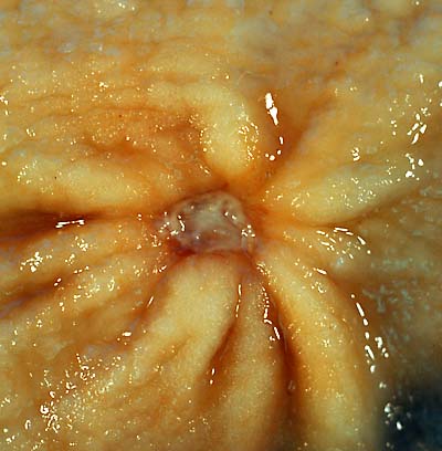 A benign gastric ulcer (from the antrum) of a gastrectomy Source:https://commons.wikimedia.org/wiki/File:Benign_gastric_ulcer_1.jpg#/media/File:Benign_gastric_ulcer_1.jpg
 -
 Duodenal ulcer specimen. Source: https://commons.wikimedia.org/wiki/File:Duodenal_ulcer01.jpg#/media/File:Duodenal_ulcer01.jpg
 -
 Gastric ulcer specimen Source:https://commons.wikimedia.org/wiki/File:Gastric_ulcer_3.jpg#/media/File:Gastric_ulcer_3.jpg


Microscopic Pathology
- A peptic ulcer is a mucosal defect produced by acid-pepsin aggression which penetrates the muscularis mucosae and muscularis propria
- There is increased plasma cells, neutrophilic infiltrate, villous blunting
- The surface epithelium usually shows mucous cell (pseudopyloric) metaplasia
- During the active phase, the base of the ulcer shows 4 zones:
- Inflammatory exudate: polymorphonuclear infiltration which along with bacterial products stimulate the production of IL-8 and tumor necrosis factor alpha (TNF-α) and IL-1 released by macrophages in response to bacterial lipopolysaccharide
- Fibrinoid necrosis
- Granulation tissue
- Fibrous tissue. The fibrous base of the ulcer may contain vessels with thickened wall or with thrombosis[38]

References
- ↑ Butler BD, Lichtenberger LM |title=Gastric mucosal barrier: hydrophobic lining to the lumen of the stomach |journal=Am. J. Physiol. |volume=244 |issue=5 |pages=G561–8 |year=1983 |pmid=6846549 |doi= |url=}}
- ↑ Clamp JR, Ene D (1989). “The gastric mucosal barrier”. Methods Find Exp Clin Pharmacol. 11 Suppl 1: 19–25. PMID 2657286.
- ↑ Werther JL (2000). “The gastric mucosal barrier”. Mt. Sinai J. Med. 67 (1): 41–53. PMID 10677782.
- ↑ Forssell H (1988). “Gastric mucosal defence mechanisms: a brief review”. Scand. J. Gastroenterol. Suppl. 155: 23–8. PMID 3072665.
- ↑ By M•Komorniczak(http://creativecommons.org/licenses/by/3.0)
- ↑ url=http://www.nejm.org/doi/full/10.1056/NEJM199003293221307 |title=Pathogenesis of Peptic Ulcer and Implications for Therapy — NEJM |format= |work= |accessdate=}}
- ↑ 7.0 7.1 “Pathogenesis of Peptic Ulcer and Implications for Therapy — NEJM”.
- ↑ Hawkey CJ (1999). “Personal review: Helicobacter pylori, NSAIDs and cognitive dissonance”. Aliment. Pharmacol. Ther. 13 (6): 695–702. PMID 10383497.
- ↑ Kusters JG, van Vliet AH, Kuipers EJ (2006). “Pathogenesis of Helicobacter pylori infection”. Clin. Microbiol. Rev. 19 (3): 449–90. doi:10.1128/CMR.00054-05. PMC 1539101. PMID 16847081.
- ↑ Huang JQ, Sridhar S, Hunt RH (2002). “Role of Helicobacter pylori infection and non-steroidal anti-inflammatory drugs in peptic-ulcer disease: a meta-analysis”. Lancet. 359 (9300): 14–22. doi:10.1016/S0140-6736(02)07273-2. PMID 11809181.
- ↑ Kim JS, Jung HC, Kim JM, Song IS, Kim CY (1998). “Interleukin-8 expression by human neutrophils activated by Helicobacter pylori soluble proteins”. Scand. J. Gastroenterol. 33 (12): 1249–55. PMID 9930387.
- ↑ Kimura K (1972). “Chronological transition of the fundic-pyloric border determined by stepwise biopsy of the lesser and greater curvatures of the stomach”. Gastroenterology. 63 (4): 584–92. PMID 5077145.
- ↑ 13.0 13.1 Malaty HM, Graham DY, Isaksson I, Engstrand L, Pedersen NL (2000). “Are genetic influences on peptic ulcer dependent or independent of genetic influences for Helicobacter pylori infection?”. Arch. Intern. Med. 160 (1): 105–9. PMID 10632311.
- ↑ url=http://www.nejm.org/doi/full/10.1056/NEJM199003293221307 |title=Pathogenesis of Peptic Ulcer and Implications for Therapy — NEJM |format= |work= |accessdate=}}
- ↑ Logan RP (1996). “Adherence of Helicobacter pylori”. Aliment. Pharmacol. Ther. 10 Suppl 1: 3–15. PMID 8730255.
- ↑ Crabtree JE (1996). “Gastric mucosal inflammatory responses to Helicobacter pylori”. Aliment. Pharmacol. Ther. 10 Suppl 1: 29–37. PMID 8730257.
- ↑ Ernst PB, Jin Y, Reyes VE, Crowe SE (1994). “The role of the local immune response in the pathogenesis of peptic ulcer formation”. Scand. J. Gastroenterol. Suppl. 205: 22–8. PMID 7863238.
- ↑ Mobley HL (1996). “The role of Helicobacter pylori urease in the pathogenesis of gastritis and peptic ulceration”. Aliment. Pharmacol. Ther. 10 Suppl 1: 57–64. PMID 8730260.
- ↑ Nilius M, Malfertheiner P (1996). “Helicobacter pylori enzymes”. Aliment. Pharmacol. Ther. 10 Suppl 1: 65–71. PMID 8730261.
- ↑ Slomiany BL, Kasinathan C, Slomiany A (1989). “Lipolytic activity of Campylobacter pylori: effect of colloidal bismuth subcitrate (De-Nol)”. Am. J. Gastroenterol. 84 (10): 1273–7. PMID 2801678.
- ↑ Sipponen P, Varis K, Fräki O, Korri UM, Seppälä K, Siurala M (1990). “Cumulative 10-year risk of symptomatic duodenal and gastric ulcer in patients with or without chronic gastritis. A clinical follow-up study of 454 outpatients”. Scand. J. Gastroenterol. 25 (10): 966–73. PMID 2263883.
- ↑ Wyatt JI, Rathbone BJ, Dixon MF, Heatley RV (1987). “Campylobacter pyloridis and acid induced gastric metaplasia in the pathogenesis of duodenitis”. J. Clin. Pathol. 40 (8): 841–8. PMC 1141122. PMID 3654985.
- ↑ el-Omar EM, Penman ID, Ardill JE, Chittajallu RS, Howie C, McColl KE (1995). “Helicobacter pylori infection and abnormalities of acid secretion in patients with duodenal ulcer disease”. Gastroenterology. 109 (3): 681–91. PMID 7657096.
- ↑ el-Omar E, Penman I, Dorrian CA, Ardill JE, McColl KE (1993). “Eradicating Helicobacter pylori infection lowers gastrin mediated acid secretion by two thirds in patients with duodenal ulcer”. Gut. 34 (8): 1060–5. PMC 1374354. PMID 8174954.
- ↑ Borody TJ, George LL, Brandl S, Andrews P, Ostapowicz N, Hyland L, Devine M (1991). “Helicobacter pylori-negative duodenal ulcer”. Am. J. Gastroenterol. 86 (9): 1154–7. PMID 1882793.
- ↑ Huang JQ, Sridhar S, Hunt RH (2002). “Role of Helicobacter pylori infection and non-steroidal anti-inflammatory drugs in peptic-ulcer disease: a meta-analysis”. Lancet. 359 (9300): 14–22. doi:10.1016/S0140-6736(02)07273-2. PMID 11809181.
- ↑ Holvoet J, Terriere L, Van Hee W, Verbist L, Fierens E, Hautekeete ML (1991). “Relation of upper gastrointestinal bleeding to non-steroidal anti-inflammatory drugs and aspirin: a case-control study”. Gut. 32 (7): 730–4. PMC 1378985. PMID 1855677.
- ↑ Müller-Lissner SA (1986). “Bile reflux is increased in cigarette smokers”. Gastroenterology. 90 (5 Pt 1): 1205–9. PMID 3956939.
- ↑ Räihä I, Kemppainen H, Kaprio J, Koskenvuo M, Sourander L (1998). “Lifestyle, stress, and genes in peptic ulcer disease: a nationwide twin cohort study”. Arch. Intern. Med. 158 (7): 698–704. PMID 9554675.
- ↑ FakhreYaseri H, Shakaraby M, Bradaran HR, Soltani Arabshahi SK, Fakhre Yaseri AM (2014). “CagA and VacA genotypes in peptic ulcer disease and non-ulcer dyspepsia: a case-control study”. Med J Islam Repub Iran. 28: 104. PMC 4301206. PMID 25664305.
- ↑ Jung DH, Kim JH, Chung HS, Park JC, Shin SK, Lee SK, Lee YC (2015). “Helicobacter pylori Eradication on the Prevention of Metachronous Lesions after Endoscopic Resection of Gastric Neoplasm: A Meta-Analysis”. PLoS ONE. 10 (4): e0124725. doi:10.1371/journal.pone.0124725. PMC 4411104. PMID 25915048.
- ↑ Nakamura S, Sugiyama T, Matsumoto T, Iijima K, Ono S, Tajika M, Tari A, Kitadai Y, Matsumoto H, Nagaya T, Kamoshida T, Watanabe N, Chiba T, Origasa H, Asaka M (2012). “Long-term clinical outcome of gastric MALT lymphoma after eradication of Helicobacter pylori: a multicentre cohort follow-up study of 420 patients in Japan”. Gut. 61 (4): 507–13. doi:10.1136/gutjnl-2011-300495. PMID 21890816.
- ↑ {{cite journal |vauthors=Suzuki T, Matsushima M, Masui A, Watanabe K, Takagi A, Ogawa Y, Shirai T, Mine T |title=Effect of Helicobacter pylori eradication in patients with chronic idiopathic thrombocytopenic purpura-a randomized controlled trial |journal=Am. J. Gastroenterol. |volume=100 |issue=6 |pages=1265–70 |year=2005 <ref name=”pmid18945961″>Stasi R, Sarpatwari A, Segal JB, Osborn J, Evangelista ML, Cooper N, Provan D, Newland A, Amadori S, Bussel JB (2009). “Effects of eradication of Helicobacter pylori infection in patients with immune thrombocytopenic purpura: a systematic review”. Blood. 113 (6): 1231–40. doi:10.1182/blood-2008-07-167155. PMID 18945961.
- ↑ {{cite journal |vauthors=Neunert C, Lim W, Crowther M, Cohen A, Solberg L, Crowther MA |title=The American Society of Hematology 2011 evidence-based practice guideline for immune thrombocytopenia |journal=Blood |volume=117 |issue=16 |pages=4190–207 |year=2011 |
- ↑ name=”urlAmerican Journal of Gastroenterology – ACG Clinical Guideline: Treatment of Helicobacter pylori Infection”>“American Journal of Gastroenterology – ACG Clinical Guideline: Treatment of Helicobacter pylori Infection”.
- ↑ “ATLAS OF PATHOLOGY”. Retrieved 2007-08-26.
Causes
Editor-In-Chief: C. Michael Gibson, M.S., M.D. [1] ;Associate Editor(s)-in-Chief: Manpreet Kaur, MD [2]
Overview
Common causes of peptic ulcer disease include Helicobacter pylori infection and NSAID use. Less common causes of peptic ulcer disease include Crohn’s disease, Zollinger-Ellison syndrome, Cushing and Curling ulcers, Carcinoid tumors, and carcinoid syndrome.
Causes
Less common causes
- Hormonal or mediator-induced including secondary acid hypersecretory states
- Antral G-cell hyperfunction
- Post-surgical antral exclusion and post gastric bypass surgery
- Cancers and lymphomas
- Cameron ulcer (gastric ulcer where a hiatus hernia passes through the diaphragmatic hiatus)
- True idiopathic ulcer
Rare causes of peptic ulcer disease
- Crohn’s disease of the stomach or duodenum
- Eosinophilic gastroduodenitis
- Systemic mastocytosis
- Radiation damage
- Viral infections (eg cytomegalovirus or herpes simplex infection, in particular in immunocompromised patients)
- Colonization of stomach with H heilmanii
- Severe systemic disease
Genetic causes[4]
- Peptic ulcer disease caused by gastrinomas (Zollinger-Ellison syndrome) caused by a mutation in MEN gene present on chromosome 11q13
Causes by Organ System
Causes in Alphabetical Order
References
- ↑ Malfertheiner P, Chan FK, McColl KE (2009). “Peptic ulcer disease”. Lancet. 374 (9699): 1449–61. doi:10.1016/S0140-6736(09)60938-7. PMID 19683340.
- ↑ Hirschowitz BI, Lanas A (2002). “Atypical and aggressive upper gastrointestinal ulceration associated with aspirin abuse”. J. Clin. Gastroenterol. 34 (5): 523–8. PMID 11960062.
- ↑ Dovjak P (2017). “[Duodenal ulcers, gastric ulcers and Helicobacter pylori]”. Z Gerontol Geriatr (in German). 50 (2): 159–169. doi:10.1007/s00391-017-1190-x. PMID 28150170.
- ↑ Jensen RT, Niederle B, Mitry E, Ramage JK, Steinmuller T, Lewington V; et al. (2006). “Gastrinoma (duodenal and pancreatic)”. Neuroendocrinology. 84 (3): 173–82. doi:10.1159/000098009. PMID 17312377.
- ↑ Kutlubay Z, Zara T, Engin B, Serdaroğlu S, Tüzün Y, Yilmaz E; et al. (2014). “Helicobacter pylori infection and skin disorders”. Hong Kong Med J. 20 (4): 317–24. doi:10.12809/hkmj134174. PMID 25045884.
- ↑ Invalid
<ref>tag; no text was provided for refs namedpmid28384332
Differentiating Peptic ulcer from other Diseases
Editor-In-Chief: C. Michael Gibson, M.S., M.D. [1]; Associate Editor(s)-in-Chief: Guillermo Rodriguez Nava, M.D. [2] Manpreet Kaur, MD [3]
Overview
Peptic ulcer disease must be differentiated from other causes of acute upper gastrointestinal bleeding such as esophageal varices, Mallory-Weiss syndrome, gastrointestinal cancer, arteriovenous malformations, esophagitis, and esophageal ulcer. Peptic ulcer disease must also be differentiated from gastroesophageal reflux disease (GERD,pancreatitis, Zollinger-Ellison Syndrome,cholelithiasis,gastric outlet syndrome,myocardial infaraction ,pleural empyema and appendicitis
Differentiating Peptic Ulcer from other Diseases
Peptic ulcer disease must be differentiated from other diseases that presents with epigastric pain such as gastritis, gastroesophageal reflux disease,acute pancreatitis,prmary biliary cirrhosis,cholelithiasis,gastric outlet syndrome,myocardial infaraction ,pleural empyema,acute appendicitis [1][2][3][4][5][6][7][8][9][10][11][12][13][14][15][16][17][18]
| Classification of pain in the abdomen based on etiology | Disease | Clinical manifestations | Diagnosis | Comments | |||||||||||
|---|---|---|---|---|---|---|---|---|---|---|---|---|---|---|---|
| Symptoms | Signs | ||||||||||||||
| Fever | Rigors and chills | Abdominal Pain | Jaundice | GI Bleed | Hypo-
tension |
Guarding | Rebound Tenderness | Bowel sounds | Lab Findings | Imaging | |||||
| Abdominal causes | Inflammatory causes | Pancreato-biliary disorders | |||||||||||||
| Acute pancreatitis | + | − | Epigastric | ± | − | ± | − | − | N | Increased amylase / lipase | Ultrasound shows evidence of inflammation | Pain radiation to back | |||
| Primary biliary cirrhosis | − | − | RUQ/Epigastric | + | − | − | − | − | N | Increased AMA level, abnormal LFTs | |||||
| Cholelithiasis | ± | − | RUQ/Epigastric | ± | − | − | + | + | N to hyperactive for dislodged stone | Leukocytosis | Ultrasound shows gallstone | Murphy’s sign | |||
| Gastric causes | Peptic ulcer disease | ± | − | EpisodicEpigastric | − |
|
+ in perforated | + | + | N | Air under diaphragm in upright CXR | Upper GI endoscopy for diagnosis | |||
| Gastritis | ± | − | Epigastric | + in chronic gastritis | − | ||||||||||
| Gastroesophageal reflux disease | − | − | Epigastric | − | − | − | − | − | |||||||
| Gastric outlet obstruction | − | − | Epigastric | − | − | ± | Hyperactive | ||||||||
| Intestinal causes | Acute appendicitis | + | +in pyogenic appendicitis | Starts in epigastrium, migrates to RLQ | − | − | + in perforated appendicitis | + | + | Hypoactive | Leukocytosis | Ultrasound shows evidence of inflammation | Nausea & vomiting, decreased appetite | ||
| Extra-abdominal causes | Pulmonary disorders | Pleural empyema | + | ± | RUQ/Epigastric | − | − | − | − | − | N | ||||
| Cardiovascular disorders | Myocardial Infarction | − | − | Epigastric | − | − | + in cardiogenic shock | − | − | N | |||||
| Abbreviations: RUQ= Right upper quadrant of the abdomen, LUQ= Left upper quadrant, LLQ= Left lower quadrant, RLQ= Right lower quadrant, LFT= Liver function test, SIRS= Systemic inflammatory response syndrome, ERCP= Endoscopic retrograde cholangiopancreatography, IV= Intravenous, N= Normal, AMA= Anti mitochondrial antibodies, LDH= Lactate dehydrogenase, GI= Gastrointestinal, CXR= Chest X ray, IgA= Immunoglobulin A, IgG= Immunoglobulin G, IgM=Immunoglobulin M, CT= Computed tomography, PMN= Polymorphonuclear cells, ESR= Erythrocyte sedimentation rate, CRP= C-reactive protein | |||||||||||||||
| Disease | Cause | Symptoms | Diagnosis | Other findings | ||||||||
|---|---|---|---|---|---|---|---|---|---|---|---|---|
| Pain | Nausea
& Vomiting |
Heartburn | Belching or
Bloating |
Weight loss | Loss of
Appetite |
Stools | Endoscopy findings | |||||
| Location | Aggravating Factors | Alleviating Factors | ||||||||||
| Acute gastritis |
|
Food | Antacids | ✔ | ✔ | ✔ | – | ✔ | Black stools | – | ||
| Chronic gastritis |
|
Food | Antacids | ✔ | ✔ | ✔ | ✔ | ✔ | – | H. pylori gastritis
Lymphocytic gastritis
|
– | |
| Atrophic gastritis | Epigastric pain | – | – | ✔ | – | ✔ | ✔ | – | H. pylori
|
Autoimmune gastritis diagnosis include:
| ||
| Crohn’s disease | – | – | – | – | – | ✔ | ✔ |
|
|
|||
| GERD |
|
|
|
✔
(Suspect delayed gastric emptying) |
✔ | – | – | – | – |
|
Other symptoms:
Complications
| |
| Peptic ulcer disease |
|
|
|
|
✔ | ✔ | – | – | – | Gastric ulcers
Duodenal ulcers
|
Other diagnostic tests | |
| Gastrinoma |
|
– | – | ✔
(suspect gastric outlet obstruction) |
✔ | – | – | – | Useful in collecting the tissue for biopsy |
Diagnostic tests
| ||
| Gastric Adenocarcinoma |
|
– | – | ✔ | ✔ | ✔ | ✔ | ✔ |
|
Esophagogastroduodenoscopy
|
Other symptoms | |
| Primary gastric lymphoma |
|
– | – | – | – | – | ✔ | – | – | Useful in collecting the tissue for biopsy | Other symptoms
| |
References
- ↑ Gralnek IM, Barkun AN, Bardou M (2008). “Management of acute bleeding from a peptic ulcer”. N Engl J Med. 359 (9): 928–37. doi:10.1056/NEJMra0706113. PMID 18753649.
- ↑ Dallal HJ, Palmer KR (2001). “ABC of the upper gastrointestinal tract: Upper gastrointestinal haemorrhage”. BMJ. 323 (7321): 1115–7. PMC 1121602. PMID 11701581.
- ↑ Nelson DR, Teckman J, Di Bisceglie AM, Brenner DA (2012). “Diagnosis and management of patients with α1-antitrypsin (A1AT) deficiency”. Clin Gastroenterol Hepatol. 10 (6): 575–80. doi:10.1016/j.cgh.2011.12.028. PMC 3360829. PMID 22200689.
- ↑ Tsochatzis EA, Bosch J, Burroughs AK (2014). “Liver cirrhosis”. Lancet. 383 (9930): 1749–61. doi:10.1016/S0140-6736(14)60121-5. PMID 24480518.
- ↑ Schuppan D, Afdhal NH (2008). “Liver cirrhosis”. Lancet. 371 (9615): 838–51. doi:10.1016/S0140-6736(08)60383-9. PMC 2271178. PMID 18328931.
- ↑ Kahrilas PJ (2008). “Clinical practice. Gastroesophageal reflux disease”. N Engl J Med. 359 (16): 1700–7. doi:10.1056/NEJMcp0804684. PMC 3058591. PMID 18923172.
- ↑ Kahrilas PJ, Shaheen NJ, Vaezi MF, Hiltz SW, Black E, Modlin IM; et al. (2008). “American Gastroenterological Association Medical Position Statement on the management of gastroesophageal reflux disease”. Gastroenterology. 135 (4): 1383–1391, 1391.e1–5. doi:10.1053/j.gastro.2008.08.045. PMID 18789939.
- ↑ Bredenoord AJ, Pandolfino JE, Smout AJ (2013). “Gastro-oesophageal reflux disease”. Lancet. 381 (9881): 1933–42. doi:10.1016/S0140-6736(12)62171-0. PMID 23477993.
- ↑ Fox M, Forgacs I (2006). “Gastro-oesophageal reflux disease”. BMJ. 332 (7533): 88–93. doi:10.1136/bmj.332.7533.88. PMC 1326932. PMID 16410582.
- ↑ Sugimachi K, Inokuchi K, Kuwano H, Ooiwa T (1984). “Acute gastritis clinically classified in accordance with data from both upper GI series and endoscopy”. Scand J Gastroenterol. 19 (1): 31–7. PMID 6710074.
- ↑ Sipponen P, Maaroos HI (2015). “Chronic gastritis”. Scand J Gastroenterol. 50 (6): 657–67. doi:10.3109/00365521.2015.1019918. PMC 4673514. PMID 25901896.
- ↑ Sartor RB (2006). “Mechanisms of disease: pathogenesis of Crohn’s disease and ulcerative colitis”. Nat Clin Pract Gastroenterol Hepatol. 3 (7): 390–407. doi:10.1038/ncpgasthep0528. PMID 16819502.
- ↑ Sipponen P (1989). “Atrophic gastritis as a premalignant condition”. Ann Med. 21 (4): 287–90. PMID 2789799.
- ↑ Badillo R, Francis D (2014). “Diagnosis and treatment of gastroesophageal reflux disease”. World J Gastrointest Pharmacol Ther. 5 (3): 105–12. doi:10.4292/wjgpt.v5.i3.105. PMC 4133436. PMID 25133039.
- ↑ Ramakrishnan K, Salinas RC (2007). “Peptic ulcer disease”. Am Fam Physician. 76 (7): 1005–12. PMID 17956071.
- ↑ Banasch M, Schmitz F (2007). “Diagnosis and treatment of gastrinoma in the era of proton pump inhibitors”. Wien Klin Wochenschr. 119 (19–20): 573–8. doi:10.1007/s00508-007-0884-2. PMID 17985090.
- ↑ Dicken BJ, Bigam DL, Cass C, Mackey JR, Joy AA, Hamilton SM (2005). “Gastric adenocarcinoma: review and considerations for future directions”. Ann Surg. 241 (1): 27–39. PMC 1356843. PMID 15621988.
- ↑ Ghimire P, Wu GY, Zhu L (2011). “Primary gastrointestinal lymphoma”. World J Gastroenterol. 17 (6): 697–707. doi:10.3748/wjg.v17.i6.697. PMC 3042647. PMID 21390139.
Epidemiology and Demographics
Editor-In-Chief: C. Michael Gibson, M.S., M.D. [1] ;Associate Editor(s)-in-Chief: Manpreet Kaur, MD [2]
Overview
The incidence and prevalence of Helicobacter pylori infection are generally higher among people born outside North America. Within North America, the prevalence of the infection is higher in certain racial and ethnic groups, and people who have immigrated to North America. Peptic ulcer disease is acquired during childhood. The incidence of Peptic ulcer disease increases with age; the median age at diagnosis is 18-30 years. Peptic ulcer disease affects both sexes equally in childhood. Men are more commonly affected by peptic ulcer disease than women in adulthood.
Epidemiology and Demographics
Incidence
- The incidence of peptic ulcer disease is approximately 10-19 per 100,000 individuals worldwide[1]
- In 2011, the incidence rate of peptic ulcer disease was estimated to be one case per 1000 individuals and its complications was 0.7 per 1000 individuals[2]
- The incidence and prevalence of Helicobacter pylori infection are generally higher among people born outside North America. Within North America, the prevalence of the infection is higher in certain racial and ethnic groups, and people who have immigrated to North America[3]
Prevalence
- The prevalence of peptic ulcer disease is approximately 23.1 per 100,000 individuals but higher in men 29.4 per 100,000 than women 14.9 per 100,000
- The prevalence of peptic ulcer disease is estimated to be 4.5 million cases annually
- Lifetime prevalence is 11-14% in men and 8-11% in women[4][5][1][6][7][8][9][10]
Mortality rate
- The mortality rate of peptic ulcer disease is approximately 1 per 100,000 individuals
Age
- Peptic ulcer disease is acquired during childhood
- The incidence of Peptic ulcer disease increases with age; the median age at diagnosis is 18-30 years
Race
- Peptic ulcer disease usually affects individuals of the African Americans with a higher proportion of African ancestry race and Mexican Americans[11][12]
- Non-Hispanic whites individuals are less likely to develop the peptic ulcer disease
Gender
- Peptic ulcer disease affect equally in childhood[13]
- Men are more commonly affected by peptic ulcer disease than women in adulthood[5]
References
- ↑ 1.0 1.1 Sung JJ, Kuipers EJ, El-Serag HB (2009). “Systematic review: the global incidence and prevalence of peptic ulcer disease”. Aliment. Pharmacol. Ther. 29 (9): 938–46. doi:10.1111/j.1365-2036.2009.03960.x. PMID 19220208.
- ↑ Lin KJ, García Rodríguez LA, Hernández-Díaz S (2011). “Systematic review of peptic ulcer disease incidence rates: do studies without validation provide reliable estimates?”. Pharmacoepidemiol Drug Saf. 20 (7): 718–28. doi:10.1002/pds.2153. PMID 21626606.
- ↑ Wang AY, Peura DA (2011). “The prevalence and incidence of Helicobacter pylori-associated peptic ulcer disease and upper gastrointestinal bleeding throughout the world”. Gastrointest. Endosc. Clin. N. Am. 21 (4): 613–35. doi:10.1016/j.giec.2011.07.011. PMID 21944414.
- ↑ Ohmann C, Imhof M, Ruppert C, Janzik U, Vogt C, Frieling T, Becker K, Neumann F, Faust S, Heiler K, Haas K, Jurisch R, Wenzel EG, Normann S, Bachmann O, Delgadillo J, Seidel F, Franke C, Lüthen R, Yang Q, Reinhold C (2005). “Time-trends in the epidemiology of peptic ulcer bleeding”. Scand. J. Gastroenterol. 40 (8): 914–20. doi:10.1080/00365520510015809. PMID 16165708.
- ↑ 5.0 5.1 Naja F, Kreiger N, Sullivan T (2007). “Helicobacter pylori infection in Ontario: prevalence and risk factors”. Can. J. Gastroenterol. 21 (8): 501–6. PMC 2657974. PMID 17703249.
- ↑ Kuipers EJ, Thijs JC, Festen HP (1995). “The prevalence of Helicobacter pylori in peptic ulcer disease”. Aliment. Pharmacol. Ther. 9 Suppl 2: 59–69. PMID 8547530.
- ↑ Pounder RE, Ng D (1995). “The prevalence of Helicobacter pylori infection in different countries”. Aliment. Pharmacol. Ther. 9 Suppl 2: 33–9. PMID 8547526.
- ↑ Frenck RW, Clemens J (2003). “Helicobacter in the developing world”. Microbes Infect. 5 (8): 705–13. PMID 12814771.
- ↑ Dutta AK, Chacko A, Balekuduru A, Sahu MK, Gangadharan SK (2012). “Time trends in epidemiology of peptic ulcer disease in India over two decades”. Indian J Gastroenterol. 31 (3): 111–5. doi:10.1007/s12664-012-0201-5. PMID 22766645.
- ↑ Goh KL (2009). “Epidemiology of Helicobacter pylori infection in Malaysia–observations in a multiracial Asian population”. Med. J. Malaysia. 64 (3): 187–92. PMID 20527265.
- ↑ Nguyen T, Ramsey D, Graham D, Shaib Y, Shiota S, Velez M, Cole R, Anand B, Vela M, El-Serag HB (2015). “The Prevalence of Helicobacter pylori Remains High in African American and Hispanic Veterans”. Helicobacter. 20 (4): 305–15. doi:10.1111/hel.12199. PMID 25689684.
- ↑ Everhart JE, Kruszon-Moran D, Perez-Perez GI, Tralka TS, McQuillan G (2000). “Seroprevalence and ethnic differences in Helicobacter pylori infection among adults in the United States”. J. Infect. Dis. 181 (4): 1359–63. doi:10.1086/315384. PMID 10762567.
- ↑ de Martel C, Parsonnet J (2006). “Helicobacter pylori infection and gender: a meta-analysis of population-based prevalence surveys”. Dig. Dis. Sci. 51 (12): 2292–301. doi:10.1007/s10620-006-9210-5. PMID 17089189.
Risk Factors
Editor-In-Chief: C. Michael Gibson, M.S., M.D. [1] ;Associate Editor(s)-in-Chief: :Manpreet Kaur, MD [2]
Overview
Common risk factors in the development of peptic ulcer disease include infection from Helicobacter pylori, chronic use of NSAIDs, cigarette smoking, alcolhol intake, family history of peptic ulcer, and age >50 years. Less common risk factors in the development of peptic ulcer disease include psychological stress, nosocomial stress ulcers, and coagulopathy. Rare conditions associated with gastric acid hypersecretion, such as zollinger-ellison syndrome, mastocytosis, or a retained antrum following partial gastrectomy, gastrinoma or multiple endocrine neoplasia types I (MEN-I), antral G cell hyperplasia, basophilic leukemias or short bowel syndrome.
Risk Factors
The most potent risk factor leading to the development of peptic ulcer disease is an infection of Helicobacter pylori. Other risk factors include chronic use of NSAIDs, family history of peptic ulcer, tobacco smoking, and psychological and nosocomial stress.[1][2][3][4][5][6][7][8]
Common risk factors
Common risk factors in the development of peptic ulcer disease include:
- Helicobacter pylori infection
- Chronic use of NSAIDs
- Family history of peptic ulcer
Less Common Risk Factors
Less common risk factors in the development of peptic ulcer disease include:
- Tobacco
- Alcohol
- Psychological stress
- Nosocomial stress ulcers due the to the use of mechanical ventilation for more than 48 hours, and coagulopathy
- Rare conditions associated with gastric acid hypersecretion such as:
- Zollinger-Ellison syndrome, mastocytosis, or a retained antrum following partial gastrectomy
- gastrinoma or multiple endocrine neoplasia types I (MEN-I), antral G cell hyperplasia, basophilic leukemias, short bowel syndrome
References
- ↑ Huang JQ, Sridhar S, Hunt RH (2002). “Role of Helicobacter pylori infection and non-steroidal anti-inflammatory drugs in peptic-ulcer disease: a meta-analysis”. Lancet. 359 (9300): 14–22. doi:10.1016/S0140-6736(02)07273-2. PMID 11809181.
- ↑ Ballinger A, Smith G (2001). “COX-2 inhibitors vs. NSAIDs in gastrointestinal damage and prevention”. Expert Opin Pharmacother. 2 (1): 31–40. doi:10.1517/14656566.2.1.31. PMID 11336566.
- ↑ Holvoet J, Terriere L, Van Hee W, Verbist L, Fierens E, Hautekeete ML (1991). “Relation of upper gastrointestinal bleeding to non-steroidal anti-inflammatory drugs and aspirin: a case-control study”. Gut. 32 (7): 730–4. PMC 1378985. PMID 1855677.
- ↑ Laporte JR, Carné X, Vidal X, Moreno V, Juan J (1991). “Upper gastrointestinal bleeding in relation to previous use of analgesics and non-steroidal anti-inflammatory drugs. Catalan Countries Study on Upper Gastrointestinal Bleeding”. Lancet. 337 (8733): 85–9. PMID 1670734.
- ↑ Wachirawat W, Hanucharurnkul S, Suriyawongpaisal P, Boonyapisit S, Levenstein S, Jearanaisilavong J, Atisook K, Boontong T, Theerabutr C (2003). “Stress, but not Helicobacter pylori, is associated with peptic ulcer disease in a Thai population”. J Med Assoc Thai. 86 (7): 672–85. PMID 12948263.
- ↑ Rosenstock S, Jørgensen T, Bonnevie O, Andersen L (2003). “Risk factors for peptic ulcer disease: a population based prospective cohort study comprising 2416 Danish adults”. Gut. 52 (2): 186–93. PMC 1774958. PMID 12524398.
- ↑ Stack WA, Atherton JC, Hawkey GM, Logan RF, Hawkey CJ (2002). “Interactions between Helicobacter pylori and other risk factors for peptic ulcer bleeding”. Aliment. Pharmacol. Ther. 16 (3): 497–506. PMID 11876703.
- ↑ Everhart JE, Byrd-Holt D, Sonnenberg A (1998). “Incidence and risk factors for self-reported peptic ulcer disease in the United States”. Am. J. Epidemiol. 147 (6): 529–36. PMID 9521179.
Screening
Editor-In-Chief: C. Michael Gibson, M.S., M.D. [1] ; Associate Editor(s)-in-Chief: Manpreet Kaur, MD [2]
Overview
According to the American Society of Gastroenterology, screening for PUD is not recommended.
Screening
According to the American Society of Gastroenterology, screening for PUD is not recommended.
References
Natural History, Complications and Prognosis
Editor-In-Chief: C. Michael Gibson, M.S., M.D. [1]; Associate Editor(s)-in-Chief: Manpreet Kaur, MD [2]
Overview
Helicobacter pylori infection is a common cause of peptic ulcer disease which is acquired usually during childhood but presents in second to fifth decade of life. Patient presents with episodic epigastric pain, indigestion, bloating,hematemesis and melena. If not treated, patients can develop complications like bleeding, perforation, obstruction or stricture. Chronic infection of helicobacter pylori leads to gastric cancer, MALT lymphoma, iron deficiency anemia, Idiopathic thrombocytopenic purpura. Peptic ulcers tend to recur if left untreated. Prognosis is good if the eradication therapy of Helicobacter pylori is taken.The recurrence rate of patients with peptic ulcer disease is less than 20%.
Natural History
- The infection of Helicobacter pylori , common cause of peptic ulcer disease is acquired usually during the childhood, mainly in those who have low socioeconomic status, increased number of siblings and similar infection in the mother.[1][2][3]
- The duodenal ulcer occurs in second to fifth decade of life. where as gastric ulcer occurs in the 4th decade of life.
- Peptic ulcer disease starts with symptoms such as episodic epigastric pain, indigestion, bloating.
- Without treatment, the patient will develop symptoms of perforation like severe abdominal pain. Chronic infection leads to gastric atrophy, gastric metaplasia and ultimately leads to gastric cancer and MALT lymphoma.[4]
Complications
Acute complications :
- Bleeding is the commonest complication.
- Perforation
- Perforation is most common occur at the duodenal bulb (62%), followed by the pyloric region (20%), and the gastric body (18%).
- Erosion of the gastro-intestinal wall by the ulcer leads to spillage of stomach or intestinal content into abdominal cavity leading to fatal consequences.
- Perforation at the anterior surface of stomach leads to acute peritonitis, initially chemical and later bacterial peritonitis.
- Often, the first symptom is a sudden intense abdominal pain.
- Posterior wall perforation leads to pancreatitis, pain in this situation often radiates to back.
- Penetration is also a form of perforation, when the ulcer continues into adjacent organs such as liver and pancreas.[7][8]
- Obstruction
- Occurs as a result of stricture and scarring narrowing in the duodenum leading to gastric outlet obstruction.
Chronic complications:
Chronic Helicobacter pylori infection leads to:
Prognosis
- Prognosis is good if the eradication therapy of Helicobacter pylori is taken.
- The recurrence rate of patients with peptic ulcer disease is less than 20%.
References
- ↑ Opekun AR, Gilger MA, Denyes SM, Nirken MH, Philip SP, Osato MS, Malaty HM, Hicks J, Graham DY (2000). “Helicobacter pylori infection in children of Texas”. J. Pediatr. Gastroenterol. Nutr. 31 (4): 405–10. PMID 11045838.
- ↑ Parkinson AJ, Gold BD, Bulkow L, Wainwright RB, Swaminathan B, Khanna B, Petersen KM, Fitzgerald MA (2000). “High prevalence of Helicobacter pylori in the Alaska native population and association with low serum ferritin levels in young adults”. Clin. Diagn. Lab. Immunol. 7 (6): 885–8. PMC 95979. PMID 11063492.
- ↑ Malaty HM, El-Kasabany A, Graham DY, Miller CC, Reddy SG, Srinivasan SR, Yamaoka Y, Berenson GS (2002). “Age at acquisition of Helicobacter pylori infection: a follow-up study from infancy to adulthood”. Lancet. 359 (9310): 931–5. doi:10.1016/S0140-6736(02)08025-X. PMID 11918912.
- ↑ Kusters JG, van Vliet AH, Kuipers EJ (2006). “Pathogenesis of Helicobacter pylori infection”. Clin. Microbiol. Rev. 19 (3): 449–90. doi:10.1128/CMR.00054-05. PMC 1539101. PMID 16847081.
- ↑ Milosavljevic T, Kostić-Milosavljević M, Jovanović I, Krstić M (2011). “Complications of peptic ulcer disease”. Dig Dis. 29 (5): 491–3. doi:10.1159/000331517. PMID 22095016.
- ↑ Cullen DJ, Hawkey GM, Greenwood DC; et al. (1997). “Peptic ulcer bleeding in the elderly: relative roles of Helicobacter pylori and non-steroidal anti-inflammatory drugs”. Gut. 41 (4): 459–62. PMID 9391242.
- ↑ “Peptic Ulcer: Peptic Disorders: Merck Manual Home Edition”. Retrieved 2007-10-10.
- ↑ “Peptic Ulcer: Peptic Disorders: Merck Manual Home Edition”. Retrieved 2007-10-10.
Diagnosis
History and Symptoms | Physical Examination | Echocardiogram | Laboratory Findings | X Ray | CT | Ultrasound | Other Imaging Findings | Other Diagnostic Studies
Looking for the patient version?
© 2026 MyEClinic – IFTM Institut für Telematik in der Medizin GmbH