
Acute cholecystitis
For patient information, click here
Editor-In-Chief: C. Michael Gibson, M.S., M.D. [1]; Associate Editor(s)-in-Chief: Furqan M M. M.B.B.S[2] Dildar Hussain, MBBS [3]
Synonyms and keywords: Acute calculous cholecystitis; Acute acalculous cholecystitis.
Overview
Editor-In-Chief: C. Michael Gibson, M.S., M.D. [1]; Associate Editor(s)-in-Chief: Furqan M M. M.B.B.S[2]
Overview
Cholecystitis is the inflammation of the gallbladder. Acute cholecystitis may be classified according to causes into two major subtypes: Acute calculous cholecystitis and acute acalculous cholecystitis. Gallstones are found in 3500 years old Egyptian mummies during the autopsies. Common risk factors in the development of acute calculous cholecystitis include advanced age, female gender, obesity, and positive family history. Long periods of fasting, total parental nutrition (TPN), and weight loss are the common risk factors for the development of acute acalculous cholecystitis. Acute calculous cholecystitis is usually caused by the mechanical obstruction of the gallbladder due to gallstones. Acute acalculous cholecystitis is caused predominantly by the gallbladder stasis. Gallstones are the most common cause of physical obstruction of the gallbladder usually at the neck or in the cystic duct. Cholesterol gallstones are the most common type of gallstones. The obstruction causes an increased pressure as the gallbladder mucosa continues to produce mucus. This raised pressure may cause the venous congestion which is followed by the arterial congestion. Eventually, the raised pressure and stasis leads to the gallbladder ischemia and necrosis. Mechanical obstruction of the gallbladder as a result of polyps, malignancy, an infestation of the gallbladder with parasites, foreign bodies, and trauma may also lead to the acute cholecystitis. Acute cholecystitis is more common in siblings and first degree relatives of affected persons. Lith gene is involved in the pathogenesis of cholecystitis. Acute cholecystitis is associated with diabetes, insulin resistance, cardiovascular diseases, non-alcoholic fatty liver disease (NAFLD) and gastrointestinal malignancies. Microscopic histopathology shows edematous and hemorrhagic gallbladder wall, mucosal necrosis with neutrophil infiltration. Bile infiltration of the gallbladder wall and bile and leukocyte margination of blood vessels are specific findings for acalculous cholecystitis. Acute cholecystitis must be differentiated from other diseases that cause right upper quadrant abdominal pain and nausea/vomiting such as biliary colic, acute cholangitis, viral hepatitis, alcoholic hepatitis, acute pancreatitis, acute appendicitis, and irritable bowel syndrome. The incidence of acute cholecystitis is approximately 6,300 per 100,000 in individuals under 50 years age and 20,900 per 100,000 in individuals over 50 years age worldwide. The prevalence of acute cholecystitis is approximately 369 per 100,000 individuals in the United states. It is estimated from the population-based statistics, based on a comprehensive survey in the U.S. Acute cholecystitis is comparatively less prevalent in the developing countries. The mortality rate of acute cholecystitis is approximately 0.6%. Acute Cholecystitis most commonly occurs as a result of the prolonged obstruction of the cystic duct leading to inflammation of the gallbladder. The obstruction further contributes to the development of the complications associated with acute cholecystitis such as gangrene, empyema, perforation, cholecysto-enteric fistula, emphysematous cholecystitis, and gallstone ileus. Prognosis is generally good if the patient receives treatment. Acute cholecystitis occurs as a result of prolonged gallstone obstruction in the bile duct, one to four patients develop biliary colic and about 20% of these patients develop acute cholecystitis annually. The hallmark of acute cholecystitis is biliary colic. A positive history of biliary colic, nausea and vomiting is suggestive of acute cholecystitis. Patients with acute cholecystitis usually appear ill. Physical examination of patients with acute cholecystitis is remarkable for right upper quadrant abdominal tenderness, positive murphy’s sign, and fever. The presence of murphy’s sign on physical examination is highly suggestive of acute cholecystitis. Laboratory findings consistent with the diagnosis of acute cholecystitis include leukocytosis and elevated CRP. Transabdominal ultrasound is the initial test of choice for the diagnosis of acute cholecystitis. Findings on an ultrasound diagnostic of acute cholecystitis include thickened gallbladder, gallstones or sludge, and pericholecystic fluid. CT scan is usually used for the diagnosis of the complications of acute cholecystitis. These complications include emphysematous cholecystitis and gangrenous cholecystitis. Cholescintigraphy is the gold standard for the diagnosis of acute cholecystitis. Cholescintigraphy is an alternative method of imaging and uses technetium-labeled hepatic 2,6-dimethyl-iminodiacetic acid (HIDA) in difficult cases or uncertain diagnosis. Findings on a cholescintigraphy diagnostic of acute cholecystitis include lack of visualization of the gallbladder. The mainstay of treatment for acute cholecystitis (calculous and acalculous) is surgery. Laparoscopic cholecystectomy is the gold standard for the treatment of acute cholecystitis and is usually preferred over the open cholecystectomy. Percutaneous cholecystostomy (PC) is an alternative to emergency cholecystectomy in complicated cases of high-risk patients. Pharmacologic medical therapy is recommended for cases of acute cholecystitis in which surgery is delayed. Empiric pharmacologic medical therapies for acute cholecystitis include either amoxicillin-clavulanic acid, cefoxitin, cefotaxime, or ceftriaxone with metronidazole, and ciprofloxacin or levofloxacin with metronidazole. The duration of medical therapy after the cholecystectomy depends on the severity of the disease.
Historical Perspective
Gallstones are found in 3500 years old Egyptian mummies during the autopsies. In 1420, Antonio Benivieni was the first to describe gallstones. Carl Langenbuch performed the first cholecystectomy of a 43-year-old man who had suffered from biliary colic for sixteen years. Historically, open cholecystectomy was the treatment employed for the treatment of acute cholecystitis. Laparoscopic cholecystectomy was developed to treat acute cholecystitis and the shift from open to laparoscopic cholecystectomy occurred in the late 1980s.
Classification
Acute cholecystitis may be classified according to causes into two major subtypes: Acute calculous cholecystitis and acute acalculous cholecystitis.
Pathophysiology
Acute calculous cholecystitis is usually caused by the mechanical obstruction of the gallbladder due to gallstones. Acute acalculous cholecystitis is caused predominantly by the gallbladder stasis. Gallstones are the most common cause of physical obstruction of the gallbladder usually at the neck or in the cystic duct. Cholesterol gallstones are the most common type of gallstones. The obstruction causes an increased pressure as the gallbladder mucosa continues to produce mucus. This raised pressure may cause the venous congestion which is followed by the arterial congestion. Eventually, the raised pressure and stasis leads to the gallbladder ischemia and necrosis. Mechanical obstruction of the gallbladder as a result of polyps, malignancy, an infestation of the gallbladder with parasites, foreign bodies, and trauma may also lead to the acute cholecystitis. Acute cholecystitis is more common in siblings and first degree relatives of affected persons. Lith gene is involved in the pathogenesis of cholecystitis. Mutations in the hepatic cholesterol transporter ABCG8 also predispose an individual to the develop gallstones. Acute cholecystitis is associated with diabetes, insulin resistance, cardiovascular diseases, non-alcoholic fatty liver disease (NAFLD) and gastrointestinal malignancies. Microscopic histopathology shows edematous and hemorrhagic gallbladder wall, mucosal necrosis with neutrophil infiltration. Bile infiltration of the gallbladder wall and bile and leucocyte margination of blood vessels are specific findings for acalculous cholecystitis.
Causes
The most common cause of acute cholecystitis is gallstones. Less common causes of acute cholecystitis include gallbladder stasis, gallbladder polyp, gallbladder malignancy, parasites, and foreign
Differentiating Acute cholecystitis from Other Diseases
Acute cholecystitis must be differentiated from other diseases that cause right upper quadrant abdominal pain and nausea/vomiting such as biliary colic, acute cholangitis, viral hepatitis, alcoholic hepatitis, acute pancreatitis, acute appendicitis, and irritable bowel syndrome.
Epidemiology and Demographics
The incidence of acute cholecystitis is approximately 6,300 per 100,000 in individuals under 50 years age and 20,900 per 100,000 in individuals over 50 years age worldwide. The prevalence of acute cholecystitis is approximately 369 per 100,000 individuals in the United states. It is estimated from the population-based statistics, based on a comprehensive survey in the U.S. Acute cholecystitis is comparatively less prevalent in the developing countries. The mortality rate of acute cholecystitis is approximately 0.6%. Acute cholecystitis usually affects individuals of the North American Indian race. Females are more commonly affected by acute cholecystitis than males. Acute cholecystitis cases are reported worldwide. Acute cholecystitis accounts for 700,000 cholecystectomies and costs of ∼$6.5 billion annually only in the United States.
Risk Factors
Common risk factors in the development of acute calculous cholecystitis include advancing age, female gender, obesity, and family history. Long periods of fasting, total parental nutrition (TPN), weight loss are the common risk factors for the development of acute acalculous cholecystitis.
Screening
There is insufficient evidence to recommend routine screening for acute cholecystitis.
Natural History, Complications, and Prognosis
Acute Cholecystitis most commonly occurs as a result of the prolonged obstruction of the cystic duct leading to inflammation of the gallbladder. The obstruction further contributes to the development of the complications associated with acute cholecystitis such as gangrene, empyema, perforation, cholecysto-enteric fistula, emphysematous cholecystitis, and gallstone ileus. Prognosis is generally good if the patient receives treatment. The majority of the patients undergo cholecystectomy.
Diagnosis
Diagnostic Study of Choice
Cholescintigraphy is the gold standard for the diagnosis of acute cholecystitis. Transabdominal ultrasonography is the initial study of choice for the diagnosis of acute cholecystitis and gallstones. Thickened gallbladder, gallstones or sludge, and pericholecystic fluid are the findings associated with transabdominal ultrasound in patients with acute cholecystitis.
History and Symptoms
The majority of patients with cholelithiasis are asymptomatic. Acute cholecystitis occurs as a result of prolonged gallstone obstruction in the bile duct, one to four patients develop biliary colic and about 20% of these patients develop acute cholecystitis annually. The hallmark of acute cholecystitis is biliary colic. A positive history of biliary colic, nausea and vomiting is suggestive of acute cholecystitis.
Physical Examination
Patients with acute cholecystitis usually appear ill. Physical examination of patients with acute cholecystitis is remarkable for right upper quadrant abdominal tenderness, positive murphy’s sign, and fever. The presence of murphy’s sign on physical examination is highly suggestive of acute cholecystitis.
Laboratory Findings
Laboratory findings consistent with the diagnosis of acute cholecystitis include leukocytosis and elevated CRP.
Electrocardiogram
There are no ECG findings associated with acute cholecystitis. However, acute cholecystitis presents with pain in the epigastrium, which can be confused with an acute myocardial infarction. ECG can be useful in excluding an MI.
X-ray
Abdominal X-Ray (AXR) does not aid diagnosis of acute cholecystitis. AXR is performed as an initial evaluation to diagnose the complicated gallbladder disease.
Ultrasound
Transabdominal ultrasound is the initial test of choice for the diagnosis of acute cholecystitis. Findings on an ultrasound diagnostic of acute cholecystitis include thickened gallbladder, gallstones or sludge, and pericholecystic fluid.
CT scan
CT scan is usually used for the diagnosis of the complications of acute cholecystitis. These complications include emphysematous cholecystitis and gangrenous cholecystitis.
MRI
Abdominal MRI may be helpful in the diagnosis of acute cholecystitis. Findings on MRI suggestive of acute cholecystitis include thickening of the gallbladder and pericholecystic fluid.
Other Imaging Findings
Cholescintigraphy is the gold standard for the diagnosis of acute cholecystitis. Cholescintigraphy is an alternative method of imaging and uses technetium-labeled hepatic 2,6-dimethyl-iminodiacetic acid (HIDA) in difficult cases or uncertain diagnosis. Findings on a cholescintigraphy diagnostic of acute cholecystitis include lack of visualization of the gallbladder.
Other Diagnostic Studies
The histopathological analysis may be helpful in the diagnosis of acute cholecystitis. Findings suggestive of acute cholecystitis include edematous and hemorrhagic gallbladder wall, mucosal necrosis with neutrophil infiltration, eosinophilic infiltration of the gallbladder mucosa, and bile infiltration and leucocyte margination of blood vessels.
Treatment
Medical Therapy
The mainstay of treatment for acute cholecystitis (calculous and acalculous) is surgery. Pharmacologic medical therapy is recommended for cases of acute cholecystitis in which surgery is delayed. Empiric pharmacologic medical therapies for acute cholecystitis include either amoxicillin-clavulanic acid, cefoxitin, cefotaxime, or ceftriaxone with metronidazole, and ciprofloxacin or levofloxacin with metronidazole. The duration of medical therapy after the cholecystectomy depends on the severity of the disease.
Surgery
Surgery is the mainstay of treatment for acute cholecystitis (calculous and acalculous).Laparoscopic cholecystectomy is the gold standard for the treatment of acute cholecystitis and is usually preferred over the open cholecystectomy. Percutaneous cholecystostomy (PC) is an alternative to emergency cholecystectomy in complicated cases of high-risk patients.
Primary Prevention
Administration of NSAIDs in the patients with biliary colic prevents the progression to acute cholecystitis.
Secondary Prevention
There are no established measures for the secondary prevention of acute cholecystitis.
References
Historical Perspective
Editor-In-Chief: C. Michael Gibson, M.S., M.D. [1]; Associate Editor(s)-in-Chief: Furqan M M. M.B.B.S[2]
Overview
Gallstones are found in 3500 years old Egyptian mummies during the autopsies. In 1420, Antonio Benivieni was the first to describe gallstones. Carl Langenbuch performed the first cholecystectomy of a 43-year-old man who had suffered from biliary colic for sixteen years. Historically, open cholecystectomy was the treatment employed for the treatment acute cholecystitis. Laparoscopic cholecystectomy was developed to treat acute cholecystitis and the shift from open to laparoscopic cholecystectomy occurred in the late 1980s.
Historical Perspective
Discovery
- Gallstone related diseases have an ancient history. Gallstones are found in 3500 years old Egyptian mummies during the autopsies.[1]
- In 1420, Antonio Benivieni was the first to describe gallstones.[2]
- In 1658, Francis Glisson described his own biliary colic attacks, “from which there is no release except by death”. He also described the liver capsule that bears his name.[3]
- In 1687, Stalpert von der Wiel found gallstones accidentally during the surgery of a purulent upper abdominal abscess in a patient with a long history of abdominal pain.[4]
Landmark Events in the Development of Treatment Strategies
The landmarks in the development of treatment strategies for acute cholecystitis are:[5][6]
- In 1733, Jean-Louis Petit, a Parisian surgeon suggested that if biliary colic occurred in association with reddening of the abdominal skin, the surgeon should lance the area, remove the gallstones, and leave a gall fistula. In 1743, he performed this procedure.
- In 1859, when J. L. W. Thudichum proposed a two-stage elective cholecystostomy.
- In 1882, Langenbuch performed the first cholecystectomy of a 43-year-old man who had suffered from biliary colic for sixteen years.
- By 1890, 47 cholecystectomies were performed by twenty-seven surgeons, and in 1897 the number had risen to nearly a hundred operations with a mortality of less than 20%.
- Historically, open cholecystectomy was the treatment employed for the treatment of acute cholecystitis.
- Laparoscopic cholecystectomy was developed to treat acute cholecystitis and the shift from open to laparoscopic cholecystectomy occurred in the late 1980s.
References
- ↑ Stinton LM, Myers RP, Shaffer EA (2010). “Epidemiology of gallstones”. Gastroenterol. Clin. North Am. 39 (2): 157–69, vii. doi:10.1016/j.gtc.2010.02.003. PMID 20478480.
- ↑ Weir, J. (1953). Gallstones. Veterans Administration Technical Bulletin TB. pp. 10–92. Vancouver style error: non-Latin character (help)
- ↑ Bett, W R (1934). A short history of some common diseases, edited by W.R. Bett. Oxford university press, H. Milford. Vancouver style error: punctuation (help)
- ↑ Langenbuch C (1696). Ein Ruckblick auf die Entwicklung der Chirurgie des Callensystems. Verhandlungen der Deutschen Cesselschaft fur Chirurgie. p. 661.
- ↑ Traverso LW (1976). “Carl Langenbuch and the first cholecystectomy”. Am. J. Surg. 132 (1): 81–2. PMID 782269.
- ↑ Knab LM, Boller AM, Mahvi DM (2014). “Cholecystitis”. Surg. Clin. North Am. 94 (2): 455–70. doi:10.1016/j.suc.2014.01.005. PMID 24679431.
Classification
Editor-In-Chief: C. Michael Gibson, M.S., M.D. [1]; Associate Editor(s)-in-Chief: Furqan M M. M.B.B.S[2]
Overview
Acute cholecystitis may be classified according to causes into two major subtypes: Acute calculous cholecystitis and acute acalculous cholecystitis.
Classification
- Acute cholecystitis may be classified according to causes into two major subtypes:[1][2][3]
- Acute calculous cholecystitis
- Acute calculous cholecystitis is caused by gallstones or biliary sludge.
- Acute acalculous cholecystitis
- Acute acalculous cholecystitis is caused by the gallbladder stasis.
- Acute calculous cholecystitis
References
Pathophysiology
Editor-In-Chief: C. Michael Gibson, M.S., M.D. [1]; Associate Editor(s)-in-Chief: Furqan M M. M.B.B.S[2]
Overview
Acute calculous cholecystitis is usually caused by the mechanical obstruction of the gallbladder due to gallstones. Acute acalculous cholecystitis is caused predominantly by the gallbladder stasis. Gallstones are the most common cause of physical obstruction of the gallbladder usually at the neck or in the cystic duct. Cholesterol gallstones are the most common type of gallstones. The obstruction causes an increased pressure as the gallbladder mucosa continues to produce mucus. This raised pressure may cause the venous congestion which is followed by the arterial congestion. Eventually, the raised pressure and stasis leads to the gallbladder ischemia and necrosis. Mechanical obstruction of the gallbladder as a result of polyps, malignancy, an infestation of the gallbladder with parasites, foreign bodies, and trauma may also lead to the acute cholecystitis. Acute cholecystitis is more common in siblings and first degree relatives of affected persons. Lith gene is involved in the pathogenesis of cholecystitis. Mutations in the hepatic cholesterol transporter ABCG8 also predispose an individual to the develop gallstones. Acute cholecystitis is associated with diabetes, insulin resistance, cardiovascular diseases, non-alcoholic fatty liver disease (NAFLD) and gastrointestinal malignancies. Microscopic histopathology shows edematous and hemorrhagic gallbladder wall, mucosal necrosis with neutrophil infiltration. Bile infiltration of the gallbladder wall and bile and leucocyte margination of blood vessels are specific findings for acalculous cholecystitis.
Pathophysiology
Pathogenesis
Inflammation of the gallbladder is termed as cholecystitis. Acute calculous cholecystitis is usually caused by the mechanical obstruction due to gallstones. Acute acalculous cholecystitis is caused predominantly by the gallbladder stasis. The pathogenesis of acute cholecystitis involves the following:[1][2][3][4][5][6][7][8][9][10][11]
- Obstruction of the gallbladder:
- Gallstones are the most common cause of physical obstruction of the gallbladder usually at the neck or in the cystic duct. The obstruction causes an increased pressure as the gallbladder mucosa continues to produce mucus. This raised pressure may cause the venous congestion which is followed by the arterial congestion. Eventually, the raised pressure and stasis leads to the gallbladder ischemia and necrosis.
- There are two major types of gallstones:
- Cholesterol gallstones:
- Cholesterol gallstones are the most predominant form of the gallstones. They account for approximately 80% of the total gallstones.
- The formation of gallstones is dependant on the following factors:
- Cholesterol supersaturation in the bile
- Crystal nucleation
- Gallbladder dysmotility
- Gallbladder absorption
- Pigmented gallstones:
- There are two types of pigmented gallstones.
- Black stones
- Black stones consist of calcium bilirubinate and mucin glycoproteins.
- Black stones are predominantly associated with hemolytic conditions or cirrhosis (as a result of increased levels of unconjugated bilirubin).
- These stones are usually located in the gallbladder.
- Brown stones
- Brown stones are usually associated with bacterial infection.
- Brown stones are usually located elsewhere in the biliary tree as opposed to the gallbladder.
- Black stones
- There are two types of pigmented gallstones.
- Cholesterol gallstones:
- There are two major types of gallstones:
- Mechanical obstruction of the gallbladder as a result of polyps, malignancy, an infestation of the gallbladder with parasites, foreign bodies, and trauma may also lead to the acute cholecystitis.
- Gallstones are the most common cause of physical obstruction of the gallbladder usually at the neck or in the cystic duct. The obstruction causes an increased pressure as the gallbladder mucosa continues to produce mucus. This raised pressure may cause the venous congestion which is followed by the arterial congestion. Eventually, the raised pressure and stasis leads to the gallbladder ischemia and necrosis.
- Gallbladder stasis:
- Gallbladder stasis is usually due to the lack of gallbladder stimulation and contractility.
- This leads to concentration of the bile salts with a build-up of pressure within the organ. An increased intraluminal pressure results in ischemia and necrosis of the gallbladder.
- The gallbladder stasis also facilitates the proliferation of the bacteria such as Escherichia coli, Klebsiella, Bacteroides, Proteus, Pseudomonas, and Enterococcus faecalis. This can also cause an inflammatory reaction in the gallbladder.
Genetics
- Acute cholecystitis is more common in siblings and first degree relatives of affected persons.[12][13]
- Lith gene is involved in the pathogenesis of cholecystitis.[6]
- Mutations in the hepatic cholesterol transporter ABCG8 also predispose an individual to the develop gallstones.[9]
Associated Conditions
The following conditions are associated with acute cholecystitis:[14]
- Diabetes
- Insulin resistance
- Cardiovascular diseases
- Non-alcoholic fatty liver disease (NAFLD)
- Gastrointestinal malignancies:
Gross Pathology
- On gross pathology, acute cholecystitis has the following features:[15][16][17][18][2]
- Enlarged/distended gallbladder
- Serosal or mucosal exudates
- Thickened wall with hemorrhage and edema
- Ulcers, pus, bile, and gallstones
Microscopic Pathology
- On microscopic histopathological analysis, acute cholecystitis has the following features:[16][17][19][20]
- Edematous and hemorrhagic gallbladder wall
- Mucosal necrosis with neutrophil infiltration
- Eosinophilic infiltration of the gallbladder mucosa
- Specific microscopic features of acalculous cholecystitis:[15]
- Bile infiltration of gallbladder wall
- Bile infiltration and leucocyte margination of blood vessels
- Lymphatic dilation
 |

|
References
- ↑ Foard DE, Haber AH (1970). “Physiologically normal senescence in seedlings grown without cell division after massive gamma-irradiation of seeds”. Radiat. Res. 42 (2): 372–80. PMID 5442405.
- ↑ 2.0 2.1 Jones MW, Ferguson T. “Gallbladder, Cholecystitis, Acalculous”. PMID 29083717.
- ↑ Knab LM, Boller AM, Mahvi DM (2014). “Cholecystitis”. Surg. Clin. North Am. 94 (2): 455–70. doi:10.1016/j.suc.2014.01.005. PMID 24679431.
- ↑ Yokoe M, Takada T, Strasberg SM, Solomkin JS, Mayumi T, Gomi H, Pitt HA, Gouma DJ, Garden OJ, Büchler MW, Kiriyama S, Kimura Y, Tsuyuguchi T, Itoi T, Yoshida M, Miura F, Yamashita Y, Okamoto K, Gabata T, Hata J, Higuchi R, Windsor JA, Bornman PC, Fan ST, Singh H, de Santibanes E, Kusachi S, Murata A, Chen XP, Jagannath P, Lee S, Padbury R, Chen MF (2012). “New diagnostic criteria and severity assessment of acute cholecystitis in revised Tokyo Guidelines”. J Hepatobiliary Pancreat Sci. 19 (5): 578–85. doi:10.1007/s00534-012-0548-0. PMC 3429769. PMID 22872303.
- ↑ Yokoe M, Takada T, Strasberg SM, Solomkin JS, Mayumi T, Gomi H, Pitt HA, Garden OJ, Kiriyama S, Hata J, Gabata T, Yoshida M, Miura F, Okamoto K, Tsuyuguchi T, Itoi T, Yamashita Y, Dervenis C, Chan AC, Lau WY, Supe AN, Belli G, Hilvano SC, Liau KH, Kim MH, Kim SW, Ker CG (2013). “TG13 diagnostic criteria and severity grading of acute cholecystitis (with videos)”. J Hepatobiliary Pancreat Sci. 20 (1): 35–46. doi:10.1007/s00534-012-0568-9. PMID 23340953.
- ↑ 6.0 6.1 Wang HH, Portincasa P, Afdhal NH, Wang DQ (2010). “Lith genes and genetic analysis of cholesterol gallstone formation”. Gastroenterol. Clin. North Am. 39 (2): 185–207, vii–viii. doi:10.1016/j.gtc.2010.02.007. PMID 20478482.
- ↑ Loozen CS, Oor JE, van Ramshorst B, van Santvoort HC, Boerma D (2017). “Conservative treatment of acute cholecystitis: a systematic review and pooled analysis”. Surg Endosc. 31 (2): 504–515. doi:10.1007/s00464-016-5011-x. PMID 27317033.
- ↑ Avegno J, Carlisle M (2016). “Evaluating the Patient with Right Upper Quadrant Abdominal Pain”. Emerg. Med. Clin. North Am. 34 (2): 211–28. doi:10.1016/j.emc.2015.12.011. PMID 27133241.
- ↑ 9.0 9.1 Lammert F, Gurusamy K, Ko CW, Miquel JF, Méndez-Sánchez N, Portincasa P, van Erpecum KJ, van Laarhoven CJ, Wang DQ (2016). “Gallstones”. Nat Rev Dis Primers. 2: 16024. doi:10.1038/nrdp.2016.24. PMID 27121416.
- ↑ Kimura Y, Takada T, Kawarada Y, Nimura Y, Hirata K, Sekimoto M, Yoshida M, Mayumi T, Wada K, Miura F, Yasuda H, Yamashita Y, Nagino M, Hirota M, Tanaka A, Tsuyuguchi T, Strasberg SM, Gadacz TR (2007). “Definitions, pathophysiology, and epidemiology of acute cholangitis and cholecystitis: Tokyo Guidelines”. J Hepatobiliary Pancreat Surg. 14 (1): 15–26. doi:10.1007/s00534-006-1152-y. PMC 2784509. PMID 17252293.
- ↑ “Acute cholecystitis | The BMJ”.
- ↑ “An Increased Familial Frequency of Gallstones – Gastroenterology”.
- ↑ Weiss KM, Ferrell RE, Hanis CL, Styne PN (1984). “Genetics and epidemiology of gallbladder disease in New World native peoples”. Am. J. Hum. Genet. 36 (6): 1259–78. PMC 1684666. PMID 6517051.
- ↑ Tiderington E, Lee SP, Ko CW (2016). “Gallstones: new insights into an old story”. F1000Res. 5. doi:10.12688/f1000research.8874.1. PMC 4962289. PMID 27508070.
- ↑ 15.0 15.1 Laurila JJ, Ala-Kokko TI, Laurila PA, Saarnio J, Koivukangas V, Syrjälä H, Karttunen TJ (2005). “Histopathology of acute acalculous cholecystitis in critically ill patients”. Histopathology. 47 (5): 485–92. doi:10.1111/j.1365-2559.2005.02238.x. PMID 16241996.
- ↑ 16.0 16.1 Owen CC, Bilhartz LE (2003). “Gallbladder polyps, cholesterolosis, adenomyomatosis, and acute acalculous cholecystitis”. Semin. Gastrointest. Dis. 14 (4): 178–88. PMID 14719768.
- ↑ 17.0 17.1 McChesney JA, Northup PG, Bickston SJ (2003). “Acute acalculous cholecystitis associated with systemic sepsis and visceral arterial hypoperfusion: a case series and review of pathophysiology”. Dig. Dis. Sci. 48 (10): 1960–7. PMID 14627341.
- ↑ Huang SM, Yao CC, Pan H, Hsiao KM, Yu JK, Lai TJ, Huang SD (2010). “Pathophysiological significance of gallbladder volume changes in gallstone diseases”. World J. Gastroenterol. 16 (34): 4341–7. PMC 2937116. PMID 20818819.
- ↑ Kasprzak A, Malkowski W, Biczysko W, Seraszek A, Sterzyńska K, Zabel M (2011). “Histological alterations of gallbladder mucosa and selected clinical data in young patients with symptomatic gallstones”. Pol J Pathol. 62 (1): 41–9. PMID 21574105.
- ↑ Yaylak F, Deger A, Bayhan Z, Kocak C, Zeren S, Kocak FE, Ekici MF, Algın MC (2016). “Histopathological gallbladder morphometric measurements in geriatric patients with symptomatic chronic cholecystitis”. Ir J Med Sci. 185 (4): 871–876. doi:10.1007/s11845-015-1385-3. PMID 26602767.
- ↑ 21.0 21.1 “Acute cholecystitis – Libre Pathology”.
Causes
Editor-In-Chief: C. Michael Gibson, M.S., M.D. [1]; Associate Editor(s)-in-Chief: Furqan M M. M.B.B.S[2]
Overview
The most common cause of acute cholecystitis is gallstones. Less common causes of acute cholecystitis include gallbladder stasis, gallbladder polyp, gallbladder malignancy, parasites, and foreign bodies.
Causes
The most common cause of acute cholecystitis is gallstones. Less common causes of acute cholecystitis include gallbladder stasis, gallbladder polyp, gallbladder malignancy, parasites, and foreign bodies..[1][2][3]
Common Causes
Acute cholecystitis may be caused by:
Less Common Causes
Less common causes of acute cholecystitis include:
- Gallbladder stasis
- Gallbladder polyp
- Gallbladder malignancy
- Genetic causes
- Parasites
- Foreign bodies
For the risk factors involved in the development of Acute cholecystitis, please click here.
Causes by Organ System
| Cardiovascular | No underlying causes |
| Chemical/Poisoning | No underlying causes |
| Dental | No underlying causes |
| Dermatologic | No underlying causes |
| Drug Side Effect | No underlying causes |
| Ear Nose Throat | No underlying causes |
| Endocrine | No underlying causes |
| Environmental | No underlying causes |
| Gastroenterologic | Gallstones, gallbladder stasis |
| Genetic | Lith gene, mutations in the hepatic cholesterol transporter ABCG8 |
| Hematologic | No underlying causes |
| Iatrogenic | No underlying causes |
| Infectious Disease | Parasitic infiltration of gallbladder |
| Musculoskeletal/Orthopedic | No underlying causes |
| Neurologic | No underlying causes |
| Nutritional/Metabolic | No underlying causes |
| Obstetric/Gynecologic | No underlying causes |
| Oncologic | Gallbladder polyp, gallbladder malignancy |
| Ophthalmologic | No underlying causes |
| Overdose/Toxicity | No underlying causes |
| Psychiatric | No underlying causes |
| Pulmonary | No underlying causes |
| Renal/Electrolyte | No underlying causes |
| Rheumatology/Immunology/Allergy | No underlying causes |
| Sexual | No underlying causes |
| Trauma | Foreign body |
| Urologic | No underlying causes |
| Miscellaneous | No underlying causes |
Causes in Alphabetical Order
List the causes of the disease in alphabetical order.
- Foreign body
- Gallbladder malignancy
- Gallbladder polyp
- Gallbladder stasis
- Gallstones
- Genetic causes
- Parasites
References
Differentiating Acute cholecystitis from other Diseases
Editor-In-Chief: C. Michael Gibson, M.S., M.D. [1]; Associate Editor(s)-in-Chief: Furqan M M. M.B.B.S[2]
Overview
Acute cholecystitis must be differentiated from other diseases that cause right upper quadrant abdominal pain and nausea/vomiting such as biliary colic, acute cholangitis, viral hepatitis, alcoholic hepatitis, acute pancreatitis, acute appendicitis, and irritable bowel syndrome.
Differentiating Acute cholecystitis from other Diseases
Acute cholecystitis must be differentiated from other diseases that cause right upper quadrant pain and nausea/vomiting such as:[1][2][3]
- Biliary colic
- Acute cholangitis
- Viral hepatitis
- Alcoholic hepatitis
- Acute pancreatitis
- Acute appendicitis
- Irritable bowel syndrome
Abbreviations:
RUQ= Right upper quadrant of the abdomen, LUQ= Left upper quadrant, LLQ= Left lower quadrant, RLQ= Right lower quadrant, LFT= Liver function test, SIRS= Systemic inflammatory response syndrome, ERCP= Endoscopic retrograde cholangiopancreatography, IV= Intravenous, N= Normal, AMA= Anti mitochondrial antibodies, LDH= Lactate dehydrogenase, GI= Gastrointestinal, CXR= Chest X ray, IgA= Immunoglobulin A, IgG= Immunoglobulin G, IgM= Immunoglobulin M, CT= Computed tomography, PMN= Polymorphonuclear cells, ESR= Erythrocyte sedimentation rate, CRP= C-reactive protein, TS= Transferrin saturation, SF= Serum Ferritin, SMA= Superior mesenteric artery, SMV= Superior mesenteric vein, ECG= Electrocardiogram, US = Ultrasound
| |||||||||||||||||||||||||||||||||||||||||||||||||||||||||||||||||||||||||||||||||||||||||||||||||||||||||||||||||||||||||||||||||||||||||||||||||||||||||||||||||||||||||||||||||||||||||||||||||||||||||||||||||||||||||||||||||||||||||||||||||||||||||||||||||||||||||||||||||||||||||||||||||||||||||||||||||||||||||||||||||||||||||||||||||||||||||||||||||||||||||||||||||||||||||||||||||||||||||||||||
<figure-inline>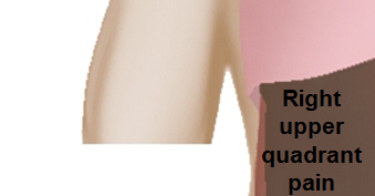 </figure-inline>|<figure-inline> </figure-inline>|<figure-inline>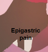 </figure-inline>|<figure-inline> </figure-inline>|<figure-inline>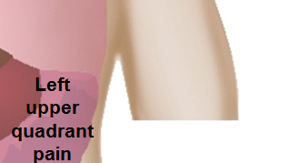 </figure-inline> </figure-inline>
|
<figure-inline>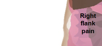 </figure-inline>|<figure-inline> </figure-inline>|<figure-inline>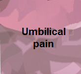 </figure-inline>|<figure-inline> </figure-inline>|<figure-inline>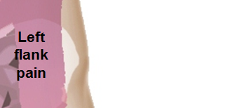 </figure-inline> </figure-inline>
|
<figure-inline>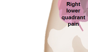 </figure-inline>|<figure-inline> </figure-inline>|<figure-inline>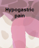 </figure-inline>|<figure-inline> </figure-inline>|<figure-inline> </figure-inline> </figure-inline>
|
References
- ↑ Bluth, Edward I.; Benson, Carol B.; Ralls, Philip W.; Siegel, Marilyn J. (2008). “1: Right Upper Quadrant Pain”. doi:10.1055/b-0034-71418.
- ↑ “Acute cholecystitis | The BMJ”.
- ↑ Knab LM, Boller AM, Mahvi DM (2014). “Cholecystitis”. Surg. Clin. North Am. 94 (2): 455–70. doi:10.1016/j.suc.2014.01.005. PMID 24679431.
Epidemiology and Demographics
Editor-In-Chief: C. Michael Gibson, M.S., M.D. [1]; Associate Editor(s)-in-Chief: Furqan M M. M.B.B.S[2]
Overview
The incidence of acute cholecystitis is approximately 6,300 per 100,000 in individuals under 50 years age and 20,900 per 100,000 in individuals over 50 years age worldwide. The prevalence of acute cholecystitis is approximately 369 per 100,000 individuals in the United States. It is estimated from the population-based statistics, based on a comprehensive survey in the U.S. Acute cholecystitis is comparatively less prevalent in the developing countries. The mortality rate of acute cholecystitis is approximately 0.6%. Acute cholecystitis usually affects individuals of the North American Indian race. Females are more commonly affected by acute cholecystitis than males. Acute cholecystitis cases are reported worldwide. Acute cholecystitis accounts for 700,000 cholecystectomies and costs of ∼$6.5 billion annually only in the United States.
Epidemiology and Demographics
Incidence
- The incidence of acute cholecystitis is approximately 6,300 per 100,000 in individuals under 50 years age and 20,900 per 100,000 in individuals over 50 years age worldwide.[1][2]
Prevalence
- The prevalence of acute cholecystitis is approximately 369 per 100,000 individuals in the United states. It is estimated from the population-based statistics, based on a comprehensive survey in the U.S.[3]
Case-fatality rate/Mortality rate
- The mortality rate of Acute cholecystitis is approximately 0.6%.[3]
Age
Race
- Gallstone diseases usually affects individuals of the North American Indian race. White Americans, Asians, African Americans, and Africans are less likely to develop acute cholecystitis.[3][4]
Gender
- Females are more commonly affected by gallstone diseases than males. The female to male ratio ranges from 10:1 in Pima Indians to 2–3:1 in Europeans women.[3][4]
Region
- Acute cholecystitis cases are reported worldwide. America and Europe have high rates of gallbladder stones as compared to Asia and Africa.[3]
Developed Countries
- Gallstone disease accounts for 700,000 cholecystectomies and costs of ∼$6.5 billion annually only in the United States.[3]
- Gallstone disease is prevalent in North America with a racial predisposition to the American Indians.
- South American countries have slightly more prevalence than North America.
- In Europe, Scandinavian countries have the highest prevalence of acute cholecystitis.
- Italy, Austria, England, Germany, and Poland have a higher prevalence among the rest of Europe.
Developing Countries
- Gallstone diseases are comparatively less prevalent in the developing countries.[3]
- India and Taiwan have a higher prevalence of acute cholecystitis in the developing countries.
References
- ↑ Kimura Y, Takada T, Kawarada Y, Nimura Y, Hirata K, Sekimoto M, Yoshida M, Mayumi T, Wada K, Miura F, Yasuda H, Yamashita Y, Nagino M, Hirota M, Tanaka A, Tsuyuguchi T, Strasberg SM, Gadacz TR (2007). “Definitions, pathophysiology, and epidemiology of acute cholangitis and cholecystitis: Tokyo Guidelines”. J Hepatobiliary Pancreat Surg. 14 (1): 15–26. doi:10.1007/s00534-006-1152-y. PMC 2784509. PMID 17252293.
- ↑ 2.0 2.1 Telfer S, Fenyö G, Holt PR, de Dombal FT (1988). “Acute abdominal pain in patients over 50 years of age”. Scand. J. Gastroenterol. Suppl. 144: 47–50. PMID 3165555.
- ↑ 3.0 3.1 3.2 3.3 3.4 3.5 3.6 3.7 Shaffer EA (2006). “Gallstone disease: Epidemiology of gallbladder stone disease”. Best Pract Res Clin Gastroenterol. 20 (6): 981–96. doi:10.1016/j.bpg.2006.05.004. PMID 17127183.
- ↑ 4.0 4.1 4.2 Knab LM, Boller AM, Mahvi DM (2014). “Cholecystitis”. Surg. Clin. North Am. 94 (2): 455–70. doi:10.1016/j.suc.2014.01.005. PMID 24679431.
Risk Factors
Editor-In-Chief: C. Michael Gibson, M.S., M.D. [1]; Associate Editor(s)-in-Chief: Furqan M M. M.B.B.S[2]
Overview
Common risk factors in the development of acute calculous cholecystitis include advancing age, female gender, obesity, and family history. Long periods of fasting, total parental nutrition (TPN), weight loss are the common risk factors for the development of acute acalculous cholecystitis.
Risk Factors
- Common risk factors in the development of acute calculous cholecystitis include advancing age, female gender, obesity, and family history. Long periods of fasting, total parental nutrition (TPN), weight loss are the common risk factors for the development of acute acalculous cholecystitis.
Common Risk Factors
- Common risk factors in the development of acute calculous cholecystitis are:[1][2][3]
- Advancing age
- Female gender
- Obesity
- Multi parity
- Family history
- Genetic factors
- Oral contraceptives
- Diabetes
- Common risk factors in the development of acute acalculous cholecystitis are:[4]
- Long lasting fasting
- Total parenteral nutrition (TPN)
- Weight loss
- Severe burns
- Sepsis
Less Common Risk Factors
For the causes of Acute cholecystitis, please click here.
References
- ↑ Knab LM, Boller AM, Mahvi DM (2014). “Cholecystitis”. Surg. Clin. North Am. 94 (2): 455–70. doi:10.1016/j.suc.2014.01.005. PMID 24679431.
- ↑ Ruhl CE, Everhart JE (2000). “Association of diabetes, serum insulin, and C-peptide with gallbladder disease”. Hepatology. 31 (2): 299–303. doi:10.1002/hep.510310206. PMID 10655249.
- ↑ Tsai CJ, Leitzmann MF, Willett WC, Giovannucci EL (2006). “Central adiposity, regional fat distribution, and the risk of cholecystectomy in women”. Gut. 55 (5): 708–14. doi:10.1136/gut.2005.076133. PMC 1856127. PMID 16478796.
- ↑ Jones MW, Ferguson T. “Gallbladder, Cholecystitis, Acalculous”. PMID 29083717.
- ↑ Kimura Y, Takada T, Kawarada Y, Nimura Y, Hirata K, Sekimoto M, Yoshida M, Mayumi T, Wada K, Miura F, Yasuda H, Yamashita Y, Nagino M, Hirota M, Tanaka A, Tsuyuguchi T, Strasberg SM, Gadacz TR (2007). “Definitions, pathophysiology, and epidemiology of acute cholangitis and cholecystitis: Tokyo Guidelines”. J Hepatobiliary Pancreat Surg. 14 (1): 15–26. doi:10.1007/s00534-006-1152-y. PMC 2784509. PMID 17252293.
Screening
Editor-In-Chief: C. Michael Gibson, M.S., M.D. [1]; Associate Editor(s)-in-Chief: Furqan M M. M.B.B.S[2]
Overview
There is insufficient evidence to recommend routine screening for acute cholecystitis.
Screening
There is insufficient evidence to recommend routine screening for acute cholecystitis.
References
Natural History, Complications and Prognosis
Editor-In-Chief: C. Michael Gibson, M.S., M.D. [1]; Associate Editor(s)-in-Chief: Dildar Hussain, MBBS [2]
Overview
Acute Cholecystitis most commonly occurs as a result of the prolonged obstruction of the cystic duct leading to inflammation of the gallbladder. The obstruction further contributes to the development of the complications associated with acute cholecystitis such as gangrene, empyema, perforation, cholecystoenteric fistula, emphysematous cholecystitis, and gallstone ileus. Prognosis is generally good if the patient receives treatment. The majority of the patients undergo cholecystectomy.
Natural History, Complications, and Prognosis
Natural History
- In calculus cholecystitis, symptoms of acute cholecystitis usually develop after the obstruction of a gallstone in the bile ducts for more than 7 to 10 days. Female sex, obesity, oral contraceptives, and an underlying disease, such as diabetes are associated with the development of acute cholecystitis. Acute cholecystitis presents with the symptoms, such as right upper quadrant abdominal pain (biliary colic), nausea, and vomiting.[1][2]
- If left untreated acute cholecystitis further leads to the development of gangrene, empyema, perforation, cholecystoenteric fistula, emphysematous cholecystitis, and gallstone ileus.
Complications
Common complications of acute cholecystitis include:[3][4][5][6][7][8]
| Complication | Explanation |
|---|---|
| Gangrene | Gangrene of gallbladder is the most common complication of acute cholecystitis, if left untreated and in elderly patients with an underlying disease of diabetes. |
| Empyema | Prolonged untreated acute cholecystitis worsens the inflammation of the gallbladder leading to the collection of pus around the gallbladder called the empyema of the gallbladder. |
| Perforation | Perforation of the gallbladder is a result of the gangrene of the gallbladder and lead to the pericholecystic abscess. Peritonitis may also occur as a result of gallbladder perforation, such patients develop septicemia and have a high mortality rate. |
| Cholecystoenteric fistula | The cholecystoenteric fistula usually occurs due to the perforation of the gallbladder associated with the long-standing pressure necrosis due to gallstones than acute cholecystitis. |
| Emphysematous cholecystitis | It is associated with secondary infection of the gall bladder wall with gas forming organisms i.e Clostridium welchii, other organisms which can be found are Escherichia coli, Staphylococci, Streptococci, Pseudomonas, and Klebsiella. |
| Gallstone Illeus | Gallstone ileus is associated with the passage of gall stone through the cholecystoenteric fistula leading to mechanical obstruction of bowel in the terminal ileum. |
Prognosis
- Prognosis is generally good after the timely treatment (cholecystectomy).[9][10]
References
- ↑ Ruhl CE, Everhart JE (2000). “Association of diabetes, serum insulin, and C-peptide with gallbladder disease”. Hepatology. 31 (2): 299–303. doi:10.1002/hep.510310206. PMID 10655249.
- ↑ Tsai CJ, Leitzmann MF, Willett WC, Giovannucci EL (2006). “Central adiposity, regional fat distribution, and the risk of cholecystectomy in women”. Gut. 55 (5): 708–14. doi:10.1136/gut.2005.076133. PMC 1856127. PMID 16478796.
- ↑ BYRNE JJ, BERGER RL (1960). “The pathogenesis of acute cholecystitis”. Arch Surg. 81: 812–6. PMID 13689586.
- ↑ Reiss R, Nudelman I, Gutman C, Deutsch AA (1990). “Changing trends in surgery for acute cholecystitis”. World J Surg. 14 (5): 567–70, discussion 570–1. PMID 2238655.
- ↑ Roslyn JJ, Thompson JE, Darvin H, DenBesten L (1987). “Risk factors for gallbladder perforation”. Am. J. Gastroenterol. 82 (7): 636–40. PMID 3605024.
- ↑ Lorenz RW, Steffen HM (1990). “Emphysematous cholecystitis: diagnostic problems and differential diagnosis of gallbladder gas accumulations”. Hepatogastroenterology. 37 Suppl 2: 103–6. PMID 2083919.
- ↑ Clavien PA, Richon J, Burgan S, Rohner A (1990). “Gallstone ileus”. Br J Surg. 77 (7): 737–42. PMID 2200556.
- ↑ Chawla A, Bosco JI, Lim TC, Srinivasan S, Teh HS, Shenoy JN (2015). “Imaging of acute cholecystitis and cholecystitis-associated complications in the emergency setting”. Singapore Med J. 56 (8): 438–43, quiz 444. doi:10.11622/smedj.2015120. PMC 4545132. PMID 26311909.
- ↑ Gurusamy KS, Koti R, Fusai G, Davidson BR (2013). “Early versus delayed laparoscopic cholecystectomy for uncomplicated biliary colic”. Cochrane Database Syst Rev (6): CD007196. doi:10.1002/14651858.CD007196.pub3. PMID 23813478.
- ↑ Gu MG, Kim TN, Song J, Nam YJ, Lee JY, Park JS (2014). “Risk factors and therapeutic outcomes of acute acalculous cholecystitis”. Digestion. 90 (2): 75–80. doi:10.1159/000362444. PMID 25196261.
Looking for the patient version?
© 2026 MyEClinic – IFTM Institut für Telematik in der Medizin GmbH