
List of subjects in Gray's Anatomy: VI. The Arteries
Editor-In-Chief: C. Michael Gibson, M.S., M.D. [1]
Introduction (Template:GraySubject)
Introduction (Template:GraySubject)
- anastomoses
- collateral circulation
- Pulmonary artery (A. Pulmonalis)
- right branch of the pulmonary artery (ramus dexter a. pulmonalis)
- left branch of the pulmonary artery (ramus sinister a. pulmonalis)
the aorta (Template:GraySubject)
the aorta (Template:GraySubject)
- Aorta
- Ascending aorta (Aorta Ascendens)
- Coronary arteries
- Right coronary artery (a. coronaria dextra)
- Coronary arteries
- Right coronary artery (a. coronaria dextra)
- Left coronary artery (a. coronaria sinistra)
- Arch of the aorta (Arcus Aortae; Transverse aorta)
- Relations
- Innominate atery (A. Anonyma; Brachiocephalic artery)
- Innominate atery (A. Anonyma; Brachiocephalic artery)
- thyreoidea ima (a. thyreoidea ima)
- thymic branch or bronchial branch
- Collateral circulation
Editor-In-Chief: C. Michael Gibson, M.S., M.D. [1]
Associate Editor-In-Chief: Cafer Zorkun, M.D., Ph.D. [2]
The aorta (generally pronounced eɪˈɔːtə or “ay-orta”) is the largest artery in the human body, originating from the left ventricle of the heart and bringing oxygenated blood to all parts of the body in the systemic circulation.
The course of the aorta
The aorta is usually divided into five segments/sections [1] [2] [3] [4] :
- Ascending aorta — the section between the heart and the arch of aorta
- Arch of aorta — the peak part that looks somewhat like an inverted “U”
- Descending aorta — the section from the arch of aorta to the point where it divides into the common iliac arteries
- Thoracic aorta — the half of the descending aorta above the diaphragm
- Abdominal aorta — the half of the descending aorta below the diaphragm
Features
The aorta is an elastic artery, and as such is quite distensible. When the left ventricle contracts to force blood into the aorta, the aorta expands. This stretching gives the potential energy that will help maintain blood pressure during diastole, as during this time the aorta contracts passively.
Diseases/pathology
- Aneurysm of sinus of Valsalva
- Aortic aneurysm – myotic, bacterial (e.g. syphilis), senile, genetic, associated with valvular heart disease
- Aortic coarctation – pre-ductal, post-ductal
- Atherosclerosis
- Marfan syndrome
- Trauma, such as traumatic aortic rupture, most often thoracic and distal to the left subclavian artery[5] and frequently quickly fatal[6]
-
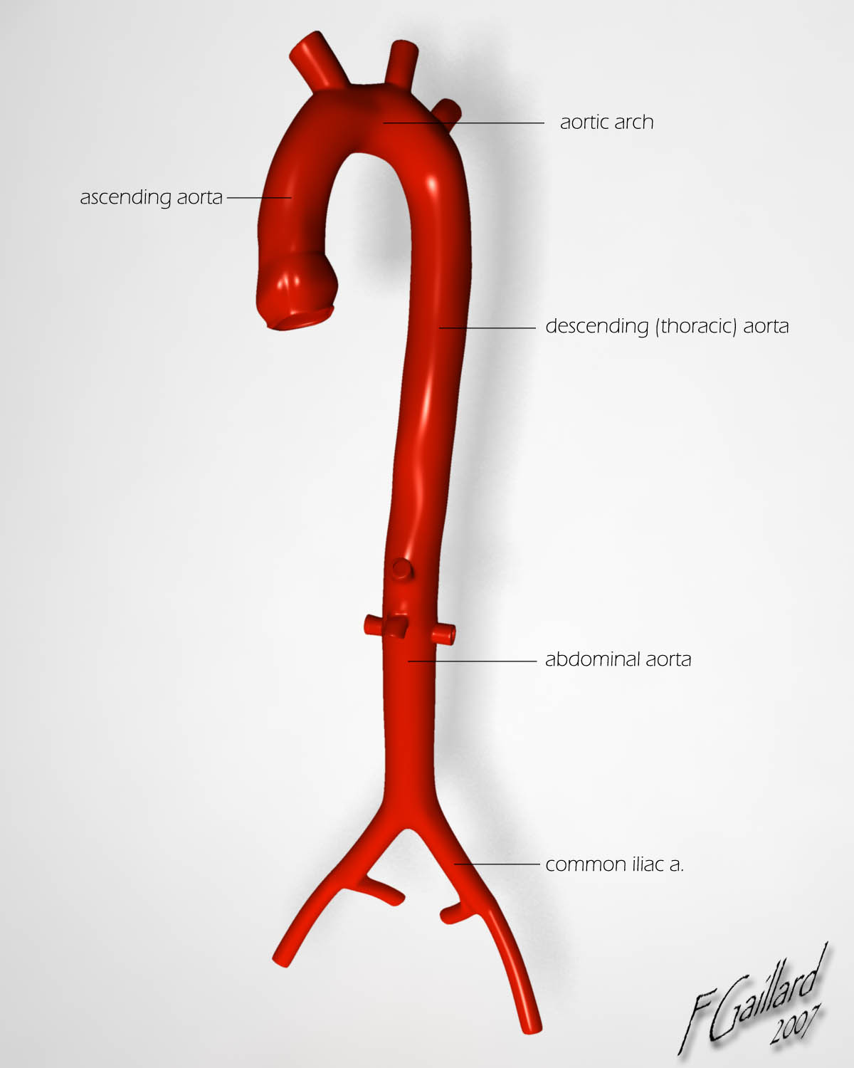 Aorta (Image courtesy of radiopaedia.org)

References
- ↑ Tortora, Gerard J: “Principles of Human W. & Karen A. Koos: Human Anatomy, second edition, page 479. Wm. C. Brown Publishing, 1994 (ISBN 0-697-12252-2)
- ↑ De Graaff, Van: “Human Anatomy, fifth edition”, pages 548-549. WCB McGraw-Hill, 1998 (ISBN 0-697-28413-1)
- ↑ Last’s Anatomy – 10th Edition – Chummy S Sinnatamby
- ↑ Clemete’s Anatomy – Regional Atlas of the Human Body – 3rd Edition
- ↑ Samett EJ. http://www.emedicine.com/radio/topic44.htm Aorta, Trauma. eMedicine.com. Accessed on: April 24, 2007.
- ↑ “Aortic Trauma in Scotland – A Population Based Study”. European Journal of Vascular and Endovascular Surgery. 32 (6): 686–689. 2006. PMID 16750920. April 24, 2007
External links
- Template:GraySubject – Descending aorta
- Template:GraySubject – Abdominal aorta
- Cleveland Clinic Webchat – Aorta Surgery Webchat with Dr. Lars Svensson
Template:Arteries of chest Template:Arteries of abdomen
ar:الشريان الأبهر
ast:Aorta
bg:Аорта
ca:Aorta
da:Aorta
de:Aorta
el:Αορτή
eo:Aorto
eu:Aorta
ko:대동맥
hr:Aorta
id:Aorta
it:Aorta
he:אב העורקים
la:Aorta
lt:Aorta
hu:Aorta
ms:Aorta
nl:Aorta
no:Aorta
nn:Livpulsåre
nds:Aorta
sq:Aorta
sk:Srdcovnica
sr:Аорта
fi:Aortta
sv:Aorta
uk:Аорта
the arteries of the head and neck
- Common carotid artery (A. Carotis Communis)
- Common carotid artery (A. Carotis Communis)
Anterior
- superior thyroid artery (a. thyreoidea superior)
- Hyoid branch (ramus hyoideus; infrahyoid branch)
- Sternocleidomastoid branch (ramus sternocleidomastoideus; sternomastoid branch)
- Superior laryngeal artery (a. laryngea superior)
- Cricothyroid branch (ramus cricothyreoideus)
- lingual artery (a. lingualis)
- Hyoid branch (ramus hyoideus; suprahyoid branch)
- Arteriæ dorsales linguæ (rami dorsales linguæ)
- Sublingual artery (a. sublingualis)
- Arteria profunda linguæ (ranine artery; deep lingual artery)
- external maxillary artery (a. maxillaris externa; facial artery)
- external maxillary artery (a. maxillaris externa; facial artery)
- Cervical Branches
- Ascending palatine artery (a. palatina ascendens)
- Tonsillar branch (ramus tonsillaris)
- Glandular branches (rami glandulares; submaxillary branches)
- Submental artery (a. submentalis)
- Muscular branches
- Facial Branches
- Inferior labial artery (a. labialis inferior; inferior coronary artery)
- Superior labial artery (a. labialis superior; superior coronary artery)
- Cervical Branches
- external maxillary artery (a. maxillaris externa; facial artery)
- Facial Branches
- Superior labial artery (a. labialis superior; superior coronary artery)
- Lateral nasal branch
- Angular artery (a. angularis)
- Muscular branches
- Facial Branches
Posterior
- occipital artery (a. occipitalis)
- Muscular branches (rami musculares)
- Sternocleidomastoid artery (a. sternocleidomastoidea; sternomastoid artery)
- Auricular branch (ramus auricularis)
- occipital artery (a. occipitalis)
- Auricular branch (ramus auricularis)
- Meningeal branch (ramus meningeus; dural branch)
- Descending branch (ramus descendens; arteria princeps cervicis)
- posterior auricular artery (a. auricularis posterior)
- Stylomastoid artery (a. stylomastoidea)
- Auricular branch (ramus auricularis)
- Occipital branch (ramus occipitalis)
Ascending
- ascending pharyngeal artery (a. pharyngea ascendens)
- ascending pharyngeal artery (a. pharyngea ascendens)
- Pharyngeal branches (rami pharyngei)
- Palatine branch
- Prevertebral branches
- Inferior tympanic artery (a. tympanica inferior)
- Meningeal branches of the ascending pharyngeal artery
Terminal
- superficial temporal artery (a. temporalis superficialis)
- Transverse facial artery (a. transversa faciei)
- Middle temporal artery (a. temporalis media)
- superficial temporal artery (a. temporalis superficialis)
- Anterior auricular branches (rami auriculares anteriores)
- Frontal branch (ramus frontalis; anterior temporal)
- Parietal branch (ramus parietalis; posterior temporal)
- internal maxillary artery (a. maxillaris interna)
- first or mandibular portion
- second or pterygoid portion
- third or pterygopalatine portion
Terminal: Branches of the First or Mandibular Portions
- Anterior tympanic artery (a. tympanica anterior; tympanic artery)
- Deep auricular artery (a. auricularis profunda)
- Middle meningeal artery (a. meningea media; medidural artery)
- Middle meningeal artery (a. meningea media; medidural artery)
- Accessory meningeal branch (ramus meningeus accessorius; small meningeal or parvidural branch)
- Inferior alveolar artery (a. alveolaris inferior; inferior dental artery)
Terminal: Branches of the Second or Pterygoid Portion
- Deep temporal branches
- Pterygoid branches (rami pterygoidei)
- Masseteric artery (a. masseterica)
- Buccinator artery (a. buccinatoria; buccal artery)
Terminal: Branches of the Third or Pterygopalatine Portion
- Posterior superior alveolar artery (a. alveolaris superior posterior; alveolar or posterior dental artery)
- Infraorbital artery (a. infraorbitalis)
- Descending palatine artery (a. palatina descendens)
- Artery of the pterygoid canal (a. canalis pterygoidei; Vidian artery)
- Pharyngeal branch
- Sphenopalatine artery (a. sphenopalatina; nasopalatine artery)
- Petrous portion
- caroticotympanic branch (ramus caroticotympanicus; tympanic branch)
- artery of the pterygoid canal (a. canilis pterygoidei; Vidian artery)
- Cavernous portion
- cavernous branches
- hypophyseal branches
- semilunar branches
- anterior meningeal branch (a. meningea anterior)
- ophthalmic artery (a. ophthalmica)
- Cavernous portion
- ophthalmic artery (a. ophthalmica)
- orbital group
- Lacrimal artery (a. lacrimalis)
- Supraorbital artery (a. supraorbitalis)
- orbital group
- ophthalmic artery (a. ophthalmica)
- Cavernous portion
- ophthalmic artery (a. ophthalmica)
- Cavernous portion
- ophthalmic artery (a. ophthalmica)
- orbital group
- Dorsal nasal artery (a. dorsalis nasi; nasal artery)
- ocular group
- Central artery of the retina (a. centralis retinœ)
- Ciliary arteries (aa. ciliares)
- Muscular branches (rami musculares)
- orbital group
- anterior cerebral artery (a. cerebri anterior)
- ophthalmic artery (a. ophthalmica)
- Cerebral portion
- anterior cerebral artery (a. cerebri anterior)
- Anterior communicating artery (a. communicans anterior)
- middle cerebral artery (a. cerebri media)
- anterior cerebral artery (a. cerebri anterior)
- Cerebral portion
- posterior communicating artery (a. communicans posterior)
- anterior choroidal artery (a. chorioidea; choroid artery)
the arteries of the brain (Template:GraySubject)

Editor-In-Chief: C. Michael Gibson, M.S., M.D. [1]
Overview
Arteries are muscular blood vessels that carry blood away from the heart.[1] All arteries, with the exception of the pulmonary and umbilical arteries, carry oxygenated blood.
The circulatory system is extremely important for sustaining life. Its proper functioning is responsible for the delivery of oxygen and nutrients to all cells, as well as the removal of carbon dioxide and waste products, maintenance of optimum pH, and the mobility of the elements, proteins and cells of the immune system. In developed countries, the two leading causes of death, myocardial infarction and stroke each may directly result from an arterial system that has been slowly and progressively compromised by years of deterioration. (See atherosclerosis).
Description
The arterial system is the higher-pressure portion of the circulatory system. Arterial pressure varies between the peak pressure during heart contraction, called the systolic pressure, and the minimum, or diastolic pressure between contractions, when the heart rests between cycles. This pressure variation within the artery produces the pulse which is observable in any artery, and reflects heart activity.
Anatomy
The outermost layer is known as the tunica externa formerly known as “tunica adventitia” and is composed of connective tissue. Inside this layer is the tunica media, or media, which is made up of smooth muscle cells and elastic tissue. The innermost layer, which is in direct contact with the flow of blood is the tunica intima, commonly called the intima. This layer is made up of mainly endothelial cells. The hollow internal cavity in which the blood flows is called the lumen.
Types of arteries
There are several types of arteries in the body:
Pulmonary arteries
The pulmonary arteries carry deoxygenated blood that has just returned from the body to the lungs, where carbon dioxide is exchanged for oxygen.
Systemic arteries
Systemic arteries deliver blood to the arterioles, and then to the capillaries, where nutrients and gasses are exchanged.
The Aorta
The aorta is the root systemic artery. It receives blood directly from the left ventricle of the heart via the aortic valve. As the aorta branches, and these arteries branch in turn, they become successively smaller in diameter, down to the arteriole. The arterioles supply capillaries which in turn empty into venules.
Arterioles
Arterioles, the smallest of the true arteries, help regulate blood pressure and deliver blood to the kidneys (capillaries).
Arterioles and blood pressure
Arterioles have the greatest collective influence on both local blood flow and on overall blood pressure. They are the primary “adjustable nozzles” in the blood system, across which the greatest pressure drop occurs. The combination of heart output (cardiac output) and systemic vascular resistance, which refers to the collective resistance of all of the body’s arterioles, are the principal determinants of arterial blood pressure at any given moment.
Capillaries
The capillaries are where all of the important exchanges happen in the circulatory system. The capillaries are a single cell thick to aid fast and easy diffusion of gases, sugars and other nutrients to surrounding tissues.
Functions of capillaries
To withstand and adapt to the pressures within, arteries are surrounded by varying thicknesses of smooth muscle which have extensive elastic and inelastic connective tissues.
The pulse pressure, i.e. Systolic vs. Diastolic difference, is determined primarily by the amount of blood ejected by each heart beat, stroke volume, versus the volume and elasticity of the major arteries.
Over time, elevated arterial blood sugar (see Diabetes Mellitus), lipoprotein cholesterol, and pressure, smoking, and other factors are all involved in damaging both the endothelium and walls of the arteries, resulting in atherosclerosis or Diabetes Mellitus.
History
Among the ancient Greeks, the arteries were considered to be “air holders” that were responsible for the transport of air to the tissues and were connected to the trachea. This theory presumably arose from the fact that the arteries are empty after death: the last beat of the heart pushes the blood through the capillaries and into the veins.
In medieval times, it was recognized that arteries carried a fluid, called “spiritual blood” or “vital spirits”, considered to be different from the contents of the veins. This theory went back to Galen. In the late medieval period, the trachea,[2] and ligaments were also called “arteries”.[3]
William Harvey described and popularized the modern concept of the circulatory system and the roles of arteries and veins in the 17th century.
Alexis Carrel at the beginning of 20th century first described the technique for vascular suturing and anastomosis and successfully performed many organ transplantations in animals; he thus actually opened the way to modern vascular surgery that was before limited to vessels permanent ligatation.
References
- ↑ Template:KMLEref
- ↑ Oxford English Dictionary.
- ↑ Shakespeare, William. Hamlet Complete, Authoritative Text with Biographical and Historical Contexts, Critical History, and Essays from Five Contemporary Critical Perspectives. Boston: Bedford Books of St. Martins Press, 1994. pg. 50.
See also
Template:Arteries of head and neck Template:Arteries of upper limbs Template:Arteries of chest Template:Arteries of abdomen Template:Arteries of lower limbs
ar:شريان
ca:Artèria
cy:Rhydweli
da:Arterie
de:Arterie
el:Αρτηρία
eo:Arterio
eu:Arteria
fa:سرخرگ
ko:동맥
hr:Arterija
id:Pembuluh nadi
is:Slagæð
it:Arteria
he:עורק
ku:Xwînber
la:Arteria
lv:Artērijas
lt:Arterija
hu:Artéria
ms:Arteri
mk:Артерија
nl:Slagader
no:Arterie
nn:Pulsåre
sq:Arteria
simple:Artery
sk:Tepna
sl:Arterija
fi:Valtimo (anatomia)
sv:Artär
ta:தமனி
uk:Артерії
the arteries of the upper extremity
the arteries of the upper extremity
- Subclavian artery (A. Subclavia)
- First Part of the Right subclavian artery
- First Part of the Left subclavian artery
- Second and Third Parts of the Subclavian artery
- branches of the subclavian artery
vertebral artery (a. vertebralis)
- branches of the vertebral artery
- Cervical Branches.
- Spinal branches (rami spinales)
- Muscular branches
- Cranial Branches
- Meningeal branch (ramus meningeus; posterior meningeal branch)
- Posterior spinal artery (a. spinalis posterior; dorsal spinal artery)
- Anterior spinal artery (a. spinalis anterior; ventral spinal artery)
- Cranial Branches
- Posterior inferior cerebellar artery (a. cerebelli inferior posterior)
- Medullary arteries (bulbar arteries)
- Basilar artery (a. basilaris)
- branches of basilar artery
- pontine branches (rami ad pontem; transverse branches)
- internal auditory artery (a. auditiva interna; auditory artery)
- anterior inferior cerebellar artery (a. cerebelli inferior anterior)
- superior cerebellar artery (a. cerebelli superior)
- posterior cerebral artery (a. cerebri posterior)
- branches of the posterior cerebral artery
- Basilar artery (a. basilaris)
- branches of basilar artery
thyrocervical trunk (truncus thyreocervicalis; thyroid axis)
- Inferior thyroid artery (a. thyreoidea inferior)
- branches of the inferior thyroid artery
- inferior laryngeal artery (a. laryngea inferior)
- tracheal branches (rami tracheales)
- esophageal branches (rami æsophagei)
- ascending cervical artery (a. cervicalis ascendens)
- muscular branches
- Transverse scapular artery (a. transversa scapulæ, suprascapular artery)
- Transverse cervical artery (a. transversa colli; transversalis colli artery)
- ascending branch (ramus ascendens; superficial cervical artery)
- descending branch (ramus descendens; posterior scapular artery)
internal mammary artery (a. mammaria interna)
- musculophrenic artery and superior epigastric artery
- branches of the internal mammary artery
- Pericardiacophrenic artery (a. pericardiacophrenica; a. comes nervi phrenici)
- Anterior mediastinal arteries (aa. mediastinales anteriores; mediastinal arteries)
- Pericardial branches
- Sternal branches (rami sternales)
- Intercostal branches (rami intercostales; anterior intercostal arteries)
- Perforating branches (rami perforantes)
- Musculophrenic artery (a. musculophrenica)
- branches of the internal mammary artery
- Superior epigastric artery (a. epigastrica superior)
costocervical trunk (truncus costocervicalis; superior intercostal artery)
- highest intercostal artery
- Profunda cervicalis (a. cervicalis profunda; deep cervical branch)
the axilla (Template:GraySubject)
the axillary artery
- Collateral circulation after Ligature of the Axillary artery (Scapular anastomosis)
- branches of the axillary artery
- first part
- highest thoracic artery (a. thoracalis suprema; superior thoracic artery)
- second part
- thoracoacromial artery (a. thoracoacromialis; acromiothoracic artery; thoracic axis)
- lateral thoracic artery (a. thoracalis lateralis; long thoracic artery; external mammary artery)
- third part
- subscapular artery (a. subscapularis)
- Scapular circumflex artery (a. circumflexa scapulæ; dorsalis scapulæ artery)
- third part
- posterior humeral circumflex artery (a. circumflexa humeri posterior; posterior circumflex artery)
- anterior humeral circumflex artery (a. circumflexa humeri anterior; anterior circumflex artery)
- branches of the brachial artery
- arteria profunda brachii (superior profunda artery)
- nutrient artery (a. nutricia humeri)
- superior ulnar collateral artery (a. collateralis ulnaris superior; inferior profunda artery)
- inferior ulnar collateral artery (a. collateralis ulnaris inferior; anastomotica magna artery)
- muscular branches (rami musculares)
- deep volar arch
- branches of the radial artery
- In the Forearm
- radial recurrent artery (a. recurrens radialis)
- muscular branches (rami musculares)
- volar carpal branch (ramus carpeus volaris; anterior radial carpal artery)
- superficial volar branch (ramus volaris superficialis; superficialis volœ artery)
- At the Wrist
- At the Wrist
- In the Hand
- arteria princeps pollicis
- arteria volaris indicis radialis (radialis indicis artery)
- first volar metacarpal artery
- deep volar arch (arcus volaris profundus; deep palmar arch)
- volar metacarpal arteries (aa. metacarpeæ volares; palmar interosseous arteries)
- perforating branches (rami perforantes)
- recurrent branches
- branches of the ulnar artery
- In the Forearm
- anterior ulnar recurrent artery (a. recurrentes ulnaris anterior)
- posterior ulnar recurrent artery (a. recurrentes ulnaris posterior)
- common interosseous artery (a. interossea communis)
- Volar interosseous artery (a. interossea volaris; anterior interosseous artery)
- Dorsal interosseous artery (a. interossea dorsalis; posterior interosseous artery)
- In the Forearm
- common interosseous artery (a. interossea communis)
- In the Forearm
- muscular branches (rami musculares)
- At the Wrist
- volar carpal branch (ramus carpeus volares; anterior ulnar carpal artery)
- dorsal carpal branch (ramus carpeus dorsalis; posterior ulnar carpal artery)
- In the Hand
- deep volar branch (ramus volaris profundus; profunda branch)
- superficial volar arch (arcus volaris superficialis; superficial palmar arch)
- Relations
- Common volar digital arteries (aa. digitales volares communes; palmar digital arteries)
- proper volar digital arteries (aa. digitales volares propriæ; collateral digital arteries)
Editor-In-Chief: C. Michael Gibson, M.S., M.D. [1]
Overview
Arteries are muscular blood vessels that carry blood away from the heart.[1] All arteries, with the exception of the pulmonary and umbilical arteries, carry oxygenated blood.
The circulatory system is extremely important for sustaining life. Its proper functioning is responsible for the delivery of oxygen and nutrients to all cells, as well as the removal of carbon dioxide and waste products, maintenance of optimum pH, and the mobility of the elements, proteins and cells of the immune system. In developed countries, the two leading causes of death, myocardial infarction and stroke each may directly result from an arterial system that has been slowly and progressively compromised by years of deterioration. (See atherosclerosis).
Description
The arterial system is the higher-pressure portion of the circulatory system. Arterial pressure varies between the peak pressure during heart contraction, called the systolic pressure, and the minimum, or diastolic pressure between contractions, when the heart rests between cycles. This pressure variation within the artery produces the pulse which is observable in any artery, and reflects heart activity.
Anatomy
The outermost layer is known as the tunica externa formerly known as “tunica adventitia” and is composed of connective tissue. Inside this layer is the tunica media, or media, which is made up of smooth muscle cells and elastic tissue. The innermost layer, which is in direct contact with the flow of blood is the tunica intima, commonly called the intima. This layer is made up of mainly endothelial cells. The hollow internal cavity in which the blood flows is called the lumen.
Types of arteries
There are several types of arteries in the body:
Pulmonary arteries
The pulmonary arteries carry deoxygenated blood that has just returned from the body to the lungs, where carbon dioxide is exchanged for oxygen.
Systemic arteries
Systemic arteries deliver blood to the arterioles, and then to the capillaries, where nutrients and gasses are exchanged.
The Aorta
The aorta is the root systemic artery. It receives blood directly from the left ventricle of the heart via the aortic valve. As the aorta branches, and these arteries branch in turn, they become successively smaller in diameter, down to the arteriole. The arterioles supply capillaries which in turn empty into venules.
Arterioles
Arterioles, the smallest of the true arteries, help regulate blood pressure and deliver blood to the kidneys (capillaries).
Arterioles and blood pressure
Arterioles have the greatest collective influence on both local blood flow and on overall blood pressure. They are the primary “adjustable nozzles” in the blood system, across which the greatest pressure drop occurs. The combination of heart output (cardiac output) and systemic vascular resistance, which refers to the collective resistance of all of the body’s arterioles, are the principal determinants of arterial blood pressure at any given moment.
Capillaries
The capillaries are where all of the important exchanges happen in the circulatory system. The capillaries are a single cell thick to aid fast and easy diffusion of gases, sugars and other nutrients to surrounding tissues.
Functions of capillaries
To withstand and adapt to the pressures within, arteries are surrounded by varying thicknesses of smooth muscle which have extensive elastic and inelastic connective tissues.
The pulse pressure, i.e. Systolic vs. Diastolic difference, is determined primarily by the amount of blood ejected by each heart beat, stroke volume, versus the volume and elasticity of the major arteries.
Over time, elevated arterial blood sugar (see Diabetes Mellitus), lipoprotein cholesterol, and pressure, smoking, and other factors are all involved in damaging both the endothelium and walls of the arteries, resulting in atherosclerosis or Diabetes Mellitus.
History
Among the ancient Greeks, the arteries were considered to be “air holders” that were responsible for the transport of air to the tissues and were connected to the trachea. This theory presumably arose from the fact that the arteries are empty after death: the last beat of the heart pushes the blood through the capillaries and into the veins.
In medieval times, it was recognized that arteries carried a fluid, called “spiritual blood” or “vital spirits”, considered to be different from the contents of the veins. This theory went back to Galen. In the late medieval period, the trachea,[2] and ligaments were also called “arteries”.[3]
William Harvey described and popularized the modern concept of the circulatory system and the roles of arteries and veins in the 17th century.
Alexis Carrel at the beginning of 20th century first described the technique for vascular suturing and anastomosis and successfully performed many organ transplantations in animals; he thus actually opened the way to modern vascular surgery that was before limited to vessels permanent ligatation.
References
- ↑ Template:KMLEref
- ↑ Oxford English Dictionary.
- ↑ Shakespeare, William. Hamlet Complete, Authoritative Text with Biographical and Historical Contexts, Critical History, and Essays from Five Contemporary Critical Perspectives. Boston: Bedford Books of St. Martins Press, 1994. pg. 50.
See also
Template:Arteries of head and neck Template:Arteries of upper limbs Template:Arteries of chest Template:Arteries of abdomen Template:Arteries of lower limbs
ar:شريان
ca:Artèria
cy:Rhydweli
da:Arterie
de:Arterie
el:Αρτηρία
eo:Arterio
eu:Arteria
fa:سرخرگ
ko:동맥
hr:Arterija
id:Pembuluh nadi
is:Slagæð
it:Arteria
he:עורק
ku:Xwînber
la:Arteria
lv:Artērijas
lt:Arterija
hu:Artéria
ms:Arteri
mk:Артерија
nl:Slagader
no:Arterie
nn:Pulsåre
sq:Arteria
simple:Artery
sk:Tepna
sl:Arterija
fi:Valtimo (anatomia)
sv:Artär
ta:தமனி
uk:Артерії
the arteries of the trunk
the descending aorta
- Branches of the Thoracic aorta
- Visceral
- pericardial branches (rami pericardiaci)
- bronchial arteries (aa. bronchiales)
- esophageal arteries (aa. æsophageæ)
- mediastinal branches (rami mediastinales)
- Parietal
- Intercostal arteries (aa. intercostales)
- Parietal
- Collateral circulation
- Visceral branches
- celiac artery (a. cæliaca; celiac axis)
- Left gastric artery (a. gastrica sinistra; gastric artery or coronary artery)
- Hepatic artery (a. hepatica)
- celiac artery (a. cæliaca; celiac axis)
- Visceral branches
- celiac artery (a. cæliaca; celiac axis)
- Hepatic artery (a. hepatica)
- right gastric artery (a. gastrica dextra; pyloric artery)
- gastroduodenal artery (a. gastroduodenalis)
- right gastroepiploic artery (a. gastroepiploica dextra)
- Hepatic artery (a. hepatica)
- celiac artery (a. cæliaca; celiac axis)
- Visceral branches
- celiac artery (a. cæliaca; celiac axis)
- Hepatic artery (a. hepatica)
- superior pancreaticoduodenal artery (a. pancreaticoduodenalis superior)
- cystic artery (a. cystica)
- Lienal artery or Splenic artery (a. lienalis)
- Hepatic artery (a. hepatica)
- celiac artery (a. cæliaca; celiac axis)
- Visceral branches
- celiac artery (a. cæliaca; celiac axis)
- Lienal artery or Splenic artery (a. lienalis)
- pancreatic branches (rami pancreatici)
- short gastric arteries (aa. gastricæ breves; vasa brevia)
- left gastroepiploic artery (a. gastroepiploica sinistra)
- Lienal artery or Splenic artery (a. lienalis)
- superior mesenteric artery (a. mesenterica superior)
- celiac artery (a. cæliaca; celiac axis)
- Visceral branches
- superior mesenteric artery (a. mesenterica superior)
- Inferior pancreaticoduodenal artery (a. pancreaticoduodenalis inferior)
- Intestinal arteries (aa. intestinales; vasa intestini tenuis)
- Ileocolic artery (a. ileocolica)
- superior mesenteric artery (a. mesenterica superior)
- Visceral branches
- superior mesenteric artery (a. mesenterica superior)
- Right colic artery (a. colica dextra)
- Middle colic artery (a. colica media)
- inferior mesenteric artery (a. mesenterica inferior)
- superior mesenteric artery (a. mesenterica superior)
- Visceral branches
- inferior mesenteric artery (a. mesenterica inferior)
- Left colic artery (a. colica sinistra)
- Sigmoid arteries (aa. sigmoideæ)
- Superior hemorrhoidal artery (a. hæmorrhoidalis superior)
- middle suprarenal arteries (aa. suprarenales media; middle capsular arteries; suprarenal arteries)
- renal arteries (aa. renales)
- inferior mesenteric artery (a. mesenterica inferior)
- Visceral branches
- internal spermatic arteries (aa. spermaticæ internæ; spermatic arteries)
- ovarian arteries (aa. ovaricæ)
- Parietal Branches
- inferior phrenic arteries (aa. phrenicæ inferiores)
- lumbar arteries (aa. lumbales)
- Parietal Branches
- middle sacral artery (a. sacralis media)
- umbilical arteries
- lateral umbilical ligament (obliterated hypogastric artery)
- branches of the hypogastric artery
- superior vesical artery (a. vesicalis superior)
- middle vesical artery (a. vesicalis medialis)
- inferior vesical artery (a. vesicalis inferior)
- middle hemorrhoidal artery (a. hæmorrhoidalis media)
- uterine artery (a. uterina)
- uterine artery (a. uterina)
- vaginal artery (a. vaginalis)
- obturator artery (a. obturatoria)
- internal pudendal artery (a. pudenda interna; internal pudic artery)
- internal pudendal artery (a. pudenda interna; internal pudic artery)
- accessory pudendal
- branches of the internal pudendal artery
- internal pudendal artery (a. pudenda interna; internal pudic artery)
- Inferior hemorrhoidal artery (a. hæmorrhoidalis inferior)
- Perineal artery (a. perinei; superficial perineal artery)
- posterior scrotal branches
- transverse perineal artery
- Artery of the urethral bulb (a. bulbi urethræ)
- Urethral artery (a. urethralis)
- internal pudendal artery (a. pudenda interna; internal pudic artery)
- Deep artery of the penis (a. profunda penis; artery to the corpus cavernosum)
- Dorsal artery of the penis (a. dorsalis penis)
- internal pudendal artery in the female
- inferior gluteal artery (a. glutæa inferior; sciatic artery)
- inferior gluteal artery (a. glutæa inferior; sciatic artery)
- iliolumbar artery (a. iliolumbalis)
- Lumbar branch (ramus lumbalis)
- Iliac branch (ramus iliacus)
- lateral sacral arteries (aa. sacrales laterales)
- superior gluteal artery (a. glutæa superior; gluteal artery)
- inferior epigastric artery (a. epigastrica inferior; deep epigastric artery)
- external spermatic artery (cremasteric artery)
- deep iliac circumflex artery (a. circumflexa ilium profunda)
Editor-In-Chief: C. Michael Gibson, M.S., M.D. [1]
Overview
Arteries are muscular blood vessels that carry blood away from the heart.[1] All arteries, with the exception of the pulmonary and umbilical arteries, carry oxygenated blood.
The circulatory system is extremely important for sustaining life. Its proper functioning is responsible for the delivery of oxygen and nutrients to all cells, as well as the removal of carbon dioxide and waste products, maintenance of optimum pH, and the mobility of the elements, proteins and cells of the immune system. In developed countries, the two leading causes of death, myocardial infarction and stroke each may directly result from an arterial system that has been slowly and progressively compromised by years of deterioration. (See atherosclerosis).
Description
The arterial system is the higher-pressure portion of the circulatory system. Arterial pressure varies between the peak pressure during heart contraction, called the systolic pressure, and the minimum, or diastolic pressure between contractions, when the heart rests between cycles. This pressure variation within the artery produces the pulse which is observable in any artery, and reflects heart activity.
Anatomy
The outermost layer is known as the tunica externa formerly known as “tunica adventitia” and is composed of connective tissue. Inside this layer is the tunica media, or media, which is made up of smooth muscle cells and elastic tissue. The innermost layer, which is in direct contact with the flow of blood is the tunica intima, commonly called the intima. This layer is made up of mainly endothelial cells. The hollow internal cavity in which the blood flows is called the lumen.
Types of arteries
There are several types of arteries in the body:
Pulmonary arteries
The pulmonary arteries carry deoxygenated blood that has just returned from the body to the lungs, where carbon dioxide is exchanged for oxygen.
Systemic arteries
Systemic arteries deliver blood to the arterioles, and then to the capillaries, where nutrients and gasses are exchanged.
The Aorta
The aorta is the root systemic artery. It receives blood directly from the left ventricle of the heart via the aortic valve. As the aorta branches, and these arteries branch in turn, they become successively smaller in diameter, down to the arteriole. The arterioles supply capillaries which in turn empty into venules.
Arterioles
Arterioles, the smallest of the true arteries, help regulate blood pressure and deliver blood to the kidneys (capillaries).
Arterioles and blood pressure
Arterioles have the greatest collective influence on both local blood flow and on overall blood pressure. They are the primary “adjustable nozzles” in the blood system, across which the greatest pressure drop occurs. The combination of heart output (cardiac output) and systemic vascular resistance, which refers to the collective resistance of all of the body’s arterioles, are the principal determinants of arterial blood pressure at any given moment.
Capillaries
The capillaries are where all of the important exchanges happen in the circulatory system. The capillaries are a single cell thick to aid fast and easy diffusion of gases, sugars and other nutrients to surrounding tissues.
Functions of capillaries
To withstand and adapt to the pressures within, arteries are surrounded by varying thicknesses of smooth muscle which have extensive elastic and inelastic connective tissues.
The pulse pressure, i.e. Systolic vs. Diastolic difference, is determined primarily by the amount of blood ejected by each heart beat, stroke volume, versus the volume and elasticity of the major arteries.
Over time, elevated arterial blood sugar (see Diabetes Mellitus), lipoprotein cholesterol, and pressure, smoking, and other factors are all involved in damaging both the endothelium and walls of the arteries, resulting in atherosclerosis or Diabetes Mellitus.
History
Among the ancient Greeks, the arteries were considered to be “air holders” that were responsible for the transport of air to the tissues and were connected to the trachea. This theory presumably arose from the fact that the arteries are empty after death: the last beat of the heart pushes the blood through the capillaries and into the veins.
In medieval times, it was recognized that arteries carried a fluid, called “spiritual blood” or “vital spirits”, considered to be different from the contents of the veins. This theory went back to Galen. In the late medieval period, the trachea,[2] and ligaments were also called “arteries”.[3]
William Harvey described and popularized the modern concept of the circulatory system and the roles of arteries and veins in the 17th century.
Alexis Carrel at the beginning of 20th century first described the technique for vascular suturing and anastomosis and successfully performed many organ transplantations in animals; he thus actually opened the way to modern vascular surgery that was before limited to vessels permanent ligatation.
References
- ↑ Template:KMLEref
- ↑ Oxford English Dictionary.
- ↑ Shakespeare, William. Hamlet Complete, Authoritative Text with Biographical and Historical Contexts, Critical History, and Essays from Five Contemporary Critical Perspectives. Boston: Bedford Books of St. Martins Press, 1994. pg. 50.
See also
Template:Arteries of head and neck Template:Arteries of upper limbs Template:Arteries of chest Template:Arteries of abdomen Template:Arteries of lower limbs
ar:شريان
ca:Artèria
cy:Rhydweli
da:Arterie
de:Arterie
el:Αρτηρία
eo:Arterio
eu:Arteria
fa:سرخرگ
ko:동맥
hr:Arterija
id:Pembuluh nadi
is:Slagæð
it:Arteria
he:עורק
ku:Xwînber
la:Arteria
lv:Artērijas
lt:Arterija
hu:Artéria
ms:Arteri
mk:Артерија
nl:Slagader
no:Arterie
nn:Pulsåre
sq:Arteria
simple:Artery
sk:Tepna
sl:Arterija
fi:Valtimo (anatomia)
sv:Artär
ta:தமனி
uk:Артерії
the arteries of the lower extremity
the arteries of the lower extremity
- femoral sheath (crural sheath)
- femoral triangle (trigonum femorale; Scarpa’s triangle)
- adductor canal (canalis adductorius; Hunter’s canal)
- Relations of the Femoral artery
branches of the femoral artery
- superficial epigastric artery (a. epigastrica superficialis)
- superficial iliac circumflex artery (a. circumflexa ilium superficialis)
- superficial external pudendal artery (a. pudenda externa superficialis; superficial external pudic artery)
- deep external pudendal artery (a. pudenda externa profunda; deep external pudic artery)
- Muscular branches (rami musculares)
- profunda femoris artery (a. profunda femoris; deep femoral artery)
- profunda femoris artery (a. profunda femoris; deep femoral artery)
- fourth perforating artery
- branches of profunda femoris artery
- Lateral femoral circumflex artery (a. circumflexa femoris lateralis; external circumflex artery)
- Medial femoral circumflex artery (a. circumflexa femoris medialis; internal circumflex artery)
- profunda femoris artery (a. profunda femoris; deep femoral artery)
- Perforating arteries
- first perforating artery (a. perforans prima)
- second perforating artery (a. perforans secunda)
- third perforating artery (a. perforans tertia)
- highest genicular artery (a. genu suprema; anastomotica magna artery)
- saphenous branch
- musculo-articular branch
- Perforating arteries
- branches of the popliteal artery
- superior muscular branches
- sural arteries (aa. surales; inferior muscular arteries)
- cutaneous branches
- superior genicular arteries (aa. genu superiores; superior articular arteries)
- middle genicular artery (a. genu media; azygos articular artery)
- inferior genicular arteries (aa. genu inferiores; inferior articular arteries)
- medial inferior genicular
- dorsalis pedis
- branches of the anterior tibial artery
- posterior tibial recurrent artery (a. recurrens tibialis posterior)
- fibular artery
- anterior tibial recurrent artery (a. recurrens tibialis anterior)
- muscular branches (rami musculares)
- anterior medial malleolar artery (a. malleolaris anterior medialis; internal malleolar artery)
- anterior lateral malleolar artery (a. malleolaris anterior lateralis; external malleolar artery)
- branches of the arteria dorsalis pedis
- lateral tarsal artery (a. tarsea lateralis; tarsal artery)
- medial tarsal arteries (aa. tarseæ mediales)
- arcuate artery (a. arcuata; metatarsal artery)
- second, third, and fourth dorsal metatarsal arteries
- first dorsal metatarsal artery (a. dorsalis hallucis)
- deep plantar artery (ramus plantaris profundus; communicating artery)
- branches of the posterior tibial artery
- peroneal artery (a. peronæa)
- branches of the peroneal
- Muscular branches
- Nutrient artery (a. nutricia fibulæ)
- Perforating branch (ramus perforans; anterior peroneal artery)
- Communicating branch (ramus communicans)
- Lateral calcaneal (ramus calcaneus lateralis; external calcaneal)
- nutrient artery (a. nutricia tibiæ)
- muscular branches
- posterior medial malleolar artery (a. malleolaris posterior medialis; internal malleolar artery)
- communicating branch (ramus communicans)
- medial calcaneal (rami calcanei mediales; internal calcaneal)
- divisions
- medial plantar artery (a. plantaris medialis; internal plantar artery)
- lateral plantar artery (a. plantaris lateralis; external plantar artery)
- divisions
- lateral plantar artery (a. plantaris lateralis; external plantar artery)
- branches of the plantar arch
- Perforating branches (rami perforantes)
- Plantar metatarsal arteries (aa. metatarseæ plantares; digital branches)
- anterior perforating branch
- first plantar metatarsal artery (arteria princeps hallucis)
- lateral plantar artery (a. plantaris lateralis; external plantar artery)
Editor-In-Chief: C. Michael Gibson, M.S., M.D. [1]
Overview
Arteries are muscular blood vessels that carry blood away from the heart.[1] All arteries, with the exception of the pulmonary and umbilical arteries, carry oxygenated blood.
The circulatory system is extremely important for sustaining life. Its proper functioning is responsible for the delivery of oxygen and nutrients to all cells, as well as the removal of carbon dioxide and waste products, maintenance of optimum pH, and the mobility of the elements, proteins and cells of the immune system. In developed countries, the two leading causes of death, myocardial infarction and stroke each may directly result from an arterial system that has been slowly and progressively compromised by years of deterioration. (See atherosclerosis).
Description
The arterial system is the higher-pressure portion of the circulatory system. Arterial pressure varies between the peak pressure during heart contraction, called the systolic pressure, and the minimum, or diastolic pressure between contractions, when the heart rests between cycles. This pressure variation within the artery produces the pulse which is observable in any artery, and reflects heart activity.
Anatomy
The outermost layer is known as the tunica externa formerly known as “tunica adventitia” and is composed of connective tissue. Inside this layer is the tunica media, or media, which is made up of smooth muscle cells and elastic tissue. The innermost layer, which is in direct contact with the flow of blood is the tunica intima, commonly called the intima. This layer is made up of mainly endothelial cells. The hollow internal cavity in which the blood flows is called the lumen.
Types of arteries
There are several types of arteries in the body:
Pulmonary arteries
The pulmonary arteries carry deoxygenated blood that has just returned from the body to the lungs, where carbon dioxide is exchanged for oxygen.
Systemic arteries
Systemic arteries deliver blood to the arterioles, and then to the capillaries, where nutrients and gasses are exchanged.
The Aorta
The aorta is the root systemic artery. It receives blood directly from the left ventricle of the heart via the aortic valve. As the aorta branches, and these arteries branch in turn, they become successively smaller in diameter, down to the arteriole. The arterioles supply capillaries which in turn empty into venules.
Arterioles
Arterioles, the smallest of the true arteries, help regulate blood pressure and deliver blood to the kidneys (capillaries).
Arterioles and blood pressure
Arterioles have the greatest collective influence on both local blood flow and on overall blood pressure. They are the primary “adjustable nozzles” in the blood system, across which the greatest pressure drop occurs. The combination of heart output (cardiac output) and systemic vascular resistance, which refers to the collective resistance of all of the body’s arterioles, are the principal determinants of arterial blood pressure at any given moment.
Capillaries
The capillaries are where all of the important exchanges happen in the circulatory system. The capillaries are a single cell thick to aid fast and easy diffusion of gases, sugars and other nutrients to surrounding tissues.
Functions of capillaries
To withstand and adapt to the pressures within, arteries are surrounded by varying thicknesses of smooth muscle which have extensive elastic and inelastic connective tissues.
The pulse pressure, i.e. Systolic vs. Diastolic difference, is determined primarily by the amount of blood ejected by each heart beat, stroke volume, versus the volume and elasticity of the major arteries.
Over time, elevated arterial blood sugar (see Diabetes Mellitus), lipoprotein cholesterol, and pressure, smoking, and other factors are all involved in damaging both the endothelium and walls of the arteries, resulting in atherosclerosis or Diabetes Mellitus.
History
Among the ancient Greeks, the arteries were considered to be “air holders” that were responsible for the transport of air to the tissues and were connected to the trachea. This theory presumably arose from the fact that the arteries are empty after death: the last beat of the heart pushes the blood through the capillaries and into the veins.
In medieval times, it was recognized that arteries carried a fluid, called “spiritual blood” or “vital spirits”, considered to be different from the contents of the veins. This theory went back to Galen. In the late medieval period, the trachea,[2] and ligaments were also called “arteries”.[3]
William Harvey described and popularized the modern concept of the circulatory system and the roles of arteries and veins in the 17th century.
Alexis Carrel at the beginning of 20th century first described the technique for vascular suturing and anastomosis and successfully performed many organ transplantations in animals; he thus actually opened the way to modern vascular surgery that was before limited to vessels permanent ligatation.
References
- ↑ Template:KMLEref
- ↑ Oxford English Dictionary.
- ↑ Shakespeare, William. Hamlet Complete, Authoritative Text with Biographical and Historical Contexts, Critical History, and Essays from Five Contemporary Critical Perspectives. Boston: Bedford Books of St. Martins Press, 1994. pg. 50.
See also
Template:Arteries of head and neck Template:Arteries of upper limbs Template:Arteries of chest Template:Arteries of abdomen Template:Arteries of lower limbs
ar:شريان
ca:Artèria
cy:Rhydweli
da:Arterie
de:Arterie
el:Αρτηρία
eo:Arterio
eu:Arteria
fa:سرخرگ
ko:동맥
hr:Arterija
id:Pembuluh nadi
is:Slagæð
it:Arteria
he:עורק
ku:Xwînber
la:Arteria
lv:Artērijas
lt:Arterija
hu:Artéria
ms:Arteri
mk:Артерија
nl:Slagader
no:Arterie
nn:Pulsåre
sq:Arteria
simple:Artery
sk:Tepna
sl:Arterija
fi:Valtimo (anatomia)
sv:Artär
ta:தமனி
uk:Артерії
Looking for the patient version?
© 2026 MyEClinic – IFTM Institut für Telematik in der Medizin GmbH