
Dyspepsia
For patient information, click here
Editor-In-Chief: C. Michael Gibson, M.S., M.D. [1] Associate Editor(s)-in-Chief: Ajay Gade MD[2]] Fahad Hasan, M.D.[3]
Synonyms and keywords: Functional dyspepsia; dyspepsia, functional; non-ulcer dyspepsia; nonulcer dyspepsia
Overview
Please help WikiDoc by adding content here. It’s easy! Click here to learn about editing.
Editor-In-Chief: C. Michael Gibson, M.S., M.D. [1]
Overview
Dyspepsia (from the Greek “δυς-” (Dys-), meaning hard or difficult, and “πέψη” (Pepse), meaning digestion) is chronic or recurrent pain or discomfort centered in the upper abdomen [1] Discomfort, in this context, includes mild pain, upper abdominal fullness and feeling full earlier than expected with eating. It can be accompanied by bloating, belching, nausea or heartburn. Heartburn is excluded from the definition of dyspesia in ICD 10, as it usually has a different cause and management pathway. Many people get dyspepsia. It is often caused by lifestyle factors, such as smoking and diet, but there are some serious causes such as cancer of the stomach, peptic ulcer disease and some medications. When people have dyspepsia but no risk factors for any of the serious causes, it can be labeled undifferentiated dyspepsia and treated without further investigations. When people have been investigated for dyspepsia but no cause has been found it can be labeled as functional dyspepsia.
Historical Perspective
The current understanding of the pathogenesis of dyspepsia began with the first description of gastric ulcer disease in 1799. The term was first used in its current form in 1916 by Walter Alvarez.
Classification
Dyspepsia is broadly classified into two major types: ulcer and non-ulcer dyspepsia. The latter is also known as functional dyspepsia.
Pathophysiology
The symptoms of functional dyspepsia are directly caused by two major pathophysiological abnormalities abnormal gastric motility and visceral hypersensitivity.These mechanisms occur in patients who have acquired excessive responsiveness to stress as a result of the environment during early life, genetic abnormalities, residual inflammation after gastrointestinal infections, or other causes, with the process modified by factors including psychophysiological abnormalities, abnormal secretion of gastric acid, Helicobacter pylori infection, diet, and lifestyle.
Causes
Life threatening causes of dyspepsia include coronary disease and ischemic bowel disease. Other common causes of dyspepsia include gastroesophageal reflux, gastritis, lactose intolerance, and peptic ulcer.
Differentiating dyspepsia from Other Diseases
Dyspepsia must be differentiated from other diseases that presents with epigastric pain such as gastritis, gastroesophageal reflux disease, acute pancreatitis, primary biliary cirrhosis, cholelithiasis, gastric outlet syndrome, myocardial infarction, pleural empyemae appendicitis
Epidemiology and Demographics
The incidence of new cases of H. pylori infection each year ranges from 3,000 to 10,000 per 100,000 individuals in developing countries. It has been observed that with advancing age, the incidence of H. pylori infection increases. In united states, 20% of adolescents are infected with H. pylori when compared to 90% by 5 years of age in the developing countries. In United States, H. pylori infection associated gastritis is more common in African Americans (54%), Hispanics (52%), and the elderly compared to Whites (21%). In acute gastritis, females are usually more affected than men. In H. pylori infection associated gastritis, males are more commonly affected than females. The incidence rates of H. pylori infection are high in Japan, Columbia, Costa Rica and China, and comparatively low in the United States. H. pylori infection is common in Southern and Eastern Europe, Mexico, South America, Africa, most Asian countries, and aboriginal people in North America.
Risk Factors
The secondary prevention strategies for gastritis following H. pylori infection to prevent recurrence of peptic ulcer disease and gastric cancer include the use of antibiotics to prevent recurrence of infection and the post-treatment confirmation of H. pylori eradication after treatment using diagnostic tests.
Screening
There is insufficient evidence to recommend routine screening for Dyspepsia.
Natural History, Complications, and Prognosis
Natural History
Dyspepsia usually persists throughout life and the chance of spontaneous healing is rare. Dyspepsia is most commonly associated with Helicobacter pylori infection. Increase in the prevalence of dyspepsia is attributed to the increasing age and the onset varies among different ethnicities. The increased risk of developing duodenal and peptic ulcers have been observed in individuals with persistent dyspepsia.
Complications
Dyspepsia is associated with complications such as peptic ulcers, anemia due to gastritis, stomach cancer, vitamin B12 deficiency, pernicious anemia.
Prognosis
Functional dyspepsia is a long-lasting disorder with an excellent prognosis regardless of H. pylori infection.
Diagnosis
History and Symptoms
The history and symptoms of dyspepsia are as follows: pain or a burning feeling in the upper portion of the stomach, nausea, bloating, sometimes uncontrollable burping, heartburn, fever, metallic taste, rumbling in the stomach, sense of fullness after eating, feeling as though something is lodged in the esophagus, pain and discomfort at the xiphoid region, sudden chills, comparable to those felt during fevers
Physical Examination
Patients with dyspepsia may appear pale. Some patients may appear fatigued and in distress, is associated with abdominal pain. Vital signs generally appear to be normal. When associated with gastrointestinal bleed, vital signs include tachycardia. Pallor may observed in patients presenting with melena and hematemesis. On examination of the eyes, conjunctival pallor may be observed. Halitosis may be observed in case of chronic gastritis. Chest tenderness may be present on palpation in case of Helicobacter pylori infection associated gastritis. Epigastric tenderness may be present. Gastritis associated with gastric ulcers may result in blood loss and the stool test may be guaiac-positive.
Laboratory Findings
There is no specific diagnostic laboratory test for dyspepsia but in the patient with the history of dyspepsia, the laboratory test is used to rule out bleeding and to document the status of eradication therapy and to test refractory ulcers
Imaging Findings
Esophagogastroduodenoscopy
People without risk factors for serious causes of dyspepsia usually do not need investigation beyond an office-based clinical examination. However, people over the age of 55 years and those with alarm features are usually investigated by esophagogastroduodenoscopy (EGD or OGD in Britain). In this painless investigation the esophagus, stomach, and duodenum are examined through an endoscope passed down through the mouth. This will rule out peptic ulcer disease, medication-related ulceration, malignancy and other rarer causes.
Other Diagnostic Studies
There are no other diagnostic studies associated with dyspepsia.
Treatment
Medical Therapy
Functional and undifferentiated dyspepsia have similar treatments. Decisions around the use of drug therapy are difficult because trials included heartburn in the definition of dyspepsia. This led to the results favoring proton pump inhibitors (PPIs), which are questionably effective for the treatment of heartburn.
Surgery
Surgical intervention is not recommended for the management of dyspepsia
Prevention
Primary prevention
Effective measures for the primary prevention of dyspepsia include avoiding long-term or extended use of medications such as NSAIDs, abstinence from alcohol, smoking cessation, coffee or acidic beverages, spicy foods and avoiding stress. Inculcating healthy eating habits, exercising regularly and maintaining healthy body weight may help in avoiding dyspepsia. Effective measures for primary prevention of the H. pylori infection include hand washing (antibacterial soaps), avoid contaminated food and water, maintain proper hygiene (hand sanitizers, antiseptic washes) and avoid close contact with infected family members ( e.g., kissing, sharing eating utensils and drinking glasses).
Secondary prevention
The secondary prevention strategies for dyspepsia following H. pylori infection to prevent recurrence of peptic ulcer disease and gastric cancer include the use of antibiotics to prevent recurrence of infection and the post-treatment confirmation of H. pylori eradication after treatment using diagnostic tests.
References
- ↑ N. Talley, et al., “Guidelines for the management of dyspepsia”, American Journal of Gastroenterology 100 (2005), pp. 2324-2337.
Historical Perspective
Editor-In-Chief: C. Michael Gibson, M.S., M.D. [1] Associate Editor(s)-in-Chief: Ajay Gade MD[2]]
Overview
The current understanding of the pathogenesis of dyspepsia began with the first description of gastric ulcer disease in 1799. The term was first used in its current form in 1916 by Walter Alvarez.
Historical Perspective
- Indigestion is an old english word meaning ‘lack of digestion’, and the symptoms of dyspepsia have known since the birth of medicine. However, the underlying pathogenesis of dyspepsia only began to be understood when Baillie in 1799 first described the pathology and symptoms of gastric ulcer disease.
- Development of barium X-ray radiology by Cannon in 1897 led to the clinical recognition of peptic ulcer disease and its relationship with symptoms.
- Walter Alvarez at the Mayo Clinic in Rochester, MN was the first to apply the term ‘functional dyspepsia’ in 1916 to describe patients with ulcer-like symptoms and a normal X-ray.
- In pre-16th century:
- Hippocrates gave a detailed describtion of the symptoms of peptic ulcer disease
- Avicenna described the relationship between abdominal pain and mealtimes in peptic ulcer patients[1]
- In 1586, Marcellus Donatus of Mantua described gastric ulcers by performing autopsies
- In 1688, Johannes von Murault gave detailed description of duodenal ulcers
- In 1812, Broussais found that if acute gastritis is left untreated, it may lead to chronic gastritis
- In 1821, Nepveu found a relationship between gastritis and gastric cancer
- In 1857, William Brintonin described ulcer of the stomach and gastric cancer in his book
- In 1875, G.Bottcher and M. Letulle hypothesized that ulcers are caused by bacteria
- In 1880, J.Cohnheim found that ulcers may be caused by chemical factors
- In 1889, Walery Jaworski found spiral-shaped organisms in sediment washings of humans and proposed that these organisms may be involved with gastric disease
- In 1910, Moynihan wrote a book on duodenal ulcer[2]
- In 1971, Howard Steer found H. pylori from biopsies of a patient with ulcers[3][4]
- In late 1970, J.R Warren, a pathologist in Perth, Australia found the appearance of spiral bacteria overlying gastric mucosa[4][5]
- In 1982 , Warren and B.J marshall cultured the organism and found a strong association between Helicobacter pylori and inflammation of gastric mucosa[4][5]
- In an act of self-experimentation Marshall drank a petri-dish containing a culture of organisms extracted from a patient and soon developed gastritis.
- His symptoms disappeared after two weeks, but he took antibiotics to kill the remaining bacteria at the urging of his wife.This experiment was published in 1984 in the Australian Medical Journal[6]
- In 1994, Parsonnet et al found an association between H. pylori and lymphomas of the gastrointestinal tract[7]
- In 1997 Tomb et al. completed sequencing of the entire 1,667,867 base pairs of the H. pylori genome. This helped in identifying new virulence factors for the infectivity of H. pylori at the molecular level[8]
- In 2001, Chan et al. showed that eradication of H. pylori prevents bleeding from ulcers that is caused by aspirin and non-steroidal anti-inflammatory drugs[9]
- In 2002, European Helicobacter pylori Study Group published the Maastricht 2-2000 Consensus Report, and found a “test-and-treat” strategy for H. pylori in young patients without typical symptoms. It suggested the use of noninvasive testing to evaluate for H. pylori [10]
- In 2005 Warren and Marshall awarded the Nobel Prize in medicine by Karolinska Institute in Stockholm for their discovery of the bacterium Helicobacter pylori and its role in gastritis and peptic ulcer disease[11]
- In 1992,Covacci discovered CagA gene, which encodes for a cytotoxin-associated surface protein, related with strains of H. pylori that caused duodenal ulcers and was discovered by molecular techniques were first involved in the pathogenesis of peptic ulcer disease [12]
References
- ↑ Kidd M, Modlin IM (1998). “A century of Helicobacter pylori: paradigms lost-paradigms regained”. Digestion. 59 (1): 1–15. PMID 9468093.
- ↑ Barry, J (2002). Helicobacter pioneers : firsthand accounts from the scientists who discovered helicobacters, 1892-1982. Victoria, Australia Malden, MA, USA: Blackwell. ISBN 0867930357.
- ↑ Barry, J (2002). Helicobacter pioneers : firsthand accounts from the scientists who discovered helicobacters, 1892-1982. Victoria, Australia Malden, MA, USA: Blackwell. ISBN 0867930357.
- ↑ 4.0 4.1 4.2 Konturek JW (2003). “Discovery by Jaworski of Helicobacter pylori and its pathogenetic role in peptic ulcer, gastritis and gastric cancer”. J. Physiol. Pharmacol. 54 Suppl 3: 23–41. PMID 15075463.
- ↑ 5.0 5.1 “Home | CDC Ulcer”.
- ↑ {{cite web url=http://www.mja.com.au/public/issues/183_11_051205/van11000_fm.html#0_i1091639| title=Research Enterprise, The 2005 Nobel Prize in Physiology or Medicine |accessdate=2007-08-26}}
- ↑ Parsonnet J, Hansen S, Rodriguez L, Gelb AB, Warnke RA, Jellum E, Orentreich N, Vogelman JH, Friedman GD (1994). “Helicobacter pylori infection and gastric lymphoma”. N. Engl. J. Med. 330 (18): 1267–71. doi:10.1056/NEJM199405053301803. PMID 8145781.
- ↑ Tomb JF, White O, Kerlavage AR, Clayton RA, Sutton GG, Fleischmann RD, Ketchum KA, Klenk HP, Gill S, Dougherty BA, Nelson K, Quackenbush J, Zhou L, Kirkness EF, Peterson S, Loftus B, Richardson D, Dodson R, Khalak HG, Glodek A, McKenney K, Fitzegerald LM, Lee N, Adams MD, Hickey EK, Berg DE, Gocayne JD, Utterback TR, Peterson JD, Kelley JM, Cotton MD, Weidman JM, Fujii C, Bowman C, Watthey L, Wallin E, Hayes WS, Borodovsky M, Karp PD, Smith HO, Fraser CM, Venter JC (1997). “The complete genome sequence of the gastric pathogen Helicobacter pylori”. Nature. 388 (6642): 539–47. doi:10.1038/41483. PMID 9252185.
- ↑ Chan FK, Chung SC, Suen BY, Lee YT, Leung WK, Leung VK, Wu JC, Lau JY, Hui Y, Lai MS, Chan HL, Sung JJ (2001). “Preventing recurrent upper gastrointestinal bleeding in patients with Helicobacter pylori infection who are taking low-dose aspirin or naproxen”. N. Engl. J. Med. 344 (13): 967–73. doi:10.1056/NEJM200103293441304. PMID 11274623.
- ↑ Malfertheiner P, Mégraud F, O’Morain C, Hungin AP, Jones R, Axon A, Graham DY, Tytgat G (2002). “Current concepts in the management of Helicobacter pylori infection-the Maastricht 2-2000 Consensus Report”. Aliment. Pharmacol. Ther. 16 (2): 167–80. PMID 11860399.
- ↑ “The Nobel Prize in Physiology or Medicine 2005”.
- ↑ Covacci A, Censini S, Bugnoli M, Petracca R, Burroni D, Macchia G, Massone A, Papini E, Xiang Z, Figura N (1993). “Molecular characterization of the 128-kDa immunodominant antigen of Helicobacter pylori associated with cytotoxicity and duodenal ulcer”. Proc. Natl. Acad. Sci. U.S.A. 90 (12): 5791–5. PMC 46808. PMID 8516329.
Classification
Editor-In-Chief: C. Michael Gibson, M.S., M.D. [1] Associate Editor(s)-in-Chief: Ajay Gade MD[2]]
Overview
Dyspepsia is broadly classified into ulcer and non-ulcer dyspepsia. The latter is also known as functional dyspepsia.
Classification
Dyspepsia may be broadly classified into two major sub-types:
Ulcer dyspepsia
- Ulcer dyspepsia accounts for 20-30 % of all dyspepsia cases and is caused by peptic ulcer disease and gastroesophageal reflux disease (GERD).
- Structural disease of the gastrointestinal tract exists in ulcer dyspepsia and hence endoscopy is abnormal.
Non-Ulcer dyspepsia
- Non-ulcer dyspepsia, also called functional dyspepsia (FD), is defined by the Rome III criteria as symptoms of epigastric pain or discomfort (found in 89-90% of cases), postprandial fullness (75-88%), and early satiety (50-82%) within the last 3 months with symptom onset at least 6 months earlier.
- Patients should not have any evidence of structural disease and predominant symptoms of gastroesophageal reflux are exclusionary.[1]
- Causes of functional dyspepsia are not clear but researchers have focused on the following factors:
- Gastric motor function dysfunction
- Visceral sensitivity
- Helicobacter pylori infection
- Psychosocial factors
References
Pathophysiology
Editor-In-Chief: C. Michael Gibson, M.S., M.D. [1] Associate Editor(s)-in-Chief: Ajay Gade MD[2]]
Overview
The symptoms of functional dyspepsia are directly caused by two major pathophysiological abnormalities in gastric motility and visceral sensitivity. These mechanisms occur in patients who have acquired excessive responsiveness to stress as a result of the environment during early life, genetic abnormalities, residual inflammation after gastrointestinal infections, or other causes. The process may be modified by factors including psychophysiological abnormalities, abnormal secretion of gastric acid, Helicobacter pylori infection, diet, and lifestyle.
Pathophysiology
The pathophysiology of dyspepsia is as follows:[1][2][3][4][5][6][7]
Physiology of digestion
- In humans, digestion begins in the mouth where food is chewed.
- Salivary amylase aids in the chemical breakdown of polysaccharides such as starch into disaccharides such as maltose.
- The chewed food is pushed down the esophagus into the stomach through peristaltic contraction of these muscles.
- Food enters the stomach where it is further broken down and thoroughly mixed with gastric acid, pepsin, and other digestive enzymes to break down proteins, fats and carbohydrates.
- After consumption of food, digestive “tonic” and peristaltic contractions begin, which helps break down the food and move it through the gastrointestinal tract. Gastric emptying is the release of food from the stomach into the duodenum.
- Gastric emptying has attracted medical interest as rapid gastric emptying is related to obesity and delayed gastric emptying syndrome is associated with diabetes mellitus, aging, gastroparesis and gastroesophageal reflux.
- After being processed in the stomach, food is passed to the small intestine. The majority of digestion and absorption occurs here after the milky chyme enters the duodenum. Here it is further mixed with three different digestive juices:
- Bile which is produced by the liver and stored in the gallbladder emulsifies fats and neutralizes the chyme.
- Pancreatic juice made by the pancreas. It secrete enzymes such as pancreatic amylase, pancreatic lipase, and trypsinogen.
- Intestinal juice secreted by the intestinal glands in the small intestine. It contains enzymes such as enteropeptidase, erepsin, trypsin, chymotrypsin, maltase, lactase, and sucrase.
Pathophysiology of functional dyspepsia
- The symptoms of functional dyspepsia (FD) are directly caused by two major physiological abnormalities
- Abnormal gastric motility. Gastroparesis and functional dyspepsia may be the same entity[8]
- Visceral hypersensitivity
- These mechanisms occur in patients who have acquired excessive responsiveness to stress as a result of the environment during early life, genetic abnormalities, residual inflammation after gastrointestinal infections, or other causes, with the process modified by factors including psychophysiological abnormalities, abnormal secretion of gastric acid, Helicobacter pylori infection, diet, and lifestyle.
- If the basis of this model of FD pathogenesis is excessive responsiveness of gastrointestinal function to stress and external stimuli, psychosomatic approaches to alter stress perception could be important treatment options.[9]
Gross Pathology
There are no significant gross or microscopic pathology associated with dyspepsia, however the gross and microscopic pathology of peptic ulcer disease should be kept in mind.
- Gastric ulcers are most often localized on the lesser curvature of the stomach
- Duodenal ulcers are more located at bulb of duodenum
- Characteristic findings of a peptic ulcer on gross pathology include:
- Round to oval
- Two to four cm diameter
- Smooth base with perpendicular borders.
- Parietal scarring with radial folds may be evident in the surrounding mucosa
-
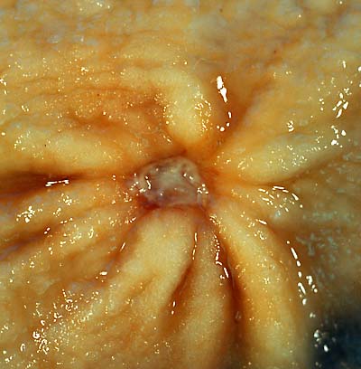 A benign gastric ulcer (from the antrum) of a gastrectomy Source:https://commons.wikimedia.org/wiki/File:Benign_gastric_ulcer_1.jpg#/media/File:Benign_gastric_ulcer_1.jpg
 -
 Duodenal ulcer specimen. Source: https://commons.wikimedia.org/wiki/File:Duodenal_ulcer01.jpg#/media/File:Duodenal_ulcer01.jpg
 -
 Gastric ulcer specimen Source:https://commons.wikimedia.org/wiki/File:Gastric_ulcer_3.jpg#/media/File:Gastric_ulcer_3.jpg


Microscopic Pathology
There are no significant gross or microscopic pathology associated with dyspepsia however, the gross and microscopic pathology of peptic ulcer disease should be kept in mind.
- A peptic ulcer is a mucosal defect produced by acid-pepsin aggression which penetrates the muscularis mucosae and muscularis propria
- There is increased plasma cells, neutrophilic infiltrate, villous blunting
- The surface epithelium usually shows mucous cell (pseudopyloric) metaplasia
- During the active phase, the base of the ulcer shows 4 zones:
- Inflammatory exudate: polymorphonuclear infiltration which along with bacterial products stimulate the production of IL-8 and tumor necrosis factor alpha (TNF-α) and IL-1 released by macrophages in response to bacterial lipopolysaccharide
- Fibrinoid necrosis
- Granulation tissue
- Fibrous tissue. The fibrous base of the ulcer may contain vessels with thickened wall or with thrombosis[10]

References
- ↑ Talley NJ, Ford AC (2015). “Functional Dyspepsia”. N. Engl. J. Med. 373 (19): 1853–63. doi:10.1056/NEJMra1501505. PMID 26535514.
- ↑ Napthali K, Koloski N, Walker MM, Talley NJ (2016). “Women and functional dyspepsia”. Womens Health (Lond). 12 (2): 241–50. doi:10.2217/whe.15.88. PMC 5375052. PMID 26901578.
- ↑ Talley NJ (2016). “Functional dyspepsia: new insights into pathogenesis and therapy”. Korean J. Intern. Med. 31 (3): 444–56. doi:10.3904/kjim.2016.091. PMC 4855108. PMID 27048251.
- ↑ Ganesh M, Nurko S (2014). “Functional dyspepsia in children”. Pediatr Ann. 43 (4): e101–5. doi:10.3928/00904481-20140325-12. PMID 24716560.
- ↑ Fock KM (2011). “Functional dyspepsia, H. pylori and post infectious FD”. J. Gastroenterol. Hepatol. 26 Suppl 3: 39–41. doi:10.1111/j.1440-1746.2011.06649.x. PMID 21443707.
- ↑ Oustamanolakis P, Tack J (2012). “Dyspepsia: organic versus functional”. J. Clin. Gastroenterol. 46 (3): 175–90. doi:10.1097/MCG.0b013e318241b335. PMID 22327302.
- ↑ Kindt S, Dubois D, Van Oudenhove L, Caenepeel P, Arts J, Bisschops R, Tack J (2009). “Relationship between symptom pattern, assessed by the PAGI-SYM questionnaire, and gastric sensorimotor dysfunction in functional dyspepsia”. Neurogastroenterol. Motil. 21 (11): 1183–e105. doi:10.1111/j.1365-2982.2009.01374.x. PMID 19663903.
- ↑ Pasricha PJ, Grover M, Yates KP, Abell TL, Bernard CE, Koch KL; et al. (2021). “Functional Dyspepsia and Gastroparesis in Tertiary Care are Interchangeable Syndromes With Common Clinical and Pathologic Features”. Gastroenterology. 160 (6): 2006–2017. doi:10.1053/j.gastro.2021.01.230. PMID 33548234 Check
|pmid=value (help). - ↑ Miwa H (2012). “Why dyspepsia can occur without organic disease: pathogenesis and management of functional dyspepsia”. J Gastroenterol. doi:10.1007/s00535-012-0625-9. PMID 22766746. Unknown parameter
|month=ignored (help) - ↑ “ATLAS OF PATHOLOGY”. Retrieved 2007-08-26.
Causes
Editor-In-Chief: C. Michael Gibson, M.S., M.D. [1]; Associate Editor(s)-in-Chief: Kiran Singh, M.D. [2] Ajay Gade MD[3]]
Overview
Life threatening causes of dyspepsia include coronary disease and ischemic bowel disease. Other common causes of dyspepsia include gastroesophageal reflux, gastritis, lactose intolerance, and peptic ulcer.
Causes
Life Threatening Causes
Common Causes
- Aerophagia
- Anxiety
- Gastroesophageal reflux
- Gastritis
- Irritable bowel syndrome
- Lactose intolerance
- Peptic ulcer
Causes by Organ system
Causes in Alphabetical Order
- 4-Chlorodehydromethyltestosterone
- Abdominal cancer
- Abdominal guarding
- Acarbose
- Acetylsalicylic acid
- Achalasia
- Achillea millefolium
- Achlorhydria
- Acid reflux
- Acute pancreatitis
- Acute viral hepatitis
- Adefovir
- Adrenal fatigue
- Adult hypertrophic pyloric stenosis
- Aerophagia
- After gastrointestinal surgery
- Alcohol
- Alendronate
- Alexithymia
- Alpha-glucosidase inhibitor
- Aluminium hydroxide
- Amanita phalloides
- Amebic dysentery
- Amitriptyline
- Amlodipine
- Ampicillin
- Amylin analogs
- Angiotensin converting enzyme inhibitors
- Angiotensin II receptor antagonist
- Anxiety
- Artemether and lumefantrin
- Ascites
- Aspirin
- Atazanavir
- Autonomic neuropathy
- Belching
- Bezafibrate
- Biguanide
- Biliary disease
- Biliary pain
- Bisphosphonates
- Bitter melon
- Brat diet
- Buprenorphine
- Burping
- Caffeine
- Calcium channel blockers
- Cancer
- Capsicum annuum
- Carbohydrate malabsorption
- Carcinoid syndrome
- Cefadroxil
- Cefixime
- Celiac artery compression syndrome
- Celiac disease
- Charles Darwin’s illness
- Chlorosis
- Chocolate
- Cholecystitis
- Cholelithiasis
- Cholestyramine
- Cholestyramine resin
- Chronic abdominal wall pain
- Chronic gastritis
- Chronic hepatitis
- Chronic intestinal ischemia
- Chronic pancreatitis
- Chronic renal disease
- Cidofovir
- Cirrhosis
- Codeine
- Coffee
- Colchicine
- Collinsonia canadensis
- Colofac
- Colon cancer
- Congestive cardiac failure
- Connective tissue disease
- Constipation
- Coptis
- Coronary disease
- Coronary ischemia
- Corticosteroids
- Couvade
- Crohn disease
- Cyclooxygenase
- Daptomycin
- Delayed gastric emptying
- Depression
- Diabetes mellitus
- Diabulimia
- Diarrhea
- Diflunisal
- Digitalis
- Digoxin
- Donepezil
- Dopamine agonist
- Duloxetine
- Duodenal cancer
- Duodenal lymphoma
- Duodenal polyps
- Duodenal ulcer
- Duodenal webs
- Duodenitis
- Eating habits
- Eating high-fiber foods
- Eflornithine
- Elvitegravir
- Emtricitabine
- Epoetin alfa injection
- Eribulin
- Erythromycin
- Esophageal cancer
- Esophageal dysmotility
- Esophageal spasm
- Esophagitis
- Esophageal disease
- Estrogens
- Ethosuximide
- Excess caffeine consumption
- Extranodal marginal zone B-cell lymphoma
- Fascioliasis
- Fasting girls
- Fentanyl skin patches
- Fibrate
- Fluoxetine
- Fluticasone
- Fluvastatin
- Food allergy
- Food intolerance
- Frovatriptan
- Functional bowel disorder
- Gallstones
- Garlic
- Gas
- Gastric cancer
- Gastric lymphoma
- Gastric motility disorder
- Gastric ulcer
- Gastritis
- Gastroenteritis
- Gastroesophageal reflux disease
- Gastrointestinal mucormycosis
- Gastrointestinal neoplasm
- Gastrointestinal zygomycosis
- Gastroparesis
- Gemfibrozil
- Giardiasis
- Ginger
- Glucocorticoids
- Gluten allergy
- Gulf war syndrome
- H2 antagonist
- Hangover
- Helicobacter pylori infection
- Hepatitis c
- Hepatitis
- Hepatocellular carcinoma
- Hepatoma
- Heroin
- Herpes simplex
- Hiatal hernia
- High-fat diet
- Hookworm
- Hydrogen potassium ATPase
- Hypercalcaemia
- Hyperkalemia
- Hyperparathyroidism
- Hyperthyroidism
- Hypertrophic gastritis
- Iberogast
- Ibuprofen
- Imatinib
- Indian gooseberry
- Indomethacin
- Inflammatory bowel disease
- Infliximab
- Interferon Beta-1b Injection
- Interferon gamma
- Intestinal motility disorder
- Intestinal obstruction
- Iron
- Irritable bowel syndrome
- Ischemic bowel disease
- Ketorolac
- Lactose intolerance
- Levodopa
- Lithium
- Macrolides
- Malabsorption
- Medications
- Meloxicam
- Ménétriér’s disease
- Meropenem injection
- Metformin
- Methylprednisolone
- Methylxanthines
- Metronidazole
- Mint
- Misoprostol
- Monoamine oxidase-b inhibitors
- Mosapride
- Multiple chemical sensitivity
- Multiple endocrine neoplasia type 1
- Myocardial ischaemia
- Nabilone
- Nalbuphine
- Narcotics
- Nausea
- Nervous
- Niacin
- Nicotine polacrilex
- Non-steroidal anti-inflammatory drug
- Non-ulcer dyspepsia
- Obesity
- Omeprazole
- Opioids
- Opisthorchiasis
- Opisthorchis infection
- Oprelvekin
- Oral allergy syndrome
- Oral contraceptives
- Oranges
- Orlistat
- Ovarian cancer
- Overeating
- Oxycodone and aspirin
- Pancreatic cancer
- Pancreatitis
- Parathyroid disorders
- PDE5 inhibitor
- Peptic ulcer
- Pergolide
- Pirfenidone
- Potassium
- Pramlintide injection
- Prednisone
- Pregnancy
- Primary hyperparathyroidism
- Prokinetic
- Proton pump inhibitor
- Quetiapine
- Quinidine
- Radish
- Reflux
- Rivastigmine
- Rome process
- S-adenosyl methionine
- Sarcoidosis
- Scleroderma
- Sertraline
- Sildenafil
- Simvastatin
- Small intestinal bacterial overgrowth
- Smoking
- Sodium dichloroisocyanurate
- Sorafenib
- Spicy foods
- SSRI discontinuation syndrome
- Steroids
- Stomach cancer
- Stomach disease
- Stress
- Strongyloidiasis
- Strychnine tree
- Suillus luteus
- Sunitinib
- Superior mesenteric artery syndrome
- Swallowed air
- Syphilis
- Tadalafil
- Tenofovir disoproxil fumarate
- Tetracycline
- Theophylline
- Thyroglossal cyst
- Thyroid medicines
- Thyroid disease
- Tiotropium Oral Inhalation
- Tobacco
- Tomatoes
- Topiramate
- Trichinosis
- Trichophagia
- Tropical sprue
- Tuberculosis
- Ulcerative colitis
- Uremia
- Valproic acid
- Venlafaxine
- Vigabatrin
- Vilazodone
- Vinca
- Visceroptosis
- Vitamin C
- Zolmitriptan
- Zopiclone
References
Differentiating Dyspepsia from other Diseases
Editor-In-Chief: C. Michael Gibson, M.S., M.D. [1] Associate Editor(s)-in-Chief: Ajay Gade MD[2]]
Overview
Dyspepsia must be differentiated from other diseases that presents with epigastric pain such as gastritis, gastroesophageal reflux disease, acute pancreatitis, primary biliary cirrhosis, cholelithiasis, gastric outlet syndrome, myocardial infarction, pleural empyemae appendicitis
Differentiating Dyspepsia from other Diseases
Dyspepsia must be differentiated from other diseases that presents with epigastric pain such as gastritis, gastroesophageal reflux disease,acute pancreatitis,prmary biliary cirrhosis,cholelithiasis,gastric outlet syndrome,myocardial infaraction ,pleural empyema,acute appendicitis [1][2][3][4][5][6][7][8][9][10][11][12][13][14][15][16][17][18]
| Classification of pain in the abdomen based on etiology | Disease | Clinical manifestations | Diagnosis | Comments | |||||||||||
|---|---|---|---|---|---|---|---|---|---|---|---|---|---|---|---|
| Symptoms | Signs | ||||||||||||||
| Fever | Rigors and chills | Abdominal Pain | Jaundice | GI Bleed | Hypo-
tension |
Guarding | Rebound Tenderness | Bowel sounds | Lab Findings | Imaging | |||||
| Abdominal causes | Inflammatory causes | Pancreato-biliary disorders | |||||||||||||
| Acute pancreatitis | + | − | Epigastric | ± | − | ± | − | − | N | Increased amylase / lipase | Ultrasound shows evidence of inflammation | Pain radiation to back | |||
| Primary biliary cirrhosis | − | − | RUQ/Epigastric | + | − | − | − | − | N | Increased AMA level, abnormal LFTs | |||||
| Cholelithiasis | ± | − | RUQ/Epigastric | ± | − | − | + | + | N to hyperactive for dislodged stone | Leukocytosis | Ultrasound shows gallstone | Murphy’s sign | |||
| Gastric causes | Peptic ulcer disease | ± | − | EpisodicEpigastric | − |
|
+ in perforated | + | + | N | Air under diaphragm in upright CXR | Upper GI endoscopy for diagnosis | |||
| Gastritis | ± | − | Epigastric | + in chronic gastritis | − | ||||||||||
| Gastroesophageal reflux disease | − | − | Epigastric | − | − | − | − | − | |||||||
| Gastric outlet obstruction | − | − | Epigastric | − | − | ± | Hyperactive | ||||||||
| Intestinal causes | Acute appendicitis | + | +in pyogenic appendicitis | Starts in epigastrium, migrates to RLQ | − | − | + in perforated appendicitis | + | + | Hypoactive | Leukocytosis | Ultrasound shows evidence of inflammation | Nausea & vomiting, decreased appetite | ||
| Extra-abdominal causes | Pulmonary disorders | Pleural empyema | + | ± | RUQ/Epigastric | − | − | − | − | − | N | ||||
| Cardiovascular disorders | Myocardial Infarction | − | − | Epigastric | − | − | + in cardiogenic shock | − | − | N | |||||
| Abbreviations: RUQ= Right upper quadrant of the abdomen, LUQ= Left upper quadrant, LLQ= Left lower quadrant, RLQ= Right lower quadrant, LFT= Liver function test, SIRS= Systemic inflammatory response syndrome, ERCP= Endoscopic retrograde cholangiopancreatography, IV= Intravenous, N= Normal, AMA= Anti mitochondrial antibodies, LDH= Lactate dehydrogenase, GI= Gastrointestinal, CXR= Chest X ray, IgA= Immunoglobulin A, IgG= Immunoglobulin G, IgM=Immunoglobulin M, CT= Computed tomography, PMN= Polymorphonuclear cells, ESR= Erythrocyte sedimentation rate, CRP= C-reactive protein | |||||||||||||||
| Disease | Cause | Symptoms | Diagnosis | Other findings | ||||||||
|---|---|---|---|---|---|---|---|---|---|---|---|---|
| Pain | Nausea
& Vomiting |
Heartburn | Belching or
Bloating |
Weight loss | Loss of
Appetite |
Stools | Endoscopy findings | |||||
| Location | Aggravating Factors | Alleviating Factors | ||||||||||
| Acute gastritis |
|
Food | Antacids | ✔ | ✔ | ✔ | – | ✔ | Black stools | – | ||
| Chronic gastritis |
|
Food | Antacids | ✔ | ✔ | ✔ | ✔ | ✔ | – | H. pylori gastritis
Lymphocytic gastritis
|
– | |
| Atrophic gastritis | Epigastric pain | – | – | ✔ | – | ✔ | ✔ | – | H. pylori
|
Autoimmune gastritis diagnosis include:
| ||
| Crohn’s disease | – | – | – | – | – | ✔ | ✔ |
|
|
|||
| GERD |
|
|
|
✔
(Suspect delayed gastric emptying) |
✔ | – | – | – | – |
|
Other symptoms:
Complications
| |
| Peptic ulcer disease |
|
|
|
|
✔ | ✔ | – | – | – | Gastric ulcers
Duodenal ulcers
|
Other diagnostic tests | |
| Gastrinoma |
|
– | – | ✔
(suspect gastric outlet obstruction) |
✔ | – | – | – | Useful in collecting the tissue for biopsy |
Diagnostic tests
| ||
| Gastric Adenocarcinoma |
|
– | – | ✔ | ✔ | ✔ | ✔ | ✔ |
|
Esophagogastroduodenoscopy
|
Other symptoms | |
| Primary gastric lymphoma |
|
– | – | – | – | – | ✔ | – | – | Useful in collecting the tissue for biopsy | Other symptoms
| |
Differentials of funtional dyspepsia
Functional dyspepsia should be differentiated from other diseases that cause chronic nausea and vomiting. The differentials include the following:[19][20][21][22][23][24][25][26][27][28][29][30][31][32][33][34][35][36][37][38][39][40][41][42][43][44][45][46][47][48][49][50]
| Disorder | Clinical features | Laboratory findings | |||||||||||||||
|---|---|---|---|---|---|---|---|---|---|---|---|---|---|---|---|---|---|
| Chronic nausea | Vomiting | Diarrhea | Retching | Lethargy | Social withdrawal | Photophobia | Epigastric pain/burning | Lanugo hair | Hypogonadism | Russel’s sign | Body mass index (normal range: 18.5 to 24.9) | Complete blood count (CBC) | Electrolyte imabalance | Lipase and amylase levels | Gastric scintigraphy | Ambulatory esophageal pH and impedance testing | |
| Gastroparesis | ✔ | ✔ (within 1 hour of eating) | – | ✔ | ✔ | – | – | ✔ | – | – | – | ↓ | ✔ |
|
|
| |
| Anorexia nervosa | ✔ | ✔ | ✔ | – | ✔ | ✔ | – | – | ✔ | ✔ | – | ↓ | ✔ |
|
|
| |
| Bulimia nervosa | ✔ | ✔ | ✔ | ✔ | ✔ | ✔ | – | – | – | ✔ | ✔ | Normal | ✔ |
|
|
| |
| Rumination syndrome | ✔ | ✔ (Regurgitation more common- within minutes of meal intake) | ✔ | – | ✔ | ✔ | ✔ | ✔ | – | – | – | ↓ |
|
✔ |
|
| |
| Functional dyspepsia | ✔ | ✔ | ✔ | ✔ | – | – | – | – | – | – | – | Normal |
|
✔ |
|
| |
| Cyclic vomiting syndrome | ✔ | ✔ | – | ✔ | ✔ | – | – | – | – | – | – | ↓ | ✔ |
|
|
| |
| Pancreatitis | ✔ | ✔ | ✔ | ✔ | ✔ | – | – | ✔ | – | – | – | Normal | ✔ |
|
|
| |
| Gastric outlet obstruction | ✔ | ✔ (within 1 hour of eating) | – | – | – | – | – | ✔ | – | – | – | ↓ | ✔ |
|
| ||
References
- ↑ Gralnek IM, Barkun AN, Bardou M (2008). “Management of acute bleeding from a peptic ulcer”. N Engl J Med. 359 (9): 928–37. doi:10.1056/NEJMra0706113. PMID 18753649.
- ↑ Dallal HJ, Palmer KR (2001). “ABC of the upper gastrointestinal tract: Upper gastrointestinal haemorrhage”. BMJ. 323 (7321): 1115–7. PMC 1121602. PMID 11701581.
- ↑ Nelson DR, Teckman J, Di Bisceglie AM, Brenner DA (2012). “Diagnosis and management of patients with α1-antitrypsin (A1AT) deficiency”. Clin Gastroenterol Hepatol. 10 (6): 575–80. doi:10.1016/j.cgh.2011.12.028. PMC 3360829. PMID 22200689.
- ↑ Tsochatzis EA, Bosch J, Burroughs AK (2014). “Liver cirrhosis”. Lancet. 383 (9930): 1749–61. doi:10.1016/S0140-6736(14)60121-5. PMID 24480518.
- ↑ Schuppan D, Afdhal NH (2008). “Liver cirrhosis”. Lancet. 371 (9615): 838–51. doi:10.1016/S0140-6736(08)60383-9. PMC 2271178. PMID 18328931.
- ↑ Kahrilas PJ (2008). “Clinical practice. Gastroesophageal reflux disease”. N Engl J Med. 359 (16): 1700–7. doi:10.1056/NEJMcp0804684. PMC 3058591. PMID 18923172.
- ↑ Kahrilas PJ, Shaheen NJ, Vaezi MF, Hiltz SW, Black E, Modlin IM; et al. (2008). “American Gastroenterological Association Medical Position Statement on the management of gastroesophageal reflux disease”. Gastroenterology. 135 (4): 1383–1391, 1391.e1–5. doi:10.1053/j.gastro.2008.08.045. PMID 18789939.
- ↑ Bredenoord AJ, Pandolfino JE, Smout AJ (2013). “Gastro-oesophageal reflux disease”. Lancet. 381 (9881): 1933–42. doi:10.1016/S0140-6736(12)62171-0. PMID 23477993.
- ↑ Fox M, Forgacs I (2006). “Gastro-oesophageal reflux disease”. BMJ. 332 (7533): 88–93. doi:10.1136/bmj.332.7533.88. PMC 1326932. PMID 16410582.
- ↑ Sugimachi K, Inokuchi K, Kuwano H, Ooiwa T (1984). “Acute gastritis clinically classified in accordance with data from both upper GI series and endoscopy”. Scand J Gastroenterol. 19 (1): 31–7. PMID 6710074.
- ↑ Sipponen P, Maaroos HI (2015). “Chronic gastritis”. Scand J Gastroenterol. 50 (6): 657–67. doi:10.3109/00365521.2015.1019918. PMC 4673514. PMID 25901896.
- ↑ Sartor RB (2006). “Mechanisms of disease: pathogenesis of Crohn’s disease and ulcerative colitis”. Nat Clin Pract Gastroenterol Hepatol. 3 (7): 390–407. doi:10.1038/ncpgasthep0528. PMID 16819502.
- ↑ Sipponen P (1989). “Atrophic gastritis as a premalignant condition”. Ann Med. 21 (4): 287–90. PMID 2789799.
- ↑ Badillo R, Francis D (2014). “Diagnosis and treatment of gastroesophageal reflux disease”. World J Gastrointest Pharmacol Ther. 5 (3): 105–12. doi:10.4292/wjgpt.v5.i3.105. PMC 4133436. PMID 25133039.
- ↑ Ramakrishnan K, Salinas RC (2007). “Peptic ulcer disease”. Am Fam Physician. 76 (7): 1005–12. PMID 17956071.
- ↑ Banasch M, Schmitz F (2007). “Diagnosis and treatment of gastrinoma in the era of proton pump inhibitors”. Wien Klin Wochenschr. 119 (19–20): 573–8. doi:10.1007/s00508-007-0884-2. PMID 17985090.
- ↑ Dicken BJ, Bigam DL, Cass C, Mackey JR, Joy AA, Hamilton SM (2005). “Gastric adenocarcinoma: review and considerations for future directions”. Ann Surg. 241 (1): 27–39. PMC 1356843. PMID 15621988.
- ↑ Ghimire P, Wu GY, Zhu L (2011). “Primary gastrointestinal lymphoma”. World J Gastroenterol. 17 (6): 697–707. doi:10.3748/wjg.v17.i6.697. PMC 3042647. PMID 21390139.
- ↑ Parkman HP (2015). “Idiopathic gastroparesis”. Gastroenterol. Clin. North Am. 44 (1): 59–68. doi:10.1016/j.gtc.2014.11.015. PMC 4324534. PMID 25667023.
- ↑ Werlin SL, Fish DL (2006). “The spectrum of valproic acid-associated pancreatitis”. Pediatrics. 118 (4): 1660–3. doi:10.1542/peds.2006-1182. PMID 17015559.
- ↑ Noddin L, Callahan M, Lacy BE (2005). “Irritable bowel syndrome and functional dyspepsia: different diseases or a single disorder with different manifestations?”. MedGenMed. 7 (3): 17. PMC 1681633. PMID 16369243.
- ↑ Gupta R, Kalla M, Gupta JB (2012). “Adult rumination syndrome: Differentiation from psychogenic intractable vomiting”. Indian J Psychiatry. 54 (3): 283–5. doi:10.4103/0019-5545.102434. PMC 3512372. PMID 23226859.
- ↑ “Body weight in bulimia nervosa | SpringerLink”.
- ↑ Sağlam F, Sivrikoz E, Alemdar A, Kamalı S, Arslan U, Güven H (2015). “Bouveret syndrome: A fatal diagnostic dilemma of gastric outlet obstruction”. Ulus Travma Acil Cerrahi Derg. 21 (2): 157–9. PMID 25904280.
- ↑ Talley NJ (2011). “Rumination syndrome”. Gastroenterol Hepatol (N Y). 7 (2): 117–8. PMC 3061016. PMID 21475419.
- ↑ Tutuian R, Castell DO (2004). “Rumination documented by using combined multichannel intraluminal impedance and manometry”. Clin. Gastroenterol. Hepatol. 2 (4): 340–3. PMID 15067630.
- ↑ Kessing BF, Smout AJ, Bredenoord AJ (2014). “Current diagnosis and management of the rumination syndrome”. J. Clin. Gastroenterol. 48 (6): 478–83. doi:10.1097/MCG.0000000000000142. PMID 24921208.
- ↑ Parkman HP (2009). “Assessment of gastric emptying and small-bowel motility: scintigraphy, breath tests, manometry, and SmartPill”. Gastrointest. Endosc. Clin. N. Am. 19 (1): 49–55, vi. doi:10.1016/j.giec.2008.12.003. PMID 19232280.
- ↑ Waseem S, Moshiree B, Draganov PV (2009). “Gastroparesis: current diagnostic challenges and management considerations”. World J. Gastroenterol. 15 (1): 25–37. PMC 2653292. PMID 19115465.
- ↑ Mearin F, Camilleri M, Malagelada JR (1986). “Pyloric dysfunction in diabetics with recurrent nausea and vomiting”. Gastroenterology. 90 (6): 1919–25. PMID 3699409.
- ↑ Abell TL, Camilleri M, Donohoe K, Hasler WL, Lin HC, Maurer AH, McCallum RW, Nowak T, Nusynowitz ML, Parkman HP, Shreve P, Szarka LA, Snape WJ, Ziessman HA (2008). “Consensus recommendations for gastric emptying scintigraphy: a joint report of the American Neurogastroenterology and Motility Society and the Society of Nuclear Medicine”. Am. J. Gastroenterol. 103 (3): 753–63. doi:10.1111/j.1572-0241.2007.01636.x. PMID 18028513.
- ↑ Jiang CF, Ng KW, Tan SW, Wu CS, Chen HC, Liang CT, Chen YH (2002). “Serum level of amylase and lipase in various stages of chronic renal insufficiency”. Zhonghua Yi Xue Za Zhi (Taipei). 65 (2): 49–54. PMID 12014357.
- ↑ Szmukler, G. I.; Young, G. P.; Lichtenstein, M.; Andrews, J. T. (1990). “A serial study of gastric emptying in anorexia nervosa and bulimia”. Australian and New Zealand Journal of Medicine. 20 (3): 220–225. doi:10.1111/j.1445-5994.1990.tb01023.x. ISSN 0004-8291.
- ↑ Diamanti A, Bracci F, Gambarara M, Ciofetta GC, Sabbi T, Ponticelli A, Montecchi F, Marinucci S, Bianco G, Castro M (2003). “Gastric electric activity assessed by electrogastrography and gastric emptying scintigraphy in adolescents with eating disorders”. J. Pediatr. Gastroenterol. Nutr. 37 (1): 35–41. PMID 12827003.
- ↑ Ferholt J, Provence S (1976). “Diagnosis and treatment of an infant with psychophysiological vomiting”. Psychoanal Study Child. 31: 439–59. PMID 981449.
- ↑ Lee H, Rhee PL, Park EH, Kim JH, Son HJ, Kim JJ, Rhee JC (2007). “Clinical outcome of rumination syndrome in adults without psychiatric illness: a prospective study”. J. Gastroenterol. Hepatol. 22 (11): 1741–7. doi:10.1111/j.1440-1746.2006.04617.x. PMID 17914944.
- ↑ Koskenpato J, Kairemo K, Korppi-Tommola T, Färkkilä M (1998). “Role of gastric emptying in functional dyspepsia: a scintigraphic study of 94 subjects”. Dig. Dis. Sci. 43 (6): 1154–8. PMID 9635600.
- ↑ Urbain JL, Vekemans MC, Parkman H, Van Cauteren J, Mayeur SM, Van den Maegdenbergh V, Charkes ND, Fisher RS, Malmud LS, De Roo M (1995). “Dynamic antral scintigraphy to characterize gastric antral motility in functional dyspepsia”. J. Nucl. Med. 36 (9): 1579–86. PMID 7658213.
- ↑ Hejazi RA, Lavenbarg TH, McCallum RW (2010). “Spectrum of gastric emptying patterns in adult patients with cyclic vomiting syndrome”. Neurogastroenterol. Motil. 22 (12): 1298–302, e338. doi:10.1111/j.1365-2982.2010.01584.x. PMID 20723071.
- ↑ “Gastric outlet obstruction – an overview | ScienceDirect Topics”.
- ↑ Minami H, McCallum RW (1984). “The physiology and pathophysiology of gastric emptying in humans”. Gastroenterology. 86 (6): 1592–610. PMID 6370777.
- ↑ Humphries LL, Adams LJ, Eckfeldt JH, Levitt MD, McClain CJ (1987). “Hyperamylasemia in patients with eating disorders”. Ann. Intern. Med. 106 (1): 50–2. PMID 2431640.
- ↑ Hempen I, Lehnert P, Fichter M, Teufel J (1989). “[Hyperamylasemia in anorexia nervosa and bulimia nervosa. Indication of a pancreatic disease?]”. Dtsch. Med. Wochenschr. (in German). 114 (49): 1913–6. doi:10.1055/s-2008-1066848. PMID 2480214.
- ↑ Okada R, Okada A, Okada T, Okada T, Hamajima N (2009). “Elevated serum lipase levels in patients with dyspepsia of unknown cause in general practice”. Med Princ Pract. 18 (2): 130–6. doi:10.1159/000189811. PMID 19204432.
- ↑ Sansone RA, Sansone LA (2012). “Hoarseness: a sign of self-induced vomiting?”. Innov Clin Neurosci. 9 (10): 37–41. PMC 3508961. PMID 23198276.
- ↑ Tack J, Caenepeel P, Arts J, Lee KJ, Sifrim D, Janssens J (2005). “Prevalence of acid reflux in functional dyspepsia and its association with symptom profile”. Gut. 54 (10): 1370–6. doi:10.1136/gut.2004.053355. PMC 1774686. PMID 15972301.
- ↑ “gut.bmj.com” (PDF).
- ↑ Boles RG, Williams JC (1999). “Mitochondrial disease and cyclic vomiting syndrome”. Dig. Dis. Sci. 44 (8 Suppl): 103S–107S. PMID 10490048.
- ↑ Ranasinghe WK, Smith M (2013). “Gastric outlet obstruction with an elevated serum pancreatic lipase secondary to an infraumbilical hernia”. Ann R Coll Surg Engl. 95 (7): 122–4. doi:10.1308/003588413X13629960047795. PMID 24112485.
- ↑ Ui, Takashi; Shibusawa, Hiroyuki; Tsukui, Hidenori; Sakuma, Kazuya; Takahashi, Shuhei; Lefor, Alan K.; Hosoya, Yoshinori; Sata, Naohiro; Yasuda, Yoshikazu (2015). “Pretreatment of gastric outlet obstruction with pancrelipase: Report of a case”. International Journal of Surgery Case Reports. 12: 87–89. doi:10.1016/j.ijscr.2015.05.023. ISSN 2210-2612.
Epidemiology and Demographics
Editor-In-Chief: C. Michael Gibson, M.S., M.D. [1] Associate Editor(s)-in-Chief: Ajay Gade MD[2]]
Overview
The incidence of new cases of H. pylori infection each year ranges from 3,000 to 10,000 per 100,000 individuals in developing countries. It has been observed that with advancing age, the incidence of H. pylori infection increases. In united states, 20% of adolescents are infected with H. pylori when compared to 90% by 5 years of age in the developing countries. In United States, H. pylori infection associated gastritis is more common in African Americans (54%), Hispanics (52%), and the elderly compared to Whites (21%). In acute gastritis, females are usually more affected than men. In H. pylori infection associated gastritis, males are more commonly affected than females. The incidence rates of H. pylori infection are high in Japan, Columbia, Costa Rica and China, and comparatively low in the United States. H. pylori infection is common in Southern and Eastern Europe, Mexico, South America, Africa, most Asian countries, and aboriginal people in North America.
Epidemiology and Demographics
Incidence
- The incidence of new cases of H. pylori infection each year ranges from 3,000 to 10,000 per 100,000 individuals in developing countries.[1]
- The incidence of new cases of H. pylori infection each year is around 500 per 100,000 individuals in developed countries.[1]
- It has been observed that with advancing age, the incidence of H. pylori infection increases. [2]
Prevalence
- The prevalence of eosinophilic gastritis is approximately 6.3 per 100,000 individuals worldwide. [3]
Age
- All age groups can develop dyspepsia.
- The prevalence of H. pyloriinfection increases with age.[4]
- About 30%-50% of H. pylori infections are acquired during childhood which increases to 90% during adulthood in developing countries.[5]
- H. pylori infection in developed countries is less common in children and reaches up to 60% with increasing age.[6]
- In United States, 20% of adolescents are infected with H. pylori when compared to 90% by 5 years of age in developing countries.[7]
- Children differ from adults with respect to H. pylori infection in terms of:[8][9]
- Prevalence of infection
- High rate of antibiotic resistance
- The near-absence of gastric malignancies
- Age specific problems with diagnostic tests and medications
Race
- In United States, H. pylori infection is more common in African Americans (54%), Hispanics (52%), and the elderly compared to Whites (21%).[10][11]
Gender
Region
- H. pylori infection is common in Southern and Eastern Europe, Mexico, South America, Africa, most Asian countries, and aboriginal people in North America.[13][14]
Developed Countries
- The incidence of new cases of H. pylori infection each year is 0.5 percent in developed countries.[1]
- The prevalence of H. pylori is declining in the United States.
- In developed countries such as the United States, children acquire the H. pylori infection at a rate of about less than 1% per year.
- It is estimated that 30%-40% of the US population is infected with H. pylori.[15][16]
- In United States, approximately 25% of children between 6-19 years old are infected.[17]
- The incidence rates of H. pylori infection are high in Japan, Columbia, Costa Rica and China, and comparatively low in the United States.
Developing Countries
- The prevalence of H. pylori is higher in developing countries than that in developed countries.[18]
- The incidence of new cases of H.pylori infection each year ranges from 3,000 to 10,000 per 100,000 individuals in developing countries.[1]
- In the developing countries, children in the age group of 2 to 8 years acquire the H.pylori infection at a rate of about 10% per year.
- H. pylori infection is common in Southern and Eastern Europe, Mexico, South America, Africa, most Asian countries, and aboriginal people in North America.[13][14]
Prevalence of Helicobacter pylori Infection Globally
Prevalence of H. pylori infection globally[19]
| Prevalence of Helicobacter pylori Infection Globally | |||
|---|---|---|---|
| Country | Prevalence per 100,000 | ||
| Children | Adult | ||
| Africa | Ethiopia | 48,000 | >95,000 |
| Nigeria | 82,000 | 91,000 | |
| Central America | Gautemala | 51,000 | 65,000 |
| Mexico | 43,000 | 90,000 | |
| North America | Canada | 7100 | 23,000 |
| USA and Canada | 30,000 | ||
| South America | Bolivia | 54,000 | |
| Brazil | 30,000 | 82,000 | |
| Chile | 36,000 | >70,000 | |
| Asia | Bangladesh | 60,000 | >90,000 |
| Hong Kong | 13,000 | ||
| India | 22,000 | >80,000 | |
| Japan | >70,000 | ||
| Siberia | 30,000 | 85,000 | |
| South Korea | 56,000 | 40,600 | |
| Sri Lanka | 67,000 | 72,000 | |
| Taiwan | 11,000 | >50,000 | |
| Australia | Australia | 20,000 | |
| Europe | Eastern | 70,000 | |
| Albania | 70700 | ||
| Bulgaria | 61,700 | ||
| Czech Republic | 42,000 | ||
| Estonia | 60,000 | ||
| Germany | 48,800 | ||
| Iceland | 36,000 | ||
| Netherlands | 12000 | ||
| Serbia | 36,400 | ||
| Sweden | 11,000 | ||
| Switzerland | 11,900 | ||
| Middle East | Egypt | 50,000 | 90,000 |
| Libya | 50,000 | 94,000 | |
| Saudi Arabia | 40,000 | 80,000 | |
| Turkey | 64,000 | 80,000 | |
References
- ↑ 1.0 1.1 1.2 1.3 Rosenberg JJ (2010). “Helicobacter pylori”. Pediatr Rev. 31 (2): 85–6, discussion 86. doi:10.1542/pir.31-2-85. PMID 20124281.
- ↑ Dooley CP, Cohen H, Fitzgibbons PL, Bauer M, Appleman MD, Perez-Perez GI; et al. (1989). “Prevalence of Helicobacter pylori infection and histologic gastritis in asymptomatic persons”. N Engl J Med. 321 (23): 1562–6. doi:10.1056/NEJM198912073212302. PMID 2586553.
- ↑ Jensen ET, Martin CF, Kappelman MD, Dellon ES (2016). “Prevalence of Eosinophilic Gastritis, Gastroenteritis, and Colitis: Estimates From a National Administrative Database”. J Pediatr Gastroenterol Nutr. 62 (1): 36–42. doi:10.1097/MPG.0000000000000865. PMC 4654708. PMID 25988554.
- ↑ Mégraud F, Brassens-Rabbé MP, Denis F, Belbouri A, Hoa DQ (1989). “Seroepidemiology of Campylobacter pylori infection in various populations”. J Clin Microbiol. 27 (8): 1870–3. PMC 267687. PMID 2549098.
- ↑ Cheng H, Hu F, Zhang L, Yang G, Ma J, Hu J; et al. (2009). “Prevalence of Helicobacter pylori infection and identification of risk factors in rural and urban Beijing, China”. Helicobacter. 14 (2): 128–33. doi:10.1111/j.1523-5378.2009.00668.x. PMID 19298340.
- ↑ Go MF (2002). “Review article: natural history and epidemiology of Helicobacter pylori infection”. Aliment Pharmacol Ther. 16 Suppl 1: 3–15. PMID 11849122.
- ↑ Frenck RW, Clemens J (2003). “Helicobacter in the developing world”. Microbes Infect. 5 (8): 705–13. PMID 12814771.
- ↑ Elitsur Y, Dementieva Y, Rewalt M, Lawrence Z (2009). “Helicobacter pylori infection rate decreases in symptomatic children: a retrospective analysis of 13 years (1993-2005) from a gastroenterology clinic in West Virginia”. J Clin Gastroenterol. 43 (2): 147–51. doi:10.1097/MCG.0b013e318157e4e7. PMID 18779740.
- ↑ Koletzko S, Jones NL, Goodman KJ, Gold B, Rowland M, Cadranel S; et al. (2011). “Evidence-based guidelines from ESPGHAN and NASPGHAN for Helicobacter pylori infection in children”. J Pediatr Gastroenterol Nutr. 53 (2): 230–43. doi:10.1097/MPG.0b013e3182227e90. PMID 21558964.
- ↑ Everhart JE, Kruszon-Moran D, Perez-Perez GI, Tralka TS, McQuillan G (2000). “Seroprevalence and ethnic differences in Helicobacter pylori infection among adults in the United States”. J Infect Dis. 181 (4): 1359–63. doi:10.1086/315384. PMID 10762567.
- ↑ Everhart, James E.; Kruszon‐Moran, Deanna; Perez‐Perez, Guillermo I.; Tralka, Tommie Sue; McQuillan, Geraldine (2000). “Seroprevalence and Ethnic Differences inHelicobacter pyloriInfection among Adults in the United States”. The Journal of Infectious Diseases. 181 (4): 1359–1363. doi:10.1086/315384. ISSN 0022-1899.
- ↑ de Martel C, Parsonnet J (2006). “Helicobacter pylori infection and gender: a meta-analysis of population-based prevalence surveys”. Dig. Dis. Sci. 51 (12): 2292–301. doi:10.1007/s10620-006-9210-5. PMID 17089189.
- ↑ 13.0 13.1 Kawakami E, Machado RS, Ogata SK, Langner M (2008). “Decrease in prevalence of Helicobacter pylori infection during a 10-year period in Brazilian children”. Arq Gastroenterol. 45 (2): 147–51. PMID 18622470.
- ↑ 14.0 14.1 Goh KL, Chan WK, Shiota S, Yamaoka Y (2011). “Epidemiology of Helicobacter pylori infection and public health implications”. Helicobacter. 16 Suppl 1: 1–9. doi:10.1111/j.1523-5378.2011.00874.x. PMC 3719046. PMID 21896079.
- ↑ Everhart JE (2000). “Recent developments in the epidemiology of Helicobacter pylori”. Gastroenterol Clin North Am. 29 (3): 559–78. PMID 11030073.
- ↑ Peterson WL, Fendrick AM, Cave DR, Peura DA, Garabedian-Ruffalo SM, Laine L (2000). “Helicobacter pylori-related disease: guidelines for testing and treatment”. Arch Intern Med. 160 (9): 1285–91. PMID 10809031.
- ↑ Staat MA, Kruszon-Moran D, McQuillan GM, Kaslow RA (1996). “A population-based serologic survey of Helicobacter pylori infection in children and adolescents in the United States”. J. Infect. Dis. 174 (5): 1120–3. PMID 8896521.
- ↑ Salih BA (2009). “Helicobacter pylori infection in developing countries: the burden for how long?”. Saudi J Gastroenterol. 15 (3): 201–7. doi:10.4103/1319-3767.54743. PMC 2841423. PMID 19636185.
- ↑ Hunt RH, Xiao SD, Megraud F, Leon-Barua R, Bazzoli F, van der Merwe S; et al. (2011). “Helicobacter pylori in developing countries. World Gastroenterology Organisation Global Guideline”. J Gastrointestin Liver Dis. 20 (3): 299–304. PMID 21961099.
Risk Factors
Editor-In-Chief: C. Michael Gibson, M.S., M.D. [1]
Overview
Common risk factors for the development of dyspepsia include, Helicobacter pylori infection, chronic use of NSAIDs, family history of peptic ulcer disease, emotional stress, increased intake of high-fiber diet, overconsumption of caffeine, high-fat and greasy foods. Less common risk factors for the development of dyspepsia include tobacco, alcohol consumption, psychological stress and Zollinger-Ellison syndrome.
Risk Factors
Risk factors for the development of dyspepsia can be divided into common and less common risk factors, which include the following:[1][2][3][4][5][6][7][8]
Common risk factors
Common risk factors in the development of dyspepsia include:
- Helicobacter pylori infection
- Chronic use of NSAIDs
- Family history of peptic ulcer
- Eating meals too quickly
- Emotional stress while eating
- Overabundance of high-fiber foods
- Overconsumption of caffeine
- Spicy, high-fat, and greasy foods
- Too much food at meals
Less common risk factors
Less common risk factors in the development of dyspepsia include:
- Tobacco
- Alcohol
- Psychological stress
- Nosocomial stress ulcers due the to the use of mechanical ventilation for more than 48 hours, and coagulopathy
- Rare conditions associated with gastric acid hypersecretion such as:
- Zollinger-Ellison syndrome, mastocytosis, or a retained antrum following partial gastrectomy
- Gastrinoma or multiple endocrine neoplasia type I (MEN-I), antral G cell hyperplasia, basophilic leukemias, short bowel syndrome
References
- ↑ Huang JQ, Sridhar S, Hunt RH (2002). “Role of Helicobacter pylori infection and non-steroidal anti-inflammatory drugs in peptic-ulcer disease: a meta-analysis”. Lancet. 359 (9300): 14–22. doi:10.1016/S0140-6736(02)07273-2. PMID 11809181.
- ↑ Ballinger A, Smith G (2001). “COX-2 inhibitors vs. NSAIDs in gastrointestinal damage and prevention”. Expert Opin Pharmacother. 2 (1): 31–40. doi:10.1517/14656566.2.1.31. PMID 11336566.
- ↑ Holvoet J, Terriere L, Van Hee W, Verbist L, Fierens E, Hautekeete ML (1991). “Relation of upper gastrointestinal bleeding to non-steroidal anti-inflammatory drugs and aspirin: a case-control study”. Gut. 32 (7): 730–4. PMC 1378985. PMID 1855677.
- ↑ Laporte JR, Carné X, Vidal X, Moreno V, Juan J (1991). “Upper gastrointestinal bleeding in relation to previous use of analgesics and non-steroidal anti-inflammatory drugs. Catalan Countries Study on Upper Gastrointestinal Bleeding”. Lancet. 337 (8733): 85–9. PMID 1670734.
- ↑ Wachirawat W, Hanucharurnkul S, Suriyawongpaisal P, Boonyapisit S, Levenstein S, Jearanaisilavong J, Atisook K, Boontong T, Theerabutr C (2003). “Stress, but not Helicobacter pylori, is associated with peptic ulcer disease in a Thai population”. J Med Assoc Thai. 86 (7): 672–85. PMID 12948263.
- ↑ Rosenstock S, Jørgensen T, Bonnevie O, Andersen L (2003). “Risk factors for peptic ulcer disease: a population based prospective cohort study comprising 2416 Danish adults”. Gut. 52 (2): 186–93. PMC 1774958. PMID 12524398.
- ↑ Stack WA, Atherton JC, Hawkey GM, Logan RF, Hawkey CJ (2002). “Interactions between Helicobacter pylori and other risk factors for peptic ulcer bleeding”. Aliment. Pharmacol. Ther. 16 (3): 497–506. PMID 11876703.
- ↑ Everhart JE, Byrd-Holt D, Sonnenberg A (1998). “Incidence and risk factors for self-reported peptic ulcer disease in the United States”. Am. J. Epidemiol. 147 (6): 529–36. PMID 9521179.
Screening
Editor-In-Chief: C. Michael Gibson, M.S., M.D. [1]; Associate Editor(s)-in-Chief: Ajay Gade MD[2]] Fahad Hasan, M.D.[3]
Overview
Dyspepsia is defined clinically as predominant epigastric pain or discomfort lasting at least one month, encompassing symptoms of epigastric pain, epigastric burning, postprandial fullness, and early satiety, occurring in the absence of an obvious structural cause.[1] Dyspepsia is among the most common reasons for primary care consultation. Using a broad clinical definition, the prevalence of dyspepsia approaches 30% at any one point in time in the general population, while application of the Rome IV criteria yields a more conservative global pooled prevalence of approximately 7–8.4% (95% CI 7.4–9.5%) across 40 countries.[2]
Dyspepsia is broadly categorized as:
- Uninvestigated dyspepsia (UD): Epigastric symptoms that have not yet been subject to diagnostic investigation.
- Investigated dyspepsia: Where an underlying cause has been identified (e.g., peptic ulcer disease, gastroesophageal reflux disease, Helicobacter pylori gastritis).
- Functional dyspepsia (FD): Dyspepsia without an identifiable organic, systemic, or metabolic cause on thorough evaluation, meeting Rome IV criteria for either postprandial distress syndrome (PDS) or epigastric pain syndrome (EPS).[3]
The Rome IV criteria define FD as one or more of: bothersome postprandial fullness, early satiation, epigastric pain, or epigastric burning, present for at least the last 3 months with symptom onset at least 6 months before diagnosis, with no evidence of structural disease to explain the symptoms.[3] PDS (postprandial symptoms) accounts for 66.6% of FD, EPS (pain-predominant) for 15.3%, and overlapping PDS/EPS for 18.1%.[4]
The global burden is higher in women (prevalence 9.0% vs. 7.0% in men) and higher in developing countries (9.1% vs. 8.0% in developed countries), and the overall prevalence has been declining from 12.4% (1990–2002) to 7.3% (2013–2020).[2]
Differentiating Dyspepsia from other Diseases
The diagnosis of functional dyspepsia requires exclusion of organic, systemic, or metabolic disorders that may present with overlapping epigastric symptoms. The following table summarizes key differential diagnoses:
| Disease | Distinguishing Features | Key Investigations |
|---|---|---|
| Peptic ulcer disease | Epigastric pain relieved by food (duodenal ulcer) or worsened by food (gastric ulcer); history of NSAID use or Helicobacter pylori infection | Upper endoscopy (gold standard); urea breath test; fecal antigen test |
| Gastroesophageal reflux disease | Predominant heartburn, acid regurgitation; symptoms worse when supine or postprandial; may overlap with dyspepsia | Clinical diagnosis; empirical proton pump inhibitor trial; ambulatory pH monitoring if needed |
| Gastroparesis | Postprandial fullness, nausea, vomiting, early satiety; often in diabetes mellitus or post-surgical context | Gastric emptying scintigraphy (4-hour solid-phase study); wireless motility capsule |
| Gastric cancer | Age ≥55 years, unexplained weight loss, dysphagia, persistent vomiting, GI bleeding, family history of gastric cancer, new-onset dyspepsia | Upper endoscopy with biopsy; computed tomography of abdomen/pelvis |
| Esophageal cancer | Dysphagia (progressive), odynophagia, weight loss, male sex, smoking, Barrett esophagus history | Upper endoscopy with biopsy; endoscopic ultrasound; computed tomography |
| Pancreatic cancer | Epigastric or back pain, jaundice, weight loss, new-onset diabetes mellitus, pancreatic insufficiency | Computed tomography of abdomen (triple phase); endoscopic ultrasound; CA 19-9; MRCP |
| Biliary tract disease / Cholelithiasis | Post-prandial right upper quadrant pain, particularly after fatty meals; crescendo-decrescendo character; nausea, vomiting | Abdominal ultrasound; liver function tests; lipase |
| Celiac disease | Diarrhea, weight loss, iron deficiency, bloating; may present without classic malabsorption | Tissue transglutaminase IgA antibodies; duodenal biopsy on endoscopy |
| Irritable bowel syndrome | Overlapping symptoms; lower abdominal cramping, altered bowel habit, relief with defecation; upper GI overlap in 26.1% | Rome IV criteria; clinical diagnosis of exclusion |
| Eosinophilic gastroenteritis | May mimic FD; associated with atopy, food allergy, peripheral eosinophilia | Upper endoscopy with gastric and duodenal biopsies (eosinophil counts) |
| Medication-induced dyspepsia | Temporal relationship to NSAID, aspirin, bisphosphonate, or iron use | Careful drug history; trial of medication withdrawal |
| Myocardial infarction / Ischemic heart disease | Atypical presentation especially in women, elderly, and diabetes mellitus; epigastric pain with radiation, diaphoresis, dyspnea | Electrocardiogram; cardiac troponins; risk factor assessment |
Screening for Dyspepsia
Who to Screen
There is no evidence supporting population-level mass screening for dyspepsia or functional dyspepsia in asymptomatic individuals. Screening and diagnostic workup is indicated when patients present with dyspeptic symptoms. The key clinical decision in the evaluation of uninvestigated dyspepsia is stratification by age, symptom pattern, and risk features to guide appropriate investigation.
The ACG/CAG (2017) joint guideline and the BSG (2022) guideline provide the principal evidence-based frameworks for this stratification.[1][3]
Alarm Features (“Red Flags”)
The following alarm (or “red flag”) features should prompt evaluation for organic pathology and lower the threshold for urgent upper endoscopy (esophagogastroduodenoscopy; EGD), regardless of patient age:
| Alarm Feature | Clinical Significance |
|---|---|
| Dysphagia (progressive) | Risk of esophageal or gastric cancer; requires urgent EGD |
| Unexplained significant weight loss | Risk of upper GI malignancy |
| Hematemesis or recurrent gastrointestinal bleeding | Risk of peptic ulcer, gastric cancer, or esophageal varices |
| Iron deficiency anemia (unexplained) | May signify upper GI blood loss or celiac disease |
| Persistent vomiting | May indicate obstruction or malignancy |
| Palpable upper abdominal mass or lymphadenopathy | Strongly suspicious for upper GI malignancy |
| Jaundice | Risk of pancreatic cancer, biliary obstruction, hepatocellular carcinoma |
| Family history of upper GI cancer (first-degree relative) | Increased risk for gastric cancer or esophageal cancer |
| Previous gastric surgery or known Barrett esophagus | Increased cancer risk; requires endoscopic surveillance |
Please note that the ACG/CAG guideline emphasizes that alarm features have a low positive predictive value for malignancy (each individual alarm feature, such as weight loss or anemia, carries a positive predictive value of less than 1% for malignancy in patients younger than 60 years). Alarm features confer a 2–3-fold relative increased risk of upper GI malignancy compared to dyspepsia without alarm features, but the absolute risk in individuals under 60 years remains well below 1%, making routine endoscopy for all young patients with alarm features cost-ineffective.[1] Alarm features should be assessed on a case-by-case basis and do not automatically mandate upper endoscopy in younger patients.
Age-Based Stratification for Endoscopy
Guidelines universally recommend age-based stratification as the cornerstone of the investigation strategy for uninvestigated dyspepsia:
| Age Group | Recommended Strategy | Guideline Source |
|---|---|---|
| < 60 years, no alarm features | Non-invasive Helicobacter pylori test and treat (urea breath test or fecal antigen test) as first-line. If H. pylori-negative or symptoms persist after eradication, empirical proton pump inhibitor (PPI) therapy. | ACG/CAG 2017 (Strong recommendation, High quality evidence)[1]; BSG 2022[3] |
| ≥ 60 years | Upper endoscopy (EGD) to exclude upper GI neoplasia. This is a conditional recommendation; the threshold may be lowered in higher-risk populations (e.g., high gastric cancer prevalence regions, family history). | ACG/CAG 2017 (Conditional recommendation, Very low quality evidence)[1] |
| ≥ 55 years, treatment-resistant dyspepsia or weight loss | Urgent direct-access endoscopy (within 2 weeks) per NICE NG12 guidance for suspected upper GI cancer | NICE NG12 (suspected cancer referral guidance)[5] |
| Any age with progressive dysphagia | Urgent EGD regardless of age | ACG/CAG 2017; BSG 2022; NICE NG12 |
| Any age with palpable abdominal mass | Urgent investigation (EGD and/or computed tomography) | ACG/CAG 2017; NICE NG12 |
The ACG/CAG guideline notes that the age threshold for endoscopy should be lowered in patients from high-risk gastric cancer regions (e.g., East Asia, parts of South America), and that sex may also be considered, as age-adjusted upper GI cancer risk is approximately twice as high in men as in women.[1]
Non-Invasive Testing for Helicobacter pylori
For patients under 60 years with uninvestigated dyspepsia and without alarm features, the ACG/CAG (2017) guideline provides a strong recommendation (high quality evidence) for the H. pylori test and treat strategy as initial management.[1] The BSG (2022) guideline similarly endorses non-invasive testing for H. pylori in all eligible patients with dyspepsia before initiating empirical acid suppression.[3]
Preferred non-invasive tests include:
- Urea breath test (UBT): High sensitivity (>95%) and specificity (>95%); preferred test. Requires 2-week washout from proton pump inhibitors and 4-week washout from antibiotics.
- Stool antigen test (SAT): Comparable sensitivity and specificity to UBT; useful where UBT is unavailable.
- H. pylori serology: Not recommended as an alternative to UBT or SAT due to lower specificity, inability to distinguish active from past infection, and persistence of antibody positivity after eradication.[3]
H. pylori test and treat was shown in six trials (2,399 patients) to be non-inferior to prompt endoscopy for dyspepsia symptom outcomes at one year (74% vs. 77% symptomatic at follow-up; RR 0.94, 95% CI 0.84–1.04), while substantially reducing endoscopy utilization and cost.[1]
A 2022 updated systematic review and meta-analysis (29 trials, 6,781 patients) confirmed that H. pylori eradication therapy provides statistically significant cure or improvement of functional dyspepsia symptoms, with the benefit particularly pronounced when eradication is confirmed.[6]
Repeat Testing After Eradication
Given that most patients with dyspepsia in primary care will have FD (rather than ulcer disease), the BSG (2022) guideline states that routine confirmatory testing to verify H. pylori eradication is not recommended after an initial course of eradication therapy in the typical primary care dyspepsia patient. Confirmatory testing should be reserved for patients at increased risk of gastric cancer (e.g., those from high-prevalence gastric cancer regions, those with a family history of gastric cancer, or prior documentation of peptic ulcer disease).[3]
References
- ↑ 1.0 1.1 1.2 1.3 1.4 1.5 1.6 1.7 Moayyedi PM, Lacy BE, Andrews CN, Enns RA, Howden CW, Vakil N (2017). “ACG and CAG Clinical Guideline: Management of Dyspepsia”. Am J Gastroenterol. 112 (7): 988–1013. doi:10.1038/ajg.2017.154. PMID 28376465.
- ↑ 2.0 2.1 Park JH, Kim BJ, Oh CM, Kim MW, Shin CM (2024). “Global prevalence of functional dyspepsia according to Rome criteria, 1990–2020: a systematic review and meta-analysis”. Sci Rep. 14 (1): 4396. doi:10.1038/s41598-024-54716-3. PMID 38404654 Check
|pmid=value (help). - ↑ 3.0 3.1 3.2 3.3 3.4 3.5 3.6 Black CJ, Paine PA, Agrawal A, Aziz I, Eugenicos MP, Houghton LA, Hungin P, Overshott R, Vasant DH, Rudd S, Winning RC, Corsetti M, Ford AC (2022). “British Society of Gastroenterology guidelines on the management of functional dyspepsia”. Gut. 71 (9): 1697–1723. doi:10.1136/gutjnl-2022-327737. PMID 35840388 Check
|pmid=value (help). - ↑ Sperber AD, Dumitrascu D, Fukudo S, Gerson C, Ghoshal UC, Gwee KA, Schmulson M, Valdovinos MA (2025). “Functional Dyspepsia and Its Subgroups: Prevalence and Impact in the Rome IV Global Epidemiology Study”. Aliment Pharmacol Ther. doi:10.1111/apt.70189. PMID 40547327 Check
|pmid=value (help). - ↑ “Suspected cancer: recognition and referral (NG12)”. National Institute for Health and Care Excellence. 2015 (updated 2023). Retrieved 2024-01-01. Check date values in:
|date=(help) - ↑ Ford AC, Tsipotis E, Yuan Y, Leontiadis GI, Moayyedi P. Efficacy of Helicobacter pylori eradication therapy for functional dyspepsia: updated systematic review and meta-analysis. Gut. 2022 Jan 12:gutjnl-2021-326583. doi: 10.1136/gutjnl-2021-326583. Epub ahead of print. PMID: 35022266.
Natural History, Complications and Prognosis
Editor-In-Chief: C. Michael Gibson, M.S., M.D. [1] Associate Editor(s)-in-Chief: Ajay Gade MD[2]]
Overview
Dyspepsia usually persists throughout life and the chance of spontaneous healing is rare. Dyspepsia is most commonly associated with Helicobacter pylori infection. Increase in the prevalence of dyspepsia is attributed to the increasing age and the onset varies among different ethnicities. An increased risk of developing peptic ulcers has been observed in individuals with persistent dyspepsia. Dyspepsia is associated with complications such as peptic ulcers, anemia due to gastritis, stomach cancer, vitamin B12 deficiency, pernicious anemia. Functional dyspepsia is a long-lasting disorder with an excellent prognosis regardless of H. pylori infection.
Natural History
- Dyspepsia usually persists throughout life and the chance of spontaneous healing is rare.
- Dyspepsia is most commonly associated with Helicobacter pylori infection.
- Increase in the prevalence of dyspepsia is attributed to the increasing age and the onset varies among different ethnicities.
- An increased risk of developing peptic ulcers has been observed in individuals with persistent dyspepsia.[1][2][3]
Complications
- Peptic ulcers
- Anemia due to gastritis
- Stomach cancer
- Vitamin B12 deficiency
- Pernicious anemia
- Increased risk of developing benign or malignant growths in the lining of the stomach.
Prognosis
Functional dyspepsia is a long-lasting disorder with an excellent prognosis regardless of H. pylori infection.
References
- ↑ Redéen S, Petersson F, Kechagias S, Mårdh E, Borch K (2010). “Natural history of chronic gastritis in a population-based cohort”. Scand J Gastroenterol. 45 (5): 540–9. doi:10.3109/00365521003624151. PMID 20180646.
- ↑ Sipponen P, Kekki M, Siurala M (1991). “The Sydney System: epidemiology and natural history of chronic gastritis”. J Gastroenterol Hepatol. 6 (3): 244–51. PMID 1912435.
- ↑ Sipponen P (1992). “Natural history of gastritis and its relationship to peptic ulcer disease”. Digestion. 51 Suppl 1: 70–5. PMID 1397747.
Diagnosis
Diagnosis
History and Symptoms | Physical Examination | Laboratory Findings | X Ray | CT | MRI | Ultrasound | Other Imaging Findings | Other Diagnostic Studies
Treatment
Treatment
Medical Therapy | Surgery | Primary Prevention | Secondary Prevention | Cost-Effectiveness of Therapy | Future or Investigational Therapies
Related Chapters
Related Chapters
Template:Gastroenterology de:Verdauungsstörung it:Dispepsia nl:Dyspepsie sv:Dyspepsi
Looking for the patient version?
© 2026 MyEClinic – IFTM Institut für Telematik in der Medizin GmbH