

Health Dictionary
Reliable, easy-to-read patient information on common medical conditions — written in plain language, free of jargon, and grounded in current evidence. Browse the most frequent diagnoses, understand what they mean for everyday life, and learn which steps and questions help you have a focused conversation with your doctor.
Pelvic Inflammatory Disease
Introduction
For patient information click here
Synonyms and keywords: PID
Overview
Overview
Pelvic inflammatory disease is a generic term for the infection of the female uterus, fallopian tubes, and/or ovaries as it progresses to scar formation with adhesions to nearby tissues and organs. This may lead to tissue necrosis (with or without abscess formation). Pus can be released into the peritoneum. Two-thirds of patients with laparoscopic evidence of previous PID were not aware they had ever had PID. PID is often associated with sexually transmitted diseases, as it is a common result of such infections. PID is a vague term and can refer to viral, fungal, or parasitic infections, though it most often refers to bacterial infections. PID should be classified by affected organs, the stage of the infection, and the causative organism(s). Although an STD is often the cause, other routes are possible, including lymphatic, postpartum, post-abortal (either miscarriage or abortion) or intrauterine device (IUD)-related and hematogenous spread.
Causes
PID occurs when bacteria move upward from a woman’s vagina or cervix (opening to the uterus) into her reproductive organs. Many different organisms can cause PID, but a majority of cases are associated with gonorrhea and/or chlamydia, two very common bacterial STDs.
Risk Factors
Pelvic inflammatory disease is more likely to occur in patients with a history of pelvic inflammatory disease, recent sexual contact, recent onset of menses, an IUD in place, or if a patient’s partner has a sexually transmitted disease. Acute pelvic inflammatory disease is highly unlikely when recent intercourse has not taken place and an IUD is not being used.
Natural History, Complications and Prognosis
While PID itself may be cured, effects of the infection can be permanent. This makes early identification by someone who can prescribe appropriate curative treatment critical for the prevention of damage to the reproductive system. Since early gonococcal infection may be asymptomatic, regular screening of high-risk individuals (e.g., though with a history of multiple partners, history of any unprotected sex, or people with symptoms) and patients who have undergone certain procedures (e.g., post pelvic operation, postpartum, miscarriage,or abortion). Prevention is also very important in maintaining viable reproductive capabilities. If the initial infection is mostly in the lower tract, a patient will likely not have reproductive difficulties after sufficient treatment. If the infection is in the fallopian tubes or ovaries, serious complications are more likely to occur.
Diagnosis
History and Symptoms
PID is difficult to diagnose because the symptoms are often subtle and mild. Many episodes of PID go undetected because the patient or her health care provider fails to recognize the implications of mild or non-specific symptoms. Because there are no precise tests for PID, a diagnosis is usually based on clinical findings. There may be no actual symptoms of PID for a given patient. If symptoms are present, then fever, cervical motion tenderness, lower abdominal pain, new or different discharge, painful intercourse, or irregular menstrual bleeding may be noted. It is important to note that PID can occur and cause serious harm without causing any noticeable symptoms. Other signs and symptoms include unusual vaginal discharge that may have a foul odor, painful intercourse, painful urination, and pain in the right upper abdomen (rare).
Laboratory Findings
No single test has adequate sensitivity and specificity to diagnose pelvic inflammatory disease. Laboratory findings that look for signs of infection include C-reactive protein (CRP), erythrocyte sedimentation rate (ESR), and WBC count. A sensitive serum pregnancy test should be obtained to rule out ectopic pregnancy. Gram-stain/smear becomes important in identification of rare and possibly more serious organisms.
Ultrasound
A pelvic ultrasound is a helpful procedure for diagnosing PID. An ultrasound can view the pelvic area to see whether the fallopian tubes are enlarged or whether an abscess is present. Pelvic and vaginal ultrasounds are helpful in the differential diagnosis of ectopic pregnancy of over six weeks.
Other Diagnostic Studies
Culdocentesis will differentiate hemoperitoneum (ruptured ectopic pregnancy or hemorrhagic cyst) from pelvic sepsis (salpingitis, ruptured pelvic abscess, or ruptured appendix).
Treatment
Medical Therapy
Treatment depends on the cause and generally involves use of antibiotic therapy. If the patient has not improved within two to three days after beginning treatment with the antibiotics, they should return to the hospital for further treatment. Drugs should also be given orally and/or intravaneously to the patient while in the hospital to begin treatment immediately to increase the effectiveness of antibiotic treatment. Hospitalization may be necessary if there is a tubo-ovarian abscess, the patient is very ill, immunodeficiency exists, the patient is pregnant, there is cervical incompetence, or because this or something else life threatening can not be ruled out. Treating partners for STDs is a very important part of treatment and prevention. Anyone with PID and partners of patients with PID since six months prior to diagnosis should be treated to prevent reinfection. Psychotherapy is highly recommended to women diagnosed with PID as the fear of redeveloping the disease after being cured may exist. It is important for a patient to communicate any issues and/or uncertainties they may have to a doctor, especially a specialist such as a gynecologist, and in doing so, to seek follow-up care.
Surgery
If symptoms continue or if an abscess does not go away, surgery may be needed. Complications of PID, which can include chronic pelvic pain and scarring, tend to be difficult to treat, though they sometimes improve with surgery.
References
Editor-In-Chief: C. Michael Gibson, M.S., M.D. [1]; Associate Editor(s)-in-Chief: Seyedmahdi Pahlavani, M.D. [2]
Historical Perspective
Historical perspective
Although there is no documentation that when PID discovered first but, it seems to be close to first discovery of chlamydia and gonorrheal infection, as they are the leading causes of PID.
- Chlamydia trachomatis was first discovered in 1907 by Halberstaedter and von Prowazek.[1]
- In 1879,Albert Neisser a german doctor discovered the gonococcus for the first time.
References
- ↑ Budai I (2007). “Chlamydia trachomatis: milestones in clinical and microbiological diagnostics in the last hundred years: a review”. Acta Microbiol Immunol Hung. 54 (1): 5–22. doi:10.1556/AMicr.54.2007.1.2. PMID 17523388.
Editor-In-Chief: C. Michael Gibson, M.S., M.D. [1];Associate Editor(s)-in-Chief: Seyedmahdi Pahlavani, M.D. [2]
Pathophysiology
Overview
Pelvic inflammatory disease (PID) is an infection that begins in the vulva or vagina and spreads upward to involve most of the structures in the female genital system. Inflammation and the resulting scarring may lead to adhesions and infertility.
Pathophysiology
Pathogenesis
 |
Development of PID is the result of ascension of microorganisms from the cervix or vagina to the upper genital tract including, endometrium, fallopian tubes, ovaries and contiguous pelvic structures. [1]
The endocervical canal functions as a barrier protecting the normally sterile upper genital tract from the organisms. Disturbance of this barrier provides vaginal bacteria access to the upper genital organs, infecting the endometrium, then endosalpinx, ovarian cortex, pelvic peritoneum, and their underlying stroma.
The factors determining the rate of infection ascending to the upper genital tract include:[2]
- Untreated chlamydial infections
- untreated gonoccoal infections
- Sexual intercourse
- Retrograde menstruation
Infection results in fibrinous or suppurative inflammatory damage along the epithelial surface of the fallopian tubes and the peritoneal surface which leads to scarring, adhesions, and possibly partial or total obstruction of the fallopian tubes.
Microscopic findings
Pelvic inflammatory disease causes a selective loss of ciliated epithelial cells, which interferes with intratubal ovum transport, resulting in infertility or ectopic pregnancy.[3]
- T-cell neutrophils and plasma cells accumulation are the immune system response to the infective pathogen.[4][5]
Video
{{#ev:youtube|w3D2KPPaAH4}}
References
- ↑ Soper DE (2010). “Pelvic inflammatory disease”. Obstet Gynecol. 116 (2 Pt 1): 419–28. doi:10.1097/AOG.0b013e3181e92c54. PMID 20664404.
- ↑ Wiesenfeld HC, Hillier SL, Krohn MA, Amortegui AJ, Heine RP, Landers DV, Sweet RL (2002). “Lower genital tract infection and endometritis: insight into subclinical pelvic inflammatory disease”. Obstet Gynecol. 100 (3): 456–63. PMID 12220764.
- ↑ Brunham RC, Gottlieb SL, Paavonen J (2015). “Pelvic inflammatory disease”. N. Engl. J. Med. 372 (21): 2039–48. doi:10.1056/NEJMra1411426. PMID 25992748.
- ↑ Patton DL, Kuo CC (1989). “Histopathology of Chlamydia trachomatis salpingitis after primary and repeated reinfections in the monkey subcutaneous pocket model”. J. Reprod. Fertil. 85 (2): 647–56. PMID 2704001.
- ↑ Van Voorhis WC, Barrett LK, Sweeney YT, Kuo CC, Patton DL (1997). “Repeated Chlamydia trachomatis infection of Macaca nemestrina fallopian tubes produces a Th1-like cytokine response associated with fibrosis and scarring”. Infect. Immun. 65 (6): 2175–82. PMC 175300. PMID 9169748.
Editor-In-Chief: C. Michael Gibson, M.S., M.D. [1];Associate Editor(s)-in-Chief: Seyedmahdi Pahlavani, M.D. [2]
Causes
Overview
Common causes of pelvic inflammatory disease include Neisseria gonorrhoeae and Chlamydia trachomatis. Other causes of pelvic inflammatory disease include Bacteroides, Enterococci, Staphylococci, Streptococci, Ureaplasma urealyticum, gram negative rods, and anaerobes.
Causes
Common Causes
Less common causes
- Anaerobes such as peptostreptococcus spp and prevotella spp
- Pathogens associated with BV including: atopobium vaginae and leptotrichia spp[7]
- Mixed infection: Sometimes, it is caused by mixed flora including, Bacteroides, Enterococci, Staphylococci, Streptococci, Ureaplasma urealyticum, E.coli, Klebsiella spp, Proteus mirabilis, Haemophilus spp and gram negative rods
Causes by Organ System
Causes in Alphabetical Order
- Abortion
- Acinetobacter
- Actinomyces
- Actinomycosis
- Bacterial vaginosis
- Bacteroides
- Campylobacter
- Cervicitis
- Childbirth
- Chlamydia infection
- Chlamydia trachomatis
- Clostridium
- Coagulase-negative staphylococcus
- Curettage
- Cytomegalovirus
- Diaphragm (contraceptive)
- Douche
- Endometrial biopsy
- Endometriosis
- Endometritis
- Enterococcus
- Escherichia coli
- Estrogen and progestin (oral contraceptives) (patient information)
- Fusobacterium
- Gardnerella
- Gonorrhea
- Group A streptococci
- Group B streptococcal infection
- Hemophilus influenza
- Herpes simplex virus 2
- HIV AIDS
- Hysteroscopy
- Intrauterine device
- Klebsiella
- Miscarriage
- Mycobacterium tuberculosis
- Mycoplasma
- Mycoplasma genitalium
- Mycoplasma hominis
- Nabothian cyst
- Neisseria gonorrhoeae
- Non-gonococcal urethritis
- Oophoritis
- Ovarian cyst
- Peptococcus
- Peptostreptococcus
- Polycystic ovary syndrome
- Prevotella
- Proteus mirabilis
- Salpingitis
- Sphingomonas
- Staphylococcus aureus
- Streptococcus
- Streptococcus agalactiae
- Streptococcus pneumoniae
- Streptococcus pyogenes
- The clap
- Trichomoniasis
- Ureaplasma urealyticum
- Uterine transplant
References
- ↑ 1.0 1.1 Gradison M (2012). “Pelvic inflammatory disease”. Am Fam Physician. 85 (8): 791–6. PMID 22534388.
- ↑ 2.0 2.1 Hoof K (2007). “[Pelvic inflammatory disease]”. Ther Umsch. 64 (7): 365–8. doi:10.1024/0040-5930.64.7.365. PMID 17948752.
- ↑ Heinonen PK, Miettinen A (1994). “Laparoscopic study on the microbiology and severity of acute pelvic inflammatory disease”. Eur. J. Obstet. Gynecol. Reprod. Biol. 57 (2): 85–9. PMID 7859910.
- ↑ Eschenbach DA, Buchanan TM, Pollock HM, Forsyth PS, Alexander ER, Lin JS, Wang SP, Wentworth BB, MacCormack WM, Holmes KK (1975). “Polymicrobial etiology of acute pelvic inflammatory disease”. N. Engl. J. Med. 293 (4): 166–71. doi:10.1056/NEJM197507242930403. PMID 806017.
- ↑ Bjartling C, Osser S, Persson K (2012). “Mycoplasma genitalium in cervicitis and pelvic inflammatory disease among women at a gynecologic outpatient service”. Am. J. Obstet. Gynecol. 206 (6): 476.e1–8. doi:10.1016/j.ajog.2012.02.036. PMID 22483084.
- ↑ Galask RP, Larsen B, Ohm MJ (1976). “Vaginal flora and its role in disease entities”. Clin Obstet Gynecol. 19 (1): 61–81. PMID 1253468.
- ↑ Hebb JK, Cohen CR, Astete SG, Bukusi EA, Totten PA (2004). “Detection of novel organisms associated with salpingitis, by use of 16S rDNA polymerase chain reaction”. J. Infect. Dis. 190 (12): 2109–20. doi:10.1086/425929. PMID 15551209.
Editor-In-Chief: C. Michael Gibson, M.S., M.D. [1]; Associate Editor(s)-in-Chief: Ogheneochuko Ajari, MB.BS, MS [2];Seyedmahdi Pahlavani, M.D. [3]
Differentiating Pelvic Inflammatory Disease from other Diseases
Overview
Pelvic inflammatory disease must be differentiated from ectopic pregnancy, ovarian torsion, ovarian cyst hemorrhage, ruptured ovarian cysts, appendicitis, endometriosis, diverticulitis and urinary tract infection.
Differentiating Pelvic inflammatory disease from other Diseases
| Disease | Findings |
|---|---|
| Ectopic pregnancy | History of missed menses, positive pregnancy test, ultrasound reveals an empty uterus and may show a mass in the fallopian tubes.[1] |
| Appendicitis | Pain localized to the right iliac fossa, vomiting, abdominal ultrasound sensitivity for diagnosis of acute appendicitis is 75% to 90%.[2] |
| Rupturedovarian cyst | Usually spontaneous, can follow history of trauma; mild chronic lower abdominal discomfort may suddenly intensify, ultrasound is diagnostic.[3] |
| Ovarian cyst torsion | Present with acute severe unilateral lower quadrant abdominal pain, nausea and vomiting, tender adnexal mass palpated in 90%, ultrasound is diagnostic.[4] |
| Hemorrhagic ovarian cyst | Presents with localized abdominal pain, nausea and vomiting. Hypovolemic shock may be present; abdominal tenderness and guarding are physical exam findings, ultrasound is diagnostic.[4] |
| Endometriosis | Present with cyclic pain that is exacerbated by onset of menses and during the luteal phase; dyspareunia, transvaginal ultrasound is suggestive, laparoscopic exploration is diagnostic.[4] |
| Diverticulitis | Present with bowel symptoms in older women |
| Acute cystitis | Features with increased frequency and urgency, dysuria, and suprapubic pain.[5][6] |
| Diseases | Diagnostic tests | Physical Examination | Symptoms | Past medical history | Other Findings | |||||||||
|---|---|---|---|---|---|---|---|---|---|---|---|---|---|---|
| Urinalysis | Urine Culture | Gold Standard | Fever | Tenderness | Discharge | Inguinal Lymphadenopathy | Hematuria | Pyuria | Frequency | Urgency | Dysuria | |||
| Urethritis |
|
– |
Gram stain & Mucoid or purulent discharge |
+ | – | Urethral discharge | + | – | + | – | – | + |
|
|
| Pyelonephritis |
|
Identifies causative bacteria | Imaging and culture | + | Flank or costovertebral angle | + | + | + | + | – | – | + |
|
|
| Cystitis |
|
>100,000CFU/mL | Urine culture | + | Suprapubic | – | + | + | + | + | + | + |
|
|
| Prostatitis |
|
Identifies causative bacteria (in bacterial subtypes) | + | – | – | – | – | + | + | + | + |
|
| |
| Bacterial Vulvovagintis | – | – |
Gram stain & Culture of discharge |
+ | – | Vaginal discharge | + | – | – | – | – | + |
|
|
| Cervicitis | – | – | culture for gonococcal cervicitis | + | Cervical |
endocervical exudate |
– | – | + | – | – | + |
|
|
| Epididymitis |
|
+ | Culture | + |
Testicular & Suprapubic |
+/- urethral discharge | + | + | – | + | + | + |
|
|
| Syphilis (STD) | – | – | Darkfield microscopy | +/- | – | – | + | – | – | – | – | – |
|
|
| Clinical Features | Physical Examination | Diagnostic Findings | |
|---|---|---|---|
| Endometriosis |
|
|
|
| Adenomyosis[7] |
|
|
|
| Submucous uterine leiomyomas[8] |
|
|
|
| Pelvic Inflammatory disease[9] |
|
|
|
| Pelvic congestion Syndrome[10] |
|
|
|
References
- ↑ Morin L, Cargill YM, Glanc P (2016). “Ultrasound Evaluation of First Trimester Complications of Pregnancy”. J Obstet Gynaecol Can. 38 (10): 982–988. doi:10.1016/j.jogc.2016.06.001. PMID 27720100.
- ↑ Balthazar EJ, Birnbaum BA, Yee J, Megibow AJ, Roshkow J, Gray C (1994). “Acute appendicitis: CT and US correlation in 100 patients”. Radiology. 190 (1): 31–5. doi:10.1148/radiology.190.1.8259423. PMID 8259423.
- ↑ Bottomley C, Bourne T (2009). “Diagnosis and management of ovarian cyst accidents”. Best Pract Res Clin Obstet Gynaecol. 23 (5): 711–24. doi:10.1016/j.bpobgyn.2009.02.001. PMID 19299205.
- ↑ 4.0 4.1 4.2 Bhavsar AK, Gelner EJ, Shorma T (2016). “Common Questions About the Evaluation of Acute Pelvic Pain”. Am Fam Physician. 93 (1): 41–8. PMID 26760839.
- ↑ W. E. Stamm (1981). “Etiology and management of the acute urethral syndrome”. Sexually transmitted diseases. 8 (3): 235–238. PMID 7292216. Unknown parameter
|month=ignored (help) - ↑ W. E. Stamm, K. F. Wagner, R. Amsel, E. R. Alexander, M. Turck, G. W. Counts & K. K. Holmes (1980). “Causes of the acute urethral syndrome in women”. The New England journal of medicine. 303 (8): 409–415. doi:10.1056/NEJM198008213030801. PMID 6993946. Unknown parameter
|month=ignored (help) - ↑ Parker JD, Leondires M, Sinaii N, Premkumar A, Nieman LK, Stratton P (2006). “Persistence of dysmenorrhea and nonmenstrual pain after optimal endometriosis surgery may indicate adenomyosis”. Fertil Steril. 86 (3): 711–5. doi:10.1016/j.fertnstert.2006.01.030. PMID 16782099.
- ↑ Donnez J, Donnez O, Matule D, Ahrendt HJ, Hudecek R, Zatik J; et al. (2016). “Long-term medical management of uterine fibroids with ulipristal acetate”. Fertil Steril. 105 (1): 165–173.e4. doi:10.1016/j.fertnstert.2015.09.032. PMID 26477496.
- ↑ Ross J, Judlin P, Jensen J, International Union against sexually transmitted infections (2014). “2012 European guideline for the management of pelvic inflammatory disease”. Int J STD AIDS. 25 (1): 1–7. doi:10.1177/0956462413498714. PMID 24216035.
- ↑ Rozenblit AM, Ricci ZJ, Tuvia J, Amis ES (2001). “Incompetent and dilated ovarian veins: a common CT finding in asymptomatic parous women”. AJR Am J Roentgenol. 176 (1): 119–22. doi:10.2214/ajr.176.1.1760119. PMID 11133549.
Editor-In-Chief: C. Michael Gibson, M.S., M.D. [1]; Associate Editor(s)-in-Chief: Seyedmahdi Pahlavani, M.D. [2]
Epidemiology and Demographics
Overview
It is difficult to have an accurate estimate of PID incidence and prevalence because most of this patients are sub-clinical.[1] PID is the most common reason for hospitalization among women. It is more common among young sexually active woman aged 15-29.
Epidemiology
Incidence
- It is estimated that between 750,000 and 1.2 million women are affected by PID each year in the United States.[2][3]
- Incidence and prevalence are decreasing since 1985 because of widespread chlamydia screening and treatment.[4]
- PID is the most common gynecologic reason for hospital admission in the United States, accounting for 18 per 10,000 recorded hospital discharges.[5][6]
- Approximately 50,000 women become infertile in the US each year as a consequence of PID.
- N. gonorrhoea is isolated in only 40-60% of women with acute salpingitis.[7] C. trachomatis was estimated to be the cause in about 60% of cases of salpingitis, which may lead to PID.
-
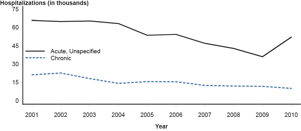 Pelvic Inflammatory Disease—Hospitalizations of Women Aged 15–44 Years, United States, 2001–2010
 -
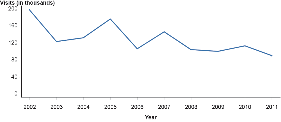 Pelvic Inflammatory Disease—Initial Visits to Physicians’ Offices by Women Aged 15–44 Years, United States, 2002–2011


Graphs adapted from https://www.cdc.gov/
Demographics
Age
PID is a common disease that tends to affect young women between the ages of 15 and 29 years.[2]
Race
In the United States, African-American or Black-Caribbean ethnicity has been associated with a higher risk of PID.[8]
References
- ↑ Grodstein F, Rothman KJ (1994). “Epidemiology of pelvic inflammatory disease”. Epidemiology. 5 (2): 234–42. PMID 8172999.
- ↑ 2.0 2.1 Ford GW, Decker CF (2016). “Pelvic inflammatory disease”. Dis Mon. 62 (8): 301–5. doi:10.1016/j.disamonth.2016.03.015. PMID 27107781.
- ↑ Rein DB, Kassler WJ, Irwin KL, Rabiee L (2000). “Direct medical cost of pelvic inflammatory disease and its sequelae: decreasing, but still substantial”. Obstet Gynecol. 95 (3): 397–402. PMID 10711551.
- ↑ Owusu-Edusei K, Bohm MK, Chesson HW, Kent CK (2010). “Chlamydia screening and pelvic inflammatory disease: Insights from exploratory time-series analyses”. Am J Prev Med. 38 (6): 652–7. doi:10.1016/j.amepre.2010.02.008. PMID 20494242.
- ↑ Gradison M (2012). “Pelvic inflammatory disease”. Am Fam Physician. 85 (8): 791–6. PMID 22534388.
- ↑ Sutton MY, Sternberg M, Zaidi A, St Louis ME, Markowitz LE (2005). “Trends in pelvic inflammatory disease hospital discharges and ambulatory visits, United States, 1985-2001”. Sex Transm Dis. 32 (12): 778–84. PMID 16314776.
- ↑ Lauren Nathan; DeCherney, Alan H.; Pernoll, Martin L. (2003). Current obstetric & gynecologic diagnosis & treatment. New York: Lange Medical Books/McGraw-Hill. ISBN 0-8385-1401-4.
- ↑ Leichliter JS, Chandra A, Aral SO (2013). “Correlates of self-reported pelvic inflammatory disease treatment in sexually experienced reproductive-aged women in the United States, 1995 and 2006-2010”. Sex Transm Dis. 40 (5): 413–8. doi:10.1097/OLQ.0b013e318285ce46. PMID 23588132.
Editor-In-Chief: C. Michael Gibson, M.S., M.D. [1]; Associate Editor(s)-in-Chief: Seyedmahdi Pahlavani, M.D. [2]
Risk Factors
Overview
Common risk factors in the development of pelvic inflammatory disease include prior history of pelvic inflammatory disease, recent sexual contact, multiple sexual partners and IUD.
Risk Factors
Important risk factors in PID development include:
- Multiple partners:
- Having multiple sexual partners within last six months increase the risk of PID.[1]
- Previous PID:
- Recurrent PID is more common among women who have a history of PID.[2]
- IUD:
References
- ↑ Lee NC, Rubin GL, Grimes DA (1991). “Measures of sexual behavior and the risk of pelvic inflammatory disease”. Obstet Gynecol. 77 (3): 425–30. PMID 1992411.
- ↑ Weström L (1975). “Effect of acute pelvic inflammatory disease on fertility”. Am. J. Obstet. Gynecol. 121 (5): 707–13. PMID 123123.
- ↑ Viberga I, Odlind V, Lazdane G, Kroica J, Berglund L, Olofsson S (2005). “Microbiology profile in women with pelvic inflammatory disease in relation to IUD use”. Infect Dis Obstet Gynecol. 13 (4): 183–90. doi:10.1080/10647440500097601. PMC 1784576. PMID 16338777.
- ↑ Grimes DA (2000). “Intrauterine device and upper-genital-tract infection”. Lancet. 356 (9234): 1013–9. doi:10.1016/S0140-6736(00)02699-4. PMID 11041414.
Editor-In-Chief: C. Michael Gibson, M.S., M.D. [1]; Associate Editor(s)-in-Chief: Seyedmahdi Pahlavani, M.D. [2]
Natural History, Complications and Prognosis
Overview
The overall prognosis of PID is good. Timely, appropriate treatment often prevents serious complications such as ectopic pregnancy, infertility, hydrosalpinx, and chronic pelvic pain.
Natural history
If left untreated, PID may lead to infertility in approximately 16% of affected women.[1] It may progress to adjacent organ involvement or even peritonitis.
Prognosis
The overall prognosis of PID is good if patients are treated within 3 days of the onset of symptoms onset, though clinical improvement cannot guarantee protection against infertility.[2]
Factors that predict poor prognosis include:[3]
- Advanced age
- History of previous open gynecological surgery
- Any cystic lesion identified by ultrasonography
- High CRP levels
Complications
Chronic pelvic pain
- Chronic pelvic pain is defined as lower abdominal pain that lasts for at least 6 months and causes functional disability. Approximately 1 in 3 of women affected by PID will experience chronic pelvic pain.[4]
- Recurrent PID is the strongest predictor for the development of chronic pelvic pain due to the scars and adhesions that result from repeated inflammation.[5]
Infertility
- PID may cause permanent damages to the fallopian tubes, this damages include loss of ciliary action, fibrosis, and occlusion that may result in infertility.[6]
- Risk factors for infertility include:
- Chlamydial infection[7]
- Delayed PID treatment[8]
- Frequent PIDs[9]
- Severity of infection[10]
Ectopic pregnancy
- Tubal damage due to PID may result in anatomical distortion, which can predispose the patient to ectopic pregnancy.[1]
Hydrosalpinx
- Tubal damage may lead to tubal blockade, sterile fluid accumulation, and/or fallopian tube enlargement, which may cause pain or infertility.[11]
Fitz Hugh Curtis syndrome
- Inflammation and adhesion formation in the liver capsule (perihepatitis) may cause right upper quadrant abdominal pain and tenderness.[12]
References
- ↑ 1.0 1.1 Weström L, Joesoef R, Reynolds G, Hagdu A, Thompson SE (1992). “Pelvic inflammatory disease and fertility. A cohort study of 1,844 women with laparoscopically verified disease and 657 control women with normal laparoscopic results”. Sex Transm Dis. 19 (4): 185–92. PMID 1411832.
- ↑ Ross J (2004). “Pelvic inflammatory disease”. Clin Evid (11): 2121–7. PMID 15652102.
- ↑ Terao M, Koga K, Fujimoto A, Wada-Hiraike O, Osuga Y, Yano T, Kozuma S (2014). “Factors that predict poor clinical course among patients hospitalized with pelvic inflammatory disease”. J. Obstet. Gynaecol. Res. 40 (2): 495–500. doi:10.1111/jog.12189. PMID 24118399.
- ↑ Ness RB, Soper DE, Holley RL, Peipert J, Randall H, Sweet RL, Sondheimer SJ, Hendrix SL, Amortegui A, Trucco G, Songer T, Lave JR, Hillier SL, Bass DC, Kelsey SF (2002). “Effectiveness of inpatient and outpatient treatment strategies for women with pelvic inflammatory disease: results from the Pelvic Inflammatory Disease Evaluation and Clinical Health (PEACH) Randomized Trial”. Am. J. Obstet. Gynecol. 186 (5): 929–37. PMID 12015517.
- ↑ Haggerty CL, Peipert JF, Weitzen S, Hendrix SL, Holley RL, Nelson DB, Randall H, Soper DE, Wiesenfeld HC, Ness RB (2005). “Predictors of chronic pelvic pain in an urban population of women with symptoms and signs of pelvic inflammatory disease”. Sex Transm Dis. 32 (5): 293–9. PMID 15849530.
- ↑ Cates W, Joesoef MR, Goldman MB (1993). “Atypical pelvic inflammatory disease: can we identify clinical predictors?”. Am. J. Obstet. Gynecol. 169 (2 Pt 1): 341–6. PMID 8362945.
- ↑ Svenstrup HF, Fedder J, Kristoffersen SE, Trolle B, Birkelund S, Christiansen G (2008). “Mycoplasma genitalium, Chlamydia trachomatis, and tubal factor infertility–a prospective study”. Fertil. Steril. 90 (3): 513–20. doi:10.1016/j.fertnstert.2006.12.056. PMID 17548070.
- ↑ Hillis SD, Joesoef R, Marchbanks PA, Wasserheit JN, Cates W, Westrom L (1993). “Delayed care of pelvic inflammatory disease as a risk factor for impaired fertility”. Am. J. Obstet. Gynecol. 168 (5): 1503–9. PMID 8498436.
- ↑ Weström L (1980). “Incidence, prevalence, and trends of acute pelvic inflammatory disease and its consequences in industrialized countries”. Am. J. Obstet. Gynecol. 138 (7 Pt 2): 880–92. PMID 7008604.
- ↑ Lepine LA, Hillis SD, Marchbanks PA, Joesoef MR, Peterson HB, Westrom L (1998). “Severity of pelvic inflammatory disease as a predictor of the probability of live birth”. Am. J. Obstet. Gynecol. 178 (5): 977–81. PMID 9609570.
- ↑ Kawwass JF, Crawford S, Kissin DM, Session DR, Boulet S, Jamieson DJ (2013). “Tubal factor infertility and perinatal risk after assisted reproductive technology”. Obstet Gynecol. 121 (6): 1263–71. doi:10.1097/AOG.0b013e31829006d9. PMC 4292839. PMID 23812461.
- ↑ Brunham RC, Gottlieb SL, Paavonen J (2015). “Pelvic inflammatory disease”. N. Engl. J. Med. 372 (21): 2039–48. doi:10.1056/NEJMra1411426. PMID 25992748.
Editor-In-Chief: C. Michael Gibson, M.S., M.D. [1]; Associate Editor(s)-in-Chief: Seyedmahdi Pahlavani, M.D. [2]
Diagnosis
Diagnostic Criteria | History and Symptoms | Physical Examination | Laboratory Findings | CT | MRI | Ultrasound | Other Imaging Findings | Other Diagnostic Studies
Treatment
Treatment
Medical Therapy | Surgery | Primary Prevention | Secondary Prevention | Cost-Effectiveness of Therapy | Future or Investigational Therapies
External Links
External Links
Template:Diseases of the pelvis, genitals and breasts nl:Eileiderontsteking
Editor-In-Chief: C. Michael Gibson, M.S., M.D. [1]; Associate Editor(s)-in-Chief: Seyedmahdi Pahlavani, M.D. [2]