
Conjunctivitis
Template:DiseaseDisorder infobox
For patient information click here
Editor-In-Chief: C. Michael Gibson, M.S., M.D. [1] Associate Editor(s)-in-Chief: Sara Mehrsefat, M.D. [2]
Synonyms and keywords: Madras eye; Pink eye; Pinkeye; Inflammation of conjunctiva; Conjunctiva inflamed; Infective conjunctivitis; Acute Hemorrhagic Conjunctivitis; Hyperacute Bacterial Conjunctivitis (HBC); Koch-Weeks conjunctivitis
Overview
Editor-In-Chief: C. Michael Gibson, M.S., M.D. [2]; Associate Editor(s)-in-Chief: Sara Mehrsefat, M.D. [3]
Overview
Conjunctivitis (con·junc·ti·vi·tis) is an inflammation of the conjunctiva (the outermost layer of the eye and the inner surface of the eyelids), most commonly due to an allergic reaction or an infection (usually bacterial or viral). Conjunctivitis may be classified based on the duration of symptoms into hyperacute, acute, or chronic. Additionally, based on the causality of the inflammation and age group, conjunctivitis may be classified into infective conjunctivitis (bacterial and viral), neonatal conjunctivitis (ophthalmia neonatorum), allergic conjunctivitis, keratoconjunctivitis sicca (dry eye syndrome), and superior limbic keratoconjunctivitis (SLK). Common causes of conjunctivitis include bacteria, viruses, and environmental factors. Viral conjunctivitis is the most common cause of infectious conjunctivitis both overall and in the adult population. Between 65% and 90% of cases of viral conjunctivitis are caused by adenoviruses. Bacterial conjunctivitis is the second most common cause. Allergic conjunctivitis is the most frequent cause, affecting 15% to 40% of the population. Noninfectious conjunctivitis includes keratoconjunctivitis sicca (dry eye syndrome) and superior limbic keratoconjunctivitis, which may caused by inflammation secondary to immune-mediated diseases. Prognosis for conjunctivitis usually good with treatment. Infective conjunctivitis resolves, in 65% of cases, within 2-5 days. Complete history will help determine the correct therapy. The symptoms of conjunctivitis differ based on the cause of the inflammation. Common symptoms to all forms of conjunctivitis include redness, excessive tearing, and irritation. Photophobia, itching, mucopurulent or non-purulent discharge, chemosis, burning eyes, blurred vision, and eyelid swelling are variable. Physical examination of patients with conjunctivitis is usually remarkable for conjunctival injections, epiphora, hyperemia, chemosis, and muco-purulent or watery discharge. However, ophthalmologic examination may be varies based on conjunctivitis subtypes. Laboratory tests are not often required in patients with mild conjunctivitis. Conjunctival cultures are generally reserved for conjunctivitis presenting with severe purulent discharge, and cases suspicious for gonococcal or chlamydial infection. Allergic conjunctivitis may be treated with artificial tears and topical antihistamines, vasoconstrictive agents, mast cell stabilizers, NSAIDs, and corticosteroids. Cool compresses are recommended to reduce eyelid and periorbital edema. Topical antimicrobial therapy is only recommended for patients with either bacterial or herpetic conjunctivitis, but not allergic or adenoviral conjunctivitis. Systemic antibiotic therapy is necessary to treat conjunctivitis due to Neisseria gonorrhoeae and Chlamydia trachomatis.
Historical Perspective
Conjunctivitis is an ancient disease. In 1750, neonatal conjunctivitis (ophthalmia neonatorum) was first described by S.T. Quellmaz. In 1883, Koch discovered the bacilli of two different forms of infectious conjunctivitis, or Egyptian ophthalmia.[1][2]
Classification
Conjunctivitis may be classified based on the duration of symptoms into hyperacute, acute, and chronic.[3][4] Additionally, conjunctivitis may be classified according to the causality of the inflammation or age groups into several subtypes: infective conjunctivitis (bacterial and viral), neonatal conjunctivitis (ophthalmia neonatorum), allergic conjunctivitis, keratoconjunctivitis sicca (dry eye syndrome), superior limbic keratoconjunctivitis, and chemical conjunctivitis.[5][6]
Pathophysiology
Conjunctivitis is defined as inflammation of bulbar and/or palpebral conjunctiva. Conjunctivitis has many etiologies, but the majority of cases can be caused by allergies or infection. Infective conjunctivitis is an infection of the conjunctiva either caused by viruses or bacteria. Airborne antigens may be involved in the pathogenesis of allergic conjunctivitis. Common airborne antigens include pollen, grass, and weeds. Keratoconjunctivitis sicca (dry eye syndrome) is a multifactorial disease and associated with different medical conditions.[7][8]
Causes
Common causes of conjunctivitis include bacteria, viruses, and environmental factors.[3] Viral conjunctivitis is the most common cause of infectious conjunctivitis both overall and in the adult population. Bacterial conjunctivitis is the second most common cause and is responsible for the majority of cases in children(Haemophilus influenzae,Moraxella catarrhalis,Streptococcus pneumoniae). Most of the cases in adults are caused by Staphylococcus aureus. Allergic conjunctivitis is the most frequent cause, affecting 15% to 40% of the population. Noninfectious conjunctivitis includes keratoconjunctivitis sicca (dry eye syndrome) and superior limbic keratoconjunctivitis caused by inflammation secondary to immune-mediated diseases or mechanical irritation.[7]
Differentiating Conjunctivitis from Other Diseases
Conjunctivitis symptoms and signs are relatively non-specific. Even after biomicroscopy, laboratory tests are often necessary to determine the underlying pathophysiology with certainty. An eye examination can help to differentiate conjunctivitis from other medical conditions.[9][10] Conjunctivitis must be differentiated from blepharitis, keratitis, and scleritis.
Epidemiology and Demographics
Conjunctivitis accounts for 1% of all primary care and emergency room visits. The incidence of viral conjunctivitis is approximately 80,000 per 100,000 cases with acute conjunctivitis. Viral conjunctivitis more commonly affects adults while bacterial conjunctivitis more commonly affects children. The prevalence of conjunctivitis varies according to the underlying cause, which may be influenced by the patient’s age, as well as the season of the year.[11]
Risk Factors
The most potent risk factor in the development of infective conjunctivitis is a direct or indirect contact with an infected person’s eye drainage. Common risk factors in the development of conjunctivitis are poor hygienic habits, contaminated personal articles, history of ocular diseases (dry eye, blepharitis, and anatomic abnormalities of the ocular surface), recent ocular surgery, medication use, and history of autoimmune disorders. Additionally, vaginal delivery is a risk factor for conjunctivitis in babies born to mothers infected with either Neisseria gonorrhoeae or Chlamydia trachomatis.[12][13]
Screening
General screening for conjunctivitis is not recommended. However, according to the Centers for Disease Control and Prevention (CDC), screening for sexually transmitted diseases (STDs) is recommended among pregnant women to prevent conjunctivitis and other medical conditions in newborns.[14]
Natural History, Complications, and Prognosis
If left untreated, viral conjunctivitis will generally clear without any complications. Bacterial conjunctivitis is often self-limited. If left untreated, bacterial conjunctivitis will clear within 1 or 2 weeks without any complications, and it is generally associated with a favorable long-term prognosis.[9] Allergic conjunctivitis improves by eliminating or significantly reducing contact with the allergen. If left untreated, most cases of allergic conjunctivitis may resolve without any long-term consequences. Keratoconjunctivitis sicca associated with Sjögren’s syndrome is associated with a particularly poor prognosis and requiring a longer course of treatment.[15][16][17] Prognosis for conjunctivitis is generally good with treatment.
Diagnosis
History and Symptoms
The symptoms of conjunctivitis differ based on the cause of the inflammation. Redness, excessive tearing, and irritation are symptoms common to all forms of conjunctivitis. Photophobia, itching, mucopurulent or non-purulent discharge, chemosis, burning eyes, blurred vision and eyelid swelling are variable. Complete history will help to determine whether the condition is associated with any specific environmental or work-related exposure.[15][9]
Physical Examination
Physical examination of patients with conjunctivitis is usually remarkable for conjunctival injections, epiphora, hyperemia, chemosis, and muco-purulent or watery discharge. However, ophthalmologic examination may be varies based on conjunctivitis subtypes.[18][7]
Laboratory Findings
Laboratory tests are not often required in patients with mild conjunctivitis. Conjunctival cultures are generally reserved for recurrent conjunctivitis, conjunctivitis recalcitrant to therapy, conjunctivitis presenting with severe mucopurulent discharge, and cases suspicious for gonococcal or chlamydial infection.[7][19]
Imaging Findings
There are no imaging findings associated with conjunctivitis. However, dynamic meibomian imaging (DMI) can be used to obtain a distinct picture of the entire everted inferior tarsal plate in a patient with keratoconjunctivitis sicca (dry eye syndrome).[20]
Other Diagnostic Studies
Other diagnostic studies for viral conjunctivitis include rapid antigen testing.[18] Other diagnostic studies for keratoconjunctivitis sicca (dry eye syndrome) conjunctivitis include corneal sensation, tear break up time, ocular surface staining, and Schirmer’s test.[21][22]
Treatment
Medical Therapy
Allergic conjunctivitis may be treated with artificial tears and topical antihistamines, vasoconstrictive agents, mast cell stabilizers, NSAIDs, and corticosteroids. Cool compresses are recommended to reduce eyelid and periorbital edema. Topical antimicrobial therapy is only recommended for patients with either bacterial or herpetic conjunctivitis, but not allergic or adenoviral conjunctivitis. Systemic antibiotic therapy is necessary to treat conjunctivitis due to Neisseria gonorrhoeae and Chlamydia trachomatis.
Surgery
Surgical intervention is not recommended for the management of infective and neonatal conjunctivitis. Allergic conjunctivitis is a self-limited disease, and extensive surgery may not be acceptable. However, surgical techniques include superficial keratectomy and penetrating keratoplasty are usually reserved for severe cases of corneal involvement. Despite the availability of efficient tear substitutes, many patients with keratoconjunctivitis sicca (dry eye syndrome) experience severe corneal injuries and a subsequent loss of vision. Surgical techniques include lateral tarsorrhaphy, punctal plugs, lens therapy, amniotic membrane transplantation, and salivary gland duct transposition.[19][1][23][24][25][26][27]
Primary Prevention
There are no formal established methods for primary prevention of allergic conjunctivitis, keratoconjunctivitis sicca (dry eye syndrome), and superior limbic keratoconjunctivitis. However, early determination of these conditions is very important in terms of morbidity.[28]
Secondary Prevention
Secondary prevention strategies following conjunctivitis include discontinued contact lens wear (infective conjunctivitis), ocular prophylaxis with 0.5% erythromycin ointment or 1% tetracycline hydrochloride (ophthalmia neonatorum), and avoiding the offending antigen (allergic conjunctivitis). Secondary prevention strategies following keratoconjunctivitis sicca (dry eye syndrome) include avoiding very dry environments, dusty areas, and prolonged visual tasks. There is no established method for secondary prevention of superior limbic keratoconjunctivitis (SLK). However, educating patients about disease process can improve compliance of patients with treatment, and help them to cope with the often prolonged symptoms.[15][28][29][30][27]
References
- ↑ 1.0 1.1 Mallika P, Asok T, Faisal H, Aziz S, Tan A, Intan G (2008). “Neonatal conjunctivitis – a review”. Malays Fam Physician. 3 (2): 77–81. PMC 4170304. PMID 25606121.
- ↑ Weeks JE (1996). “The bacillus of acute conjunctival catarrh, or ‘pink eye’. 1886”. Arch Ophthalmol. 114 (12): 1510–1. PMID 8953986.
- ↑ 3.0 3.1 National Eye Institute (2015). [1] Accessed on June 23, 2016
- ↑ Blochmichel E, Helleboid L, Corvec MP (1993). “Chronic allergic conjunctivitis”. Ocul Immunol Inflamm. 1 (1–2): 9–12. doi:10.3109/09273949309086529. PMID 22827184.
- ↑ Alfonso SA, Fawley JD, Alexa Lu X (2015). “Conjunctivitis”. Prim Care. 42 (3): 325–45. doi:10.1016/j.pop.2015.05.001. PMID 26319341.
- ↑ Leonardi A, Castegnaro A, Valerio AL, Lazzarini D (2015). “Epidemiology of allergic conjunctivitis: clinical appearance and treatment patterns in a population-based study”. Curr Opin Allergy Clin Immunol. 15 (5): 482–8. doi:10.1097/ACI.0000000000000204. PMID 26258920.
- ↑ 7.0 7.1 7.2 7.3 Azari AA, Barney NP (2013). “Conjunctivitis: a systematic review of diagnosis and treatment”. JAMA. 310 (16): 1721–9. doi:10.1001/jama.2013.280318. PMC 4049531. PMID 24150468.
- ↑ Kyei S, Koffuor GA, Ramkissoon P, Abokyi S, Owusu-Afriyie O, Wiredu EA (2015). “Possible Mechanism of Action of the Antiallergic Effect of an Aqueous Extract of Heliotropium indicum L. in Ovalbumin-Induced Allergic Conjunctivitis”. J Allergy (Cairo). 2015: 245370. doi:10.1155/2015/245370. PMC 4657065. PMID 26681960.
- ↑ 9.0 9.1 9.2 Leibowitz HM (2000). “The red eye”. N Engl J Med. 343 (5): 345–51. doi:10.1056/NEJM200008033430507. PMID 10922425.
- ↑ American Academy of ophthalmology (2016) http://eyewiki.aao.org/Bacterial_Conjunctivitis Accessed on June 27, 2016
- ↑ Høvding G (2008). “Acute bacterial conjunctivitis”. Acta Ophthalmol. 86 (1): 5–17. doi:10.1111/j.1600-0420.2007.01006.x. PMID 17970823.
- ↑ Epling J (2010). “Bacterial conjunctivitis”. BMJ Clin Evid. 2010. PMC 2907624. PMID 21718563.
- ↑ Cronau H, Kankanala RR, Mauger T (2010). “Diagnosis and management of red eye in primary care”. Am Fam Physician. 81 (2): 137–44. PMID 20082509.
- ↑ Centers for Disease Control and Prevention (2015) http://www.cdc.gov/std/tg2015/screening-recommendations.htm Accessed on June 29, 2016
- ↑ 15.0 15.1 15.2 Rose P (2007). “Management strategies for acute infective conjunctivitis in primary care: a systematic review”. Expert Opin Pharmacother. 8 (12): 1903–21. doi:10.1517/14656566.8.12.1903. PMID 17696792.
- ↑ Schaumberg DA, Dana R, Buring JE, Sullivan DA (2009). “Prevalence of dry eye disease among US men: estimates from the Physicians’ Health Studies”. Arch Ophthalmol. 127 (6): 763–8. doi:10.1001/archophthalmol.2009.103. PMC 2836718. PMID 19506195.
- ↑ Watson S, Tullo AB, Carley F (2002). “Treatment of superior limbic keratoconjunctivitis with a unilateral bandage contact lens”. Br J Ophthalmol. 86 (4): 485–6. PMC 1771108. PMID 11914237.
- ↑ 18.0 18.1 Jhanji V, Chan TC, Li EY, Agarwal K, Vajpayee RB (2015). “Adenoviral keratoconjunctivitis”. Surv Ophthalmol. 60 (5): 435–43. doi:10.1016/j.survophthal.2015.04.001. PMID 26077630.
- ↑ 19.0 19.1 Wood M (1999). “Conjunctivitis: diagnosis and management”. Community Eye Health. 12 (30): 19–20. PMC 1706007. PMID 17491982.
- ↑ Qazi Y, Aggarwal S, Hamrah P (2014). “Image-guided evaluation and monitoring of treatment response in patients with dry eye disease”. Graefes Arch Clin Exp Ophthalmol. 252 (6): 857–72. doi:10.1007/s00417-014-2618-2. PMC 4038672. PMID 24696045.
- ↑ Savini G, Prabhawasat P, Kojima T, Grueterich M, Espana E, Goto E (2008). “The challenge of dry eye diagnosis”. Clin Ophthalmol. 2 (1): 31–55. PMC 2698717. PMID 19668387.
- ↑ Beckman KA, Luchs J, Milner MS (2016). “Making the diagnosis of Sjögren’s syndrome in patients with dry eye”. Clin Ophthalmol. 10: 43–53. doi:10.2147/OPTH.S80043. PMC 4699514. PMID 26766898.
- ↑ Rajak S, Rajak J, Selva D (2015). “Performing a tarsorrhaphy”. Community Eye Health. 28 (89): 10–1. PMC 4579993. PMID 26435586.
- ↑ Baxter SA, Laibson PR (2004). “Punctal plugs in the management of dry eyes”. Ocul Surf. 2 (4): 255–65. PMID 17216100.
- ↑ Khodadoust A, Quinter AP (2003). “Microsurgical approach to the conjunctival flap”. Arch Ophthalmol. 121 (8): 1189–93. doi:10.1001/archopht.121.8.1189. PMID 12912699.
- ↑ Güerrissi JO, Belmonte J (2004). “Surgical treatment of dry eye syndrome: conjunctival graft of the minor salivary gland”. J Craniofac Surg. 15 (1): 6–10. PMID 14704553.
- ↑ 27.0 27.1 Nelson JD (1989). “Superior limbic keratoconjunctivitis (SLK)”. Eye (Lond). 3 ( Pt 2): 180–9. doi:10.1038/eye.1989.26. PMID 2695351.
- ↑ 28.0 28.1 Matejcek A, Goldman RD (2013). “Treatment and prevention of ophthalmia neonatorum”. Can Fam Physician. 59 (11): 1187–90. PMC 3828094. PMID 24235191.
- ↑ La Rosa M, Lionetti E, Reibaldi M, Russo A, Longo A, Leonardi S; et al. (2013). “Allergic conjunctivitis: a comprehensive review of the literature”. Ital J Pediatr. 39: 18. doi:10.1186/1824-7288-39-18. PMC 3640929. PMID 23497516.
- ↑ Messmer EM (2015). “The pathophysiology, diagnosis, and treatment of dry eye disease”. Dtsch Arztebl Int. 112 (5): 71–81, quiz 82. doi:10.3238/arztebl.2015.0071. PMC 4335585. PMID 25686388.
Historical Perspective
Editor-In-Chief: C. Michael Gibson, M.S., M.D. [1]; Associate Editor(s)-in-Chief: Sara Mehrsefat, M.D. [2]
Overview
Conjunctivitis is an ancient disease, and was first described by S.T. Quellmaz.[1] In 1881, Crédé introduced 2% silver nitrate for the first time as a prophylaxis treatment method for conjunctivitis in the newborns in Leipzig.[2]
Historical Perspective
- In 1750, neonatal conjunctivitis (ophthalmia neonatorum) was first described by S.T. Quellmaz.[1]
- In 1881, Crédé introduced 2% silver nitrate for the first time as a prophylaxis treatment method for conjunctivitis in the newborns in Leipzig.[2]
- In 1883, Koch discovered the bacilli of two different forms of infectious conjunctivitis or Egyptian ophthalmia.
- In 1886, Weeks discovered the same organism to be the cause of pink eye.[3]
- In 1953, Micheal Hogan first described the association between atopic dermatitis and conjunctival inflammation.[4]
- In 1963, Thygeson and Kimura first described superior limbic keratoconjunctivitis as a chronic, localized, filamentary conjunctivitis, and later this condition was given its name, superior limbic keratoconjunctivitis, by Theodore.[5]
- In 1968, Tenzel and Corwin were the first to discover the association between thyroid abnormalities and development of superior limbic keratoconjunctivitis.[6]
- In 1969, acute hemorrhagic conjunctivitis was first described as occurring in Ghana.[7]
- Between 1981-2003, four major epidemics of acute hemorrhagic conjunctivitis had occurred in the Western Hemisphere.[8]
- In 2006, an epidemic acute hemorrhagic conjunctivitis (involving more than 200,000 people) was reported as occurring in Brazil.[9]
References
- ↑ 1.0 1.1 Mallika P, Asok T, Faisal H, Aziz S, Tan A, Intan G (2008). “Neonatal conjunctivitis – a review”. Malays Fam Physician. 3 (2): 77–81. PMC 4170304. PMID 25606121.
- ↑ 2.0 2.1 “Reports from the obstetrical clinic in Leipzig. Prevention of eye inflammation in the newborn”. Am J Dis Child. 121 (1): 3–4. 1971. PMID 5543850.
- ↑ Weeks JE (1996). “The bacillus of acute conjunctival catarrh, or ‘pink eye’. 1886”. Arch Ophthalmol. 114 (12): 1510–1. PMID 8953986.
- ↑ HOGAN MJ (1952). “Atopic keratoconjunctivitis”. Trans Am Ophthalmol Soc. 50: 265–81. PMC 1313175. PMID 13102561.
- ↑ Watson S, Tullo AB, Carley F (2002). “Treatment of superior limbic keratoconjunctivitis with a unilateral bandage contact lens”. Br J Ophthalmol. 86 (4): 485–6. PMC 1771108. PMID 11914237.
- ↑ Nelson JD (1989). “Superior limbic keratoconjunctivitis (SLK)”. Eye (Lond). 3 ( Pt 2): 180–9. doi:10.1038/eye.1989.26. PMID 2695351.
- ↑ Azari AA, Barney NP (2013). “Conjunctivitis: a systematic review of diagnosis and treatment”. JAMA. 310 (16): 1721–9. doi:10.1001/jama.2013.280318. PMC 4049531. PMID 24150468.
- ↑ Centers for Disease Control and Prevention (2004) https://www.cdc.gov/mmwr/preview/mmwrhtml/mm5328a2.htm Accessed on June 24, 2016
- ↑ Workowski KA, Berman S, Centers for Disease Control and Prevention (CDC) (2010). “Sexually transmitted diseases treatment guidelines, 2010”. MMWR Recomm Rep. 59 (RR-12): 1–110. PMID 21160459.
Classification
Editor-In-Chief: C. Michael Gibson, M.S., M.D. [3]; Associate Editor(s)-in-Chief: Sara Mehrsefat, M.D. [4]
Overview
Conjunctivitis may be classified based on the duration of symptoms into hyperacute, acute, and chronic.[1][2] Additionally, conjunctivitis may be classified according to the causality of the inflammation or age groups into several subtypes: infective conjunctivitis (bacterial and viral), neonatal conjunctivitis (ophthalmia neonatorum), allergic conjunctivitis, keratoconjunctivitis sicca (dry eye syndrome), superior limbic keratoconjunctivitis, and chemical conjunctivitis.[3][4]
Classification
Based on the duration of symptoms, conjunctivitis may be classified into hyperacute, acute (less than 3 weeks), and chronic (greater than 3 weeks).
According to the etiology and age groups, conjunctivitis may be classified into:[5][6][5][7][8] [9]
- Infective conjunctivitis
- Neonatal conjunctivitis (ophthalmia neonatorum)
- Allergic conjunctivitis
- Seasonal allergic conjunctivitis
- Perennial allergic conjunctivitis
- Vernal keratoconjunctivitis
- Atopic keratoconjunctivitis
- Giant papillary conjunctivitis
- Keratoconjunctivitis sicca (dry eye syndrome)
- Superior limbic keratoconjunctivitis (SLK)
- Chemical or irritative conjunctivitis
References
- ↑ National Eye Institute (2015). [1] Accessed on June 23, 2016
- ↑ Blochmichel E, Helleboid L, Corvec MP (1993). “Chronic allergic conjunctivitis”. Ocul Immunol Inflamm. 1 (1–2): 9–12. doi:10.3109/09273949309086529. PMID 22827184.
- ↑ Alfonso SA, Fawley JD, Alexa Lu X (2015). “Conjunctivitis”. Prim Care. 42 (3): 325–45. doi:10.1016/j.pop.2015.05.001. PMID 26319341.
- ↑ Leonardi A, Castegnaro A, Valerio AL, Lazzarini D (2015). “Epidemiology of allergic conjunctivitis: clinical appearance and treatment patterns in a population-based study”. Curr Opin Allergy Clin Immunol. 15 (5): 482–8. doi:10.1097/ACI.0000000000000204. PMID 26258920.
- ↑ 5.0 5.1 Centers for Disease Control and Prevention (2016). [2] Accessed on June 23, 2016 Invalid parameter “viral” in
<ref>tag. The supported parameters are: dir, follow, group, name. - ↑ Menon BB, Zhou X, Spurr-Michaud S, Rajaiya J, Chodosh J, Gipson IK (2016). “Epidemic Keratoconjunctivitis-Causing Adenoviruses Induce MUC16 Ectodomain Release To Infect Ocular Surface Epithelial Cells”. mSphere. 1 (1). doi:10.1128/mSphere.00112-15. PMC 4863608. PMID 27303700.
- ↑ Mallika P, Asok T, Faisal H, Aziz S, Tan A, Intan G (2008). “Neonatal conjunctivitis – a review”. Malays Fam Physician. 3 (2): 77–81. PMC 4170304. PMID 25606121.
- ↑ Li Z, Chen W, Zhang Y, Jhanji V, Fan Z, Mu G (2015). “Topical Fluorometholone Versus Diclofenac Sodium in Cases With Perennial Allergic Conjunctivitis”. Eye Contact Lens. 41 (5): 310–3. doi:10.1097/ICL.0000000000000127. PMID 26322818.
- ↑ Bischoff G (2014). “[Giant papillary conjunctivitis]”. Klin Monbl Augenheilkd. 231 (5): 518–21. doi:10.1055/s-0034-1368334. PMID 24799171.
Pathophysiology
Editor-In-Chief: C. Michael Gibson, M.S., M.D. [3]; Associate Editor(s)-in-Chief: Sara Mehrsefat, M.D. [4]
Overview
Conjunctivitis is defined as inflammation of bulbar and/or palpebral conjunctiva. Conjunctivitis has many etiologies, but the majority of cases can be caused by allergies or infection. Infective conjunctivitis is an infection of the conjunctiva either caused by viruses or bacteria. Airborne antigens may be involved in the pathogenesis of allergic conjunctivitis. Common airborne antigens include pollen, grass, and weeds. Keratoconjunctivitis sicca (dry eye syndrome) is a multifactorial disease and associated with different medical conditions.[1][2]
Pathophysiology
Pathogenesis
Infective Conjunctivitis
Infective conjunctivitis is an infection of the conjunctiva either caused by viruses or bacteria, such as adenovirus, Staphylococcus aureus, Streptococcus pneumoniae, and Haemophilus influenzae. Any change in the host defense, or in the species of normal flora of the eye, can lead to clinical infection and conjunctivitis.
Change in the normal flora may occur by:[3]
- External contamination
- Contact lens wear
- Swimming
Infective conjunctivitis is usually spread by:[1][2]
- Direct contact with the infected person’s eye drainage or drainage from the person’s cough, sneeze, or runny nose
- Contact with the infected person’s fingers, hands, or objects (eye makeup applicators, towels, shared eye medications)
- Adjacent infectious sites (rubbing of the eyes)
Neonatal Conjunctivitis
Neonatal conjunctivitis is occurring in a newborn during the first month of life, and often known as ophthalmia neonatorum. Neonatal conjunctivitis is mainly caused by sexually transmitted diseases agents such as Chlamydia trachomatis, Neisseria gonorrhoeae, and herpes simplex virus (HSV). Chlamydia trachomatis is the most common cause of ophthalmia neonatorum in the developed countries because of higher prevalence of chlamydia as a sexually transmitted disease.
The recognized routes of transmission of the organisms to the newborns include:[4]
- Infected birth canal during vaginal birth
- Transmembrane transmission of the infection
- Transplacental transmission of the infection
Additionally, silver nitrate drops (ocular prophylaxis) can cause ocular irritation and result in chemical conjunctivitis in newborn.[5]
Allergic Conjunctivitis
Development of allergic conjunctivitis is the result of type 1 hypersensitivity reaction in the conjunctiva. Allergic conjunctivitis is caused by the immunoglobulin E (IgE) mediated reaction to environmental allergens. Following exposure to environmental allergens in sensitized individual, the cross-linking of IgE at the mast cell membrane level results in subsequent cell degranulation and secretion of histamine, leukotriene, and prostaglandin. The principal effects of these products are vasodilation and smooth-muscle contraction. Common airborne antigens involve in the pathogenesis of allergic conjunctivitis include pollen, grass, and weeds.[6][7]
Combination of type 1 and type 4 hypersensitivity reactions may be responsible for giant papillary conjunctivitis. Also, prolonged mechanical irritation to the superior tarsal conjunctiva of the upper lid from foreign bodies may also be a contributing factor in giant papillary conjunctivitis.[8][9]
Keratoconjunctivitis Sicca
Keratoconjunctivitis sicca (dry eye syndrome) is a multifactorial disease and associated with different medical conditions such as:[10][11]
- Sjögren’s syndrome
- Ocular surface disease
- Ocular allergy
- Dysfunction of the lacrimal gland with reduced tear production and mucin deficiency
- Anxiety disorder
- Depression
The exact pathogenesis of the keratoconjunctivitis sicca associated Sjögren’s syndrome is not fully understood. It is thought that keratoconjunctivitis sicca associated Sjögren’s syndrome is the result of lymphocytic infiltration and malfunction of the lacrimal glands. This lymphasitic infiltration (CD4+ T cells and B cells) may result in lacrimal gland dysfunction and reduced tear production (mediated by apoptosis).
Superior Limbic Keratoconjunctivitis
Superior limbic keratoconjunctivitis (SLK) is disease characterized by inflammation of the upper palpebral and superior bulbar conjunctiva. The exact pathogenesis of superior limbic keratoconjunctivitis is not fully understood, a mechanical hypothesis seems most attractive. It is thought that mechanical trauma from tight upper lids or loose redundant conjunctiva can lead to the disruption of normal epithelium. Also, the association between thyroid abnormalities (graves ophthalmopathy) and superior limbic keratoconjunctivitis has been reported.[12][13][14]
Gross Pathology
On gross pathology, the following are characteristic findings of conjunctivitis:[15]
- Conjunctival injection
- Mucopurulent discharge or non-purulent discharge
- Pseudomembrane formation
- Chemosis
- Eyelid edema
- Conjunctival hemorrhage (specific for acute hemorrhagic conjunctivitis)
Microscopic Histopathological Analysis
On microscopic histopathological analysis, the following are characteristic findings of conjunctivitis:[16]
- Mild spongiotic reaction pattern (low power view of the histology)
- Stromal infiltration by polymorphonuclear cells, leukocytes, plasma cells, mastocytes, and lymphocyte
- Eosinophils (allergic conjunctivitis)
- Numerous neutrophils (bacterial conjunctivitis)
- Hyperplastic with increase numbers of plasma cell (chronic conjunctivitis)
- Keratinization of the superior limbus (superior limbic keratoconjunctivitis (SLK))
- Dyskeratosis (superior limbic keratoconjunctivitis (SLK))
Images
The following are gross and microscopic images associated with conjunctivitis:
-
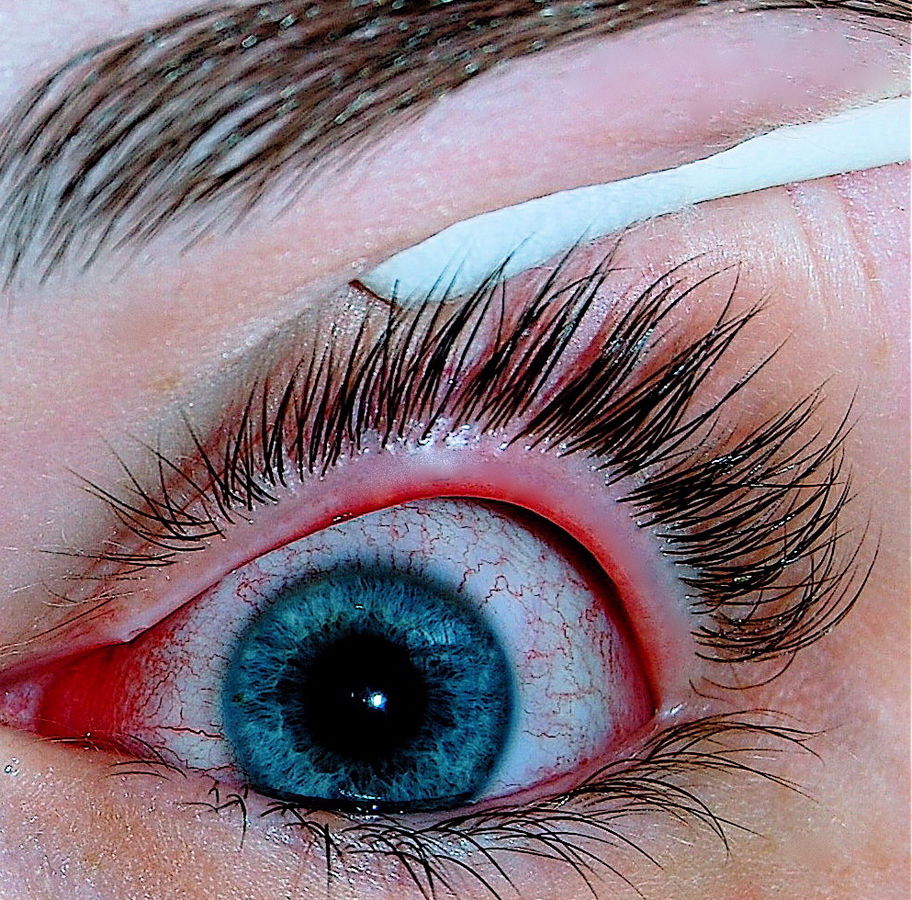 Viral conjunctivitis – By Joyhill09 – I took this photo with a Nikon D40 of my eye infected with conjunctivitis, CC BY-SA 3.0, https://commons.wikimedia.org/w/index.php?curid=18954730
 -
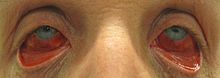 Acute hemorrhagic conjunctivitis – By James Heilman, MD – Own work, CC BY-SA 3.0, https://commons.wikimedia.org/w/index.php?curid=27401136
 -
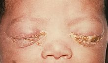 Neonatal conjunctivitis – Source: https://www.cdc.gov/


References
- ↑ 1.0 1.1 Azari AA, Barney NP (2013). “Conjunctivitis: a systematic review of diagnosis and treatment”. JAMA. 310 (16): 1721–9. doi:10.1001/jama.2013.280318. PMC 4049531. PMID 24150468.
- ↑ 2.0 2.1 Kyei S, Koffuor GA, Ramkissoon P, Abokyi S, Owusu-Afriyie O, Wiredu EA (2015). “Possible Mechanism of Action of the Antiallergic Effect of an Aqueous Extract of Heliotropium indicum L. in Ovalbumin-Induced Allergic Conjunctivitis”. J Allergy (Cairo). 2015: 245370. doi:10.1155/2015/245370. PMC 4657065. PMID 26681960.
- ↑ Everitt H, Kumar S, Little P (2003). “A qualitative study of patients’ perceptions of acute infective conjunctivitis”. Br J Gen Pract. 53 (486): 36–41. PMC 1314490. PMID 12564275.
- ↑ Treadwell P (1994). “Sexually transmitted diseases in neonates and infants”. Semin Dermatol. 13 (4): 256–61. PMID 7848819.
- ↑ Mallika P, Asok T, Faisal H, Aziz S, Tan A, Intan G (2008). “Neonatal conjunctivitis – a review”. Malays Fam Physician. 3 (2): 77–81. PMC 4170304. PMID 25606121.
- ↑ Malling HJ, Montagut A, Melac M, Patriarca G, Panzner P, Seberova E; et al. (2009). “Efficacy and safety of 5-grass pollen sublingual immunotherapy tablets in patients with different clinical profiles of allergic rhinoconjunctivitis”. Clin Exp Allergy. 39 (3): 387–93. doi:10.1111/j.1365-2222.2008.03152.x. PMC 4233960. PMID 19134019.
- ↑ Kämpe M, Stålenheim G, Janson C, Stolt I, Carlson M (2007). “Systemic and local eosinophil inflammation during the birch pollen season in allergic patients with predominant rhinitis or asthma”. Clin Mol Allergy. 5: 4. doi:10.1186/1476-7961-5-4. PMC 2174506. PMID 17967188.
- ↑ Donshik PC (1994). “Giant papillary conjunctivitis”. Trans Am Ophthalmol Soc. 92: 687–744. PMC 1298525. PMID 7886881.
- ↑ Donshik PC, Porazinski AD (1999). “Giant papillary conjunctivitis in frequent-replacement contact lens wearers: a retrospective study”. Trans Am Ophthalmol Soc. 97: 205–16, discussion 216-20. PMC 1298261. PMID 10703125.
- ↑ Sivaraman KR, Jivrajka RV, Soin K, Bouchard CS, Movahedan A, Shorter E; et al. (2016). “Superior Limbic Keratoconjunctivitis-like Inflammation in Patients with Chronic Graft-Versus-Host Disease”. Ocul Surf. doi:10.1016/j.jtos.2016.04.003. PMID 27179980.
- ↑ Messmer EM (2015). “The pathophysiology, diagnosis, and treatment of dry eye disease”. Dtsch Arztebl Int. 112 (5): 71–81, quiz 82. doi:10.3238/arztebl.2015.0071. PMC 4335585. PMID 25686388.
- ↑ Nelson JD (1989). “Superior limbic keratoconjunctivitis (SLK)”. Eye (Lond). 3 ( Pt 2): 180–9. doi:10.1038/eye.1989.26. PMID 2695351.
- ↑ Chelala E, El Rami H, Dirani A, Fakhoury H, Fadlallah A (2015). “Extensive superior limbic keratoconjunctivitis in Graves’ disease: case report and mini-review of the literature”. Clin Ophthalmol. 9: 467–8. doi:10.2147/OPTH.S79561. PMC 4362972. PMID 25792798.
- ↑ American Academy of Ophthalmology (2015) http://eyewiki.aao.org/Superior_limbic_keratoconjunctivitis Accessed on June 27, 2016
- ↑ National Eye Institute (2015) [1] Accessed on June 24, 2016
- ↑ DermNet NZ (2015)[2] Accessed on June 26, 2016
Causes
Editor-In-Chief: C. Michael Gibson, M.S., M.D. [4]; Associate Editor(s)-in-Chief: Ogheneochuko Ajari, MB.BS, MS [5], Sara Mehrsefat, M.D. [6]
Overview
Common causes of conjunctivitis include bacteria, viruses, and environmental factors.[1] Viral conjunctivitis is the most common cause of infectious conjunctivitis both overall and in the adult population. Bacterial conjunctivitis is the second most common cause and is responsible for the majority of cases in children. Allergic conjunctivitis is the most frequent cause, affecting 15% to 40% of the population. Noninfectious conjunctivitis includes keratoconjunctivitis sicca (dry eye syndrome) and superior limbic keratoconjunctivitis may cause by inflammation secondary to immune-mediated diseases or mechanical irritation.[2]
Causes
Viral conjunctivitis[3]
- Adenovirus (most common)
- Herpes simplex virus (HSV) type 1 and 2
- Varicella zoster virus
- Picornaviruses (Coxsackievirus A24 and Enterovirus 70)
- Molluscum contagiosum
- Rubella virus
- Epstein-Barr virus (EBV)
Bacterial conjunctivitis[4]
- Gram-positive bacteria
- Staphylococcus aureus
- Methicillin-resistant Staphylococcus aureus (common in nursing homes)
- Streptococcus pneumoniae
- Gram-negative bacteria
- Pseudomonas aeruginosa
- Serratia marcescens
- Haemophilus influenzae (most common pathogen in children)
- Moraxella catarrhalis
- Chlamydia trachomatis (sexually transmitted bacteria)
- Neisseria gonorrhoeae (sexually transmitted bacteria)
- Septic
- Chlamydia trachomatis
- Neisseria gonorrhoeae
- Herpes simplex virus
- Pseudomonas aeruginosa (non–sexually transmitted bacteria)
- Staphylococcus aureus (non–sexually transmitted bacteria)
- Aseptic
- Chemical (silver nitrate solution)
Allergic conjunctivitis[3]
- Allergens (pollen, animal dander, dust mites, or molds)
Keratoconjunctivitis[7]
- Lacrimal gland dysfunction (Sjögren’s syndrome) with reduced tear production
- Reflex hyposecretion
- Mucin deficiency
- Low blink rate
- Mechanical trauma
- Vitamin A deficiency
- Autoimmune diseases
- Unknown etiology
Irritative Conjunctivitis
- Contact lenses
- Lens solutions
- Chlorine in a swimming pool
- Smog
- Cosmetics
Causes by Organ System
Causes in Alphabetical Order
- 7th cranial nerve disorder
- Acanthamoeba
- Acetaldehyde
- Acetic acid
- Acetic anhydride
- Acetylcarbromal
- Acute febrile neutrophilic dermatosis
- Adenovirus
- Aesculus pollen
- Alder
- Allergic conjunctivitis
- Allergy
- Alternaria
- American feverfew
- Ammonia
- Angelucci’s syndrome
- Anisole
- Arsenic
- Arsenic trioxide
- Arsenicals
- Artemether and lumefantrin
- Ash juniper tree
- Ash tree
- Aspergillus
- Aureobasidium
- Bacterial conjunctivitis
- Beech tree
- Behcet disease
- Bermuda grass
- Beryllium
- Birch tree
- Blepharitis
- Blunt injury
- Boric acid
- Box elder tree
- Box jellyfish poisoning
- Burns
- Calcium sulfate
- Canary grass
- Candida albicans
- Capeweed
- Cat scratch disease
- Caterpillar poisoning
- Cetuximab
- Chemical irritation
- Chikungunya fever
- Chlamydia
- Chlorine dioxide
- Chloroacetophenone
- Cicatricial pemphigoid
- Cladosporium
- Clomethiazole
- Cobra poisoning
- Cocklebur
- Cogan syndrome
- Common variable immunodeficiency
- Congenital chlamydia
- Congenital gonorrhea
- Conjunctival squamous cell carcinoma
- Contact lens
- Corneal abrasion
- Coxsackie virus
- Crimean-Congo hemorrhagic fever
- Crouzon syndrome
- CS gas
- Cypress tree
- Cytarabine
- Cytosine arabinose syndrome
- Dacryocystitis
- Daisy
- Dandelion
- Desmopressin
- Dexamethasone
- Diethylene glycol monobutyl ether
- Dimethylamine
- Dipivefrin
- Doxorubicin hydrochloride
- Dust mite
- Dyskeratosis congenita
- Ectropion
- Elm tree
- Enterovirus 70
- Entropion
- Environmental pollution
- Epicoccum
- Epidemic keratoconjunctivitis
- Epirubicin hydrochloride
- Erythema multiformae
- Erythromycin
- Ether
- Ethylene oxide
- Etretinate
- Euphorbiaceae
- Facial nerve palsy
- Familial cold urticaria
- Familial hibernian fever
- Flurbiprofen
- Fluticasone (spray)
- Follicular conjunctivitis
- Foreign body
- Furfural
- Fusarium
- Gestational trophoblastic neoplasia
- Giant papillary conjunctivitis
- Gonorrhea
- Goosefoot
- Granulomatosis with polyangiitis
- Green Lynx spider poisoning
- Guayule
- Haemophilus influenzae
- Hantavirus pulmonary syndrome
- Hay fever
- Helminth infections
- Helminthosporium
- Hemorrhage
- Hemp
- Herpes simplex
- Herpes virus 2
- Herpes zoster
- Hickory tree
- Hornbeam tree
- Horse chestnut tree
- Human enterovirus A
- Hydrogen sulfide
- Hydroquinone
- Hypersensitivity to pollen
- Job syndrome
- Johnson grass
- Kawasaki disease
- Kentucky bluegrass
- Keratoconjunctivitis sicca
- Kikuchi disease
- Kindler syndrome
- Koch-Weeks conjunctivitis
- Lacrimal canaliculitis
- Lepidopterism
- Leprosy
- Leptospirosis
- Lewisite
- Ligneous conjunctivitis
- Listeria monocytogenes
- Lyme disease
- Maple tree
- Marshelder
- Mayapple poisoning
- Measles
- Medrysone
- Melarsoprol
- Metaldehyde
- Methylene diisocyanate
- Mold allergy
- Molluscum contagiosum
- Monteroy pine tree
- Moraxella catarrhalis
- Morpholine
- Mountain cedar tree
- Mucor
- Mugwort tree
- Neisseria gonorrhoeae
- Neisseria meningiditis
- Newcastle disease
- Nitisinone
- Non-specific urethritis
- Oak tree
- Ocular cicatricial pemphigoid
- Ocular rosacea
- Olea tree
- Olive tree
- Omsk hemorrhagic fever
- Onchocerciasis
- Ophthalmia neonatorum
- Orache (Atriplex)
- Oral allergy syndrome
- Orchard grass
- Osmium
- Pasteurella multocida
- Pecan trees
- Pediculosis ciliaris
- Pemetrexed
- Penetrating trauma
- Penicillium
- Pfiesteria piscicida
- Phlyctenular keratoconjunctivitis
- Phosgene oxime
- Picornavirus
- Pigweed
- Plane tree
- Plasminogen deficiency
- Pollen
- Polychondritis
- Poplar tree
- Propylene glycol dinitrate
- Pseudomonas aeruginosa
- Psittacosis
- Psoriatic arthritis
- Pyridoxine deficiency
- Quinone
- Ragweed
- Rapeseed oil
- Rasagiline
- Reactive arthritis
- Redtop grass
- Reiter’s syndrome
- Relapsing polychondritis
- Respiratory syncytial virus
- Rhizopus
- Rosacea
- Rubella
- Ryegrass
- Sabia virus
- Serratia
- Severe combined immunodeficiency
- Sicca syndrome
- Silver nitrate drops
- Sjogren’s syndrome
- Smog
- Smoke
- Smut
- Staphylococcus aureus
- Stevens-Johnson syndrome
- Streptococcus pneumoniae
- Sulfuric acid
- Sulfuryl fluoride
- Sunflower
- Sweet’s syndrome
- Tetracycline
- The clap
- Theodore’s superior limbic keratoconjunctivitis
- Tilia tree
- Timothy grass
- TNF receptor associated periodic syndrome
- Toluene diisocyanate
- Toxic-shock syndrome
- Trachoma
- Trauma
- Tularemia
- Tumble weed
- Type II tyrosinemia
- Vanadium
- Velvet grass
- Vernal conjunctivitis
- Vernal keratoconjunctivitis
- Viral conjunctivitis
- Wall pellitory
- Walnut tree
- Wegener’s granulomatosis
- White cedar tree
- Willow tree
- Xeroderma pigmentosum
- X-linked agammaglobulinaemia
- Xylene
References
- ↑ National Eye Institute (2015). [1] Accessed on June 23, 2016
- ↑ Azari AA, Barney NP (2013). “Conjunctivitis: a systematic review of diagnosis and treatment”. JAMA. 310 (16): 1721–9. doi:10.1001/jama.2013.280318. PMC 4049531. PMID 24150468.
- ↑ 3.0 3.1 Centers for Disease Control and Prevention (2016). [2] Accessed on June 23, 2016 Invalid parameter “viral” in
<ref>tag. The supported parameters are: dir, follow, group, name. - ↑ name= Bacterial Conjunctivitis > Centers for Disease Control and Prevention (2015). [3] Accessed on June 23, 2016
- ↑ Mallika P, Asok T, Faisal H, Aziz S, Tan A, Intan G (2008). “Neonatal conjunctivitis – a review”. Malays Fam Physician. 3 (2): 77–81. PMC 4170304. PMID 25606121.
- ↑ Woods, Charles R. “Gonococcal infections in neonates and young children.” Seminars in pediatric infectious diseases. Vol. 16. No. 4. WB Saunders, 2005.
- ↑ Barabino S, Dana MR (2007). “Dry eye syndromes”. Chem Immunol Allergy. 92: 176–84. doi:10.1159/000099268. PMID 17264493.
Differentiating Conjunctivitis from other Diseases
Editor-In-Chief: C. Michael Gibson, M.S., M.D. [1]; Associate Editor(s)-in-Chief: Sara Mehrsefat, M.D. [2]
Overview
Conjunctivitis must be differentiated from blepharitis, keratitis, and scleritis.[1][2] Conjunctivitis symptoms and signs are relatively non-specific. Even after eye examination, laboratory tests are often necessary to determine the underlying pathophysiology with certainty.
Differentiating Conjunctivitis from Other Diseases
Bacterial Conjunctivitis
A mucopurulent discharge strongly suggests bacterial cause, unless there is known exposure to toxins. Infection with Neisseria gonorrhoeae and Chlamydia trachomatis should be suspected if the discharge is particularly thick and copious. Bacterial conjunctivitis must be differentiated from:[1][2]
- Viral conjunctivitis
- Keratoconjunctivitis sicca (dry eye syndrome)
- Glaucoma
- Blepharitis
- Uveitis
- Iritis
- Keratitis
- Episcleritis
- Scleritis
- Neisseria meningitis (it can lead to fatal meningeal or systemic infection in the patient with hyperacute conjunctivitis due to Neisseria gonorrhoeae)
Viral Conjunctivitis
A diffuse, highly contagious, characterized by watery discharge, less injected conjunctivitis (looking pink rather than red) suggests a viral cause. Viral conjunctivitis must be differentiated from:[3]
- Bacterial conjunctivitis
- Keratoconjunctivitis
- Nasolacrimal duct obstruction
- Foreign body
- Keratitis
- Uveitis
- Pharyngoconjunctival fever
Neonatal conjunctivitis
Neonatal conjunctivitis must be differentiated from:[4][5]
- Dacrocysitis
- Congenital glaucoma
- Nasolacrimal duct obstruction
- Preseptal/Orbital cellulitis
- Keratitis
Allergic conjunctivitis
Allergic conjunctivitis has a protracted course, with the severity of symptoms waxing and waning throughout the allergy season. It is characterized by itchy eyes, tearing, bilateral eye redness, and watery discharge. Allergic conjunctivitis must be differentiated from:[6]
- Viral conjunctivitis
- Bacterial conjunctivitis
Keratoconjunctivitis sicca
Keratoconjunctivitis sicca (dry eye syndrome) must be differentiated from:[7][8]
- Allergic conjunctivitis (atopic and vernal keratoconjunctivitis)
- Blepharitis
- Bell’s palsy
- Keratoconjunctivitis sicca (dry eye syndrome)
- Thyroid ophthalmopathy
Superior limbic keratoconjunctivitis
Superior limbic keratoconjunctivitis (SLK) is chronic condition with remission and exacerbations, and it must be differentiated from:[9]
- Infective conjunctivitis
- Allergic conjunctivitis
- Keratoconjunctivitis sicca (dry eye syndrome)
- Floppy eyelid syndrome
- Thyroid ophthalmopathy
- Ocular surface squamous neoplasia
- Sebaceous gland carcinoma
- Episcleritis
- Trachoma
References
- ↑ 1.0 1.1 Leibowitz HM (2000). “The red eye”. N Engl J Med. 343 (5): 345–51. doi:10.1056/NEJM200008033430507. PMID 10922425.
- ↑ 2.0 2.1 American Academy of ophthalmology (2016) http://eyewiki.aao.org/Bacterial_Conjunctivitis Accessed on June 27, 2016
- ↑ Rose P (2007). “Management strategies for acute infective conjunctivitis in primary care: a systematic review”. Expert Opin Pharmacother. 8 (12): 1903–21. doi:10.1517/14656566.8.12.1903. PMID 17696792.
- ↑ Mallika P, Asok T, Faisal H, Aziz S, Tan A, Intan G (2008). “Neonatal conjunctivitis – a review”. Malays Fam Physician. 3 (2): 77–81. PMC 4170304. PMID 25606121.
- ↑ Woods, Charles R. “Gonococcal infections in neonates and young children.” Seminars in pediatric infectious diseases. Vol. 16. No. 4. WB Saunders, 2005.
- ↑ La Rosa M, Lionetti E, Reibaldi M, Russo A, Longo A, Leonardi S; et al. (2013). “Allergic conjunctivitis: a comprehensive review of the literature”. Ital J Pediatr. 39: 18. doi:10.1186/1824-7288-39-18. PMC 3640929. PMID 23497516.
- ↑ Zhang X, Zhao L, Deng S, Sun X, Wang N (2016). “Dry Eye Syndrome in Patients with Diabetes Mellitus: Prevalence, Etiology, and Clinical Characteristics”. J Ophthalmol. 2016: 8201053. doi:10.1155/2016/8201053. PMC 4861815. PMID 27213053.
- ↑ Sivaraman KR, Jivrajka RV, Soin K, Bouchard CS, Movahedan A, Shorter E; et al. (2016). “Superior Limbic Keratoconjunctivitis-like Inflammation in Patients with Chronic Graft-Versus-Host Disease”. Ocul Surf. doi:10.1016/j.jtos.2016.04.003. PMID 27179980.
- ↑ Watson S, Tullo AB, Carley F (2002). “Treatment of superior limbic keratoconjunctivitis with a unilateral bandage contact lens”. Br J Ophthalmol. 86 (4): 485–6. PMC 1771108. PMID 11914237.
Epidemiology and Demographics
Editor-In-Chief: C. Michael Gibson, M.S., M.D. [1]; Associate Editor(s)-in-Chief: Sara Mehrsefat, M.D. [2]
Overview
Conjunctivitis accounts for 1% of all primary care and emergency room visits. The incidence of viral conjunctivitis is approximately 80,000 per 100,000 cases with acute conjunctivitis. Viral conjunctivitis more commonly affects adults while bacterial conjunctivitis more commonly affects children.
Epidemiology and Demographics
Prevalence and Incidence
- The prevalence and incidence of conjunctivitis varies according to the underlying cause, which may be influenced by the patient’s age, as well as the season of the year.[1]
Infective Conjunctivitis
- The incidence of viral conjunctivitis is approximately 80,000 per 100,000 cases with acute conjunctivitis.
- The incidence of viral conjunctivitis caused by adenoviruses approximately ranges from 65,000 to 90,000 cases per 100,000 cases with viral conjunctivitis .
- The incidence of viral conjunctivitis caused by herpes simplex virus (HSV) ranges from 1,300 to 4,800 cases per 100,000 cases with acute conjunctivitis.[2]
- The incidence of bacterial conjunctivitis was estimated to be 1,350 cases per 100,000 cases with acute conjunctivitis.[3]
Neonatal Conjunctivitis
- Worldwide, neonatal conjunctivitis or ophthalmia neonatorum still blinds approximately 10,000 babies annually.[4]
Allergic Conjunctivitis
- The prevalence of allergic conjunctivitis is estimated to range from 6,000 to 40,000 cases per 100,000 individuals annually.[5][6]
Keratoconjunctivitis Sicca
- The prevalence of keratoconjunctivitis sicca (Dry eye syndrome) approximately ranges from 10,000 to 30,000 cases per 100,000 individuals annually.[7]
Age
- Viral conjunctivitis commonly affects adults, and bacterial conjunctivitis commonly affects children.
- The incidence of infective conjunctivitis is higher in children <1 year old (8,000 cases per 100,000 patient) than in children >4 years of age (1,200 cases per 100,000 patient).[8]
- Vernal keratoconjunctivitis (VKC), an allergic conjunctivitis subtype, commonly affects young males.[9]
- Keraroconjunctivitis sicca commonly affects patients older than 40 years.[10]
- Superior limbic keratoconjunctivitis (SLK) commonly affects middle-aged people (around the sixth decade of life).[11]
Gender
- Infective Conjunctivitis occurs equally in males and females.[12]
- The incidence of neonatal conjunctivitis does not vary by gender.[13]
- Women are more commonly affected with keratoconjunctivitis sicca associated with Sjögren’s syndrome than men. The female to male ratio is approximately 9 to 1.[14]
- Women are more commonly affected with Superior limbic keratoconjunctivitis (SLK) than men. The female to male ratio is approximately 3 to 1.[15]
Race
- The prevalence of Infective conjunctivitis does not vary by race.[16]
- Vernal keraroconjunctivitis commonly affects dark-skinned individuals from Africa and India.[17]
- Keraroconjunctivitis sicca usually affects individuals of the Hispanic and Asian populations.[18]
Developed Countries
Prevalence of neonatal conjunctivitis has decreased significantly in developed countries since the abandonment of silver nitrate as topical prophylaxis. Current prevalence of neonatal conjunctivitis in developed countries are 5 per 1,000 live births.[19]
- In Belgium and the Netherlands, the prevalence of neonatal conjunctivitis due to gonococcal infection was estimated 0.04 per 1,000 live births.[20]
- In the United States, the prevalence of neonatal conjunctivitis due to gonococcal infection was estiamted 0.3 per 1,000 live births.
- In the United States, the prevalence of neonatal conjunctivitis caused by chlamydial infection was estimated 5 to 60 cases per 1,000 live births.
- In the United Kingdom, the prevalence of neonatal conjunctivitis caused by chlamydial infection was estimated 4 cases per 1,000 live births.[21]
- In the United States, the incidence of bacterial conjunctivitis is 23,000 per 100,000 cases (in the 0-2 year age range), 28,000 per 100,000 cases (in the 3-9 year range), 13,000 per 100,000 cases (in the 10-19 year range) with the remaining 36,000 per 100,000 cases (in adults).[22]
Developing Countries
- In developing countries, the incidence of bacterial conjunctivitis is continuing to decrease.
- In the Africa, the incidence of neonatal conjunctivitis is still high.[21]
References
- ↑ Høvding G (2008). “Acute bacterial conjunctivitis”. Acta Ophthalmol. 86 (1): 5–17. doi:10.1111/j.1600-0420.2007.01006.x. PMID 17970823.
- ↑ Leibowitz HM (2000). “The red eye”. N Engl J Med. 343 (5): 345–51. doi:10.1056/NEJM200008033430507. PMID 10922425.
- ↑ Smith AF, Waycaster C (2009). “Estimate of the direct and indirect annual cost of bacterial conjunctivitis in the United States”. BMC Ophthalmol. 9: 13. doi:10.1186/1471-2415-9-13. PMC 2791746. PMID 19939250.
- ↑ Isenberg SJ, Apt L, Wood M (1996). “The influence of perinatal infective factors on ophthalmia neonatorum”. J Pediatr Ophthalmol Strabismus. 33 (3): 185–8. PMID 8771523.
- ↑ Leonardi A, Castegnaro A, Valerio AL, Lazzarini D (2015). “Epidemiology of allergic conjunctivitis: clinical appearance and treatment patterns in a population-based study”. Curr Opin Allergy Clin Immunol. 15 (5): 482–8. doi:10.1097/ACI.0000000000000204. PMID 26258920.
- ↑ Rosario N, Bielory L (2011) Epidemiology of allergic conjunctivitis. Curr Opin Allergy Clin Immunol 11 (5):471-6. DOI:10.1097/ACI.0b013e32834a9676 PMID: 21785348
- ↑ Schaumberg DA, Sullivan DA, Buring JE, Dana MR (2003). “Prevalence of dry eye syndrome among US women”. Am J Ophthalmol. 136 (2): 318–26. PMID 12888056.
- ↑ Rose P (2007). “Management strategies for acute infective conjunctivitis in primary care: a systematic review”. Expert Opin Pharmacother. 8 (12): 1903–21. doi:10.1517/14656566.8.12.1903. PMID 17696792. Unknown parameter
|month=ignored (help) - ↑ Bonini S, Coassin M, Aronni S, Lambiase A (2004). “Vernal keratoconjunctivitis”. Eye (Lond). 18 (4): 345–51. doi:10.1038/sj.eye.6700675. PMID 15069427.
- ↑ Schaumberg DA, Dana R, Buring JE, Sullivan DA (2009). “Prevalence of dry eye disease among US men: estimates from the Physicians’ Health Studies”. Arch Ophthalmol. 127 (6): 763–8. doi:10.1001/archophthalmol.2009.103. PMC 2836718. PMID 19506195.
- ↑ Watson S, Tullo AB, Carley F (2002). “Treatment of superior limbic keratoconjunctivitis with a unilateral bandage contact lens”. Br J Ophthalmol. 86 (4): 485–6. PMC 1771108. PMID 11914237.
- ↑ Fitch CP, Rapoza PA, Owens S, Murillo-Lopez F, Johnson RA, Quinn TC; et al. (1989). “Epidemiology and diagnosis of acute conjunctivitis at an inner-city hospital”. Ophthalmology. 96 (8): 1215–20. PMID 2797725.
- ↑ Moore DL, MacDonald NE, Canadian Paediatric Society, Infectious Diseases and Immunization Committee (2015). “Preventing ophthalmia neonatorum”. Can J Infect Dis Med Microbiol. 26 (3): 122–5. PMC 4507834. PMID 26236350.
- ↑ “The epidemiology of dry eye disease: report of the Epidemiology Subcommittee of the International Dry Eye WorkShop (2007)”. Ocul Surf. 5 (2): 93–107. 2007. PMID 17508117.
- ↑ Nelson JD (1989). “Superior limbic keratoconjunctivitis (SLK)”. Eye (Lond). 3 ( Pt 2): 180–9. doi:10.1038/eye.1989.26. PMID 2695351.
- ↑ O’Brien TP, Jeng BH, McDonald M, Raizman MB (2009). “Acute conjunctivitis: truth and misconceptions”. Curr Med Res Opin. 25 (8): 1953–61. doi:10.1185/03007990903038269. PMID 19552618.
- ↑ Bremond-Gignac D, Donadieu J, Leonardi A, Pouliquen P, Doan S, Chiambarretta F; et al. (2008). “Prevalence of vernal keratoconjunctivitis: a rare disease?”. Br J Ophthalmol. 92 (8): 1097–102. doi:10.1136/bjo.2007.117812. PMID 18356259.
- ↑ Moss SE, Klein R, Klein BE (2000). “Prevalence of and risk factors for dry eye syndrome”. Arch Ophthalmol. 118 (9): 1264–8. PMID 10980773.
- ↑ Azari AA, Barney NP (2013). “Conjunctivitis: a systematic review of diagnosis and treatment”. JAMA. 310 (16): 1721–9. doi:10.1001/jama.2013.280318. PMC 4049531. PMID 24150468.
- ↑ Mallika P, Asok T, Faisal H, Aziz S, Tan A, Intan G (2008). “Neonatal conjunctivitis – a review”. Malays Fam Physician. 3 (2): 77–81. PMC 4170304. PMID 25606121.
- ↑ 21.0 21.1 Schaller UC, Klauss V (2001). “Is Credé’s prophylaxis for ophthalmia neonatorum still valid?”. Bull World Health Organ. 79 (3): 262–3. PMC 2566367. PMID 11285676.
- ↑ Epling J (2010). “Bacterial conjunctivitis”. BMJ Clin Evid. 2010. PMC 2907624. PMID 21718563.
Risk Factors
Editor-In-Chief: C. Michael Gibson, M.S., M.D. [1]; Associate Editor(s)-in-Chief: Sara Mehrsefat, M.D. [2]
Overview
The most potent risk factor in the development of infective conjunctivitis is a direct or indirect contact with an infected person’s eye drainage. Common risk factors in the development of conjunctivitis are poor hygienic habits, contaminated personal articles, history of ocular diseases (dry eye, blepharitis, and anatomic abnormalities of the ocular surface), recent ocular surgery, medication use, and history of autoimmune disorders. Additionally, vaginal delivery is a risk factor for conjunctivitis in babies born to mothers infected with either Neisseria gonorrhoeae or Chlamydia trachomatis.[1][2]
Risk Factors
Infective Conjunctivitis
Common risk factors in the development of infective conjunctivitis include:[2][3][4]
- Poor hygiene
- Contact lens misuse
- Contaminated personal articles
- Crowded living or social conditions (elementary schools, military barracks)
- History of ocular diseases including dry eye, blepharitis, and anatomic abnormalities of the ocular surface and lids
- Recent ocular surgery, exposed sutures, or ocular foreign bodies
- Chronic use of topical medications
- Immune compromise
- Winter/Summer months (bacterial conjunctivitis peaks in the winter and viral conjunctivitis peaks in the summer)
Neonatal Conjunctivitis
Common risk factors in the development of neonatal conjunctivitis include:[5][6][7]
- Maternal infections harbored in the mother’s birth canal
- HIV infected mothers
- Exposure of the infant to infectious organisms
- Premature rupture of membranes (PROM)
- Inadequacy of ocular prophylaxis after birth
- Silver nitrate exposure
- Ocular trauma during delivery
- Mechanical ventilation
- Prematurity
- Poor prenatal care
- Poor hygienic delivery conditions
Allergic Conjunctivitis
Common risk factors in the development of allergic conjunctivitis include:[8][9]
- Pollen from trees and grass
- Animal skin and secretions, such as saliva
- Perfumes
- Cosmetics
- Air pollution
- Smoke
- Spring/Summer months
Keratoconjunctivitis Sicca
Common risk factors in the development of keratoconjunctivitis sicca (dry eye syndrome) include:[10][11][12]
- Allergies
- Decreased hormones (aging and pregnancy)
- Thyroid eye conditions
- Blepharitis
- Medication use (antihistamines, beta-blockers, pain relievers, sleep aid, diuretics, hormone replacement, and oral contraceptives)
- Autoimmune disorders (Sjogren’s syndrome, lupus, and rheumatoid arthritis)
- Eye surgery
- Infrequent blinking (e.g., frequent staring at computer and Parkinson’s)
- Environmental (dusty or windy)
- Contact lens use
- Neurologic conditions (stroke, Bell’s palsy, Parkinson’s, trigeminal nerve dysfunction)
- Uveitis
- Iritis
- Diabetes
- Vitamin A deficiency
Superior Limbic Keratoconjunctivitis
Common risk factors in the development of superior limbic keratoconjunctivitis (SLK) include:[13][14]
- Mechanical soft-tissue microtrauma (conjunctiva laxity)
- Morphologic changes in superior conjunctival
- Prolonged eyelid closure
- Thyroid abnormalities
References
- ↑ Epling J (2010). “Bacterial conjunctivitis”. BMJ Clin Evid. 2010. PMC 2907624. PMID 21718563.
- ↑ 2.0 2.1 Cronau H, Kankanala RR, Mauger T (2010). “Diagnosis and management of red eye in primary care”. Am Fam Physician. 81 (2): 137–44. PMID 20082509.
- ↑ Gigliotti F, Williams WT, Hayden FG, Hendley JO, Benjamin J, Dickens M; et al. (1981). “Etiology of acute conjunctivitis in children”. J Pediatr. 98 (4): 531–6. PMID 6970802.
- ↑ Tagliaferri A, Love TE, Szczotka-Flynn LB (2014). “Risk factors for contact lens-induced papillary conjunctivitis associated with silicone hydrogel contact lens wear”. Eye Contact Lens. 40 (3): 117–22. doi:10.1097/ICL.0000000000000019. PMC 4113198. PMID 24681609.
- ↑ Gichuhi S, Bosire R, Mbori-Ngacha D, Gichuhi C, Wamalwa D, Maleche-Obimbo E; et al. (2009). “Risk factors for neonatal conjunctivitis in babies of HIV-1 infected mothers”. Ophthalmic Epidemiol. 16 (6): 337–45. doi:10.3109/09286580903144746. PMC 3223245. PMID 19995198.
- ↑ Zar HJ (2005). “Neonatal chlamydial infections: prevention and treatment”. Paediatr Drugs. 7 (2): 103–10. PMID 15871630.
- ↑ Nahmias AJ, Visintine AM, Caldwell DR, Wilson LA (1976). “Eye infections with herpes simplex viruses in neonates”. Surv Ophthalmol. 21 (2): 100–5. PMID 982267.
- ↑ Hsieh VC, Liu CC, Hsiao YC, Wu TN (2016). “Risk of Allergic Rhinitis, Allergic Conjunctivitis, and Eczema in Children Born to Mothers with Gum Inflammation during Pregnancy”. PLoS One. 11 (5): e0156185. doi:10.1371/journal.pone.0156185. PMC 4880316. PMID 27224053.
- ↑ Borish L (2003). “Allergic rhinitis: systemic inflammation and implications for management”. J Allergy Clin Immunol. 112 (6): 1021–31. doi:10.1016/j.jaci.2003.09.015. PMID 14657851.
- ↑ Gumus K, Cavanagh DH (2009). “The role of inflammation and antiinflammation therapies in keratoconjunctivitis sicca”. Clin Ophthalmol. 3: 57–67. PMC 2709015. PMID 19668545.
- ↑ Stern ME, Beuerman RW, Fox RI, Gao J, Mircheff AK, Pflugfelder SC (1998). “The pathology of dry eye: the interaction between the ocular surface and lacrimal glands”. Cornea. 17 (6): 584–9. PMID 9820935.
- ↑ Zhang X, Zhao L, Deng S, Sun X, Wang N (2016). “Dry Eye Syndrome in Patients with Diabetes Mellitus: Prevalence, Etiology, and Clinical Characteristics”. J Ophthalmol. 2016: 8201053. doi:10.1155/2016/8201053. PMC 4861815. PMID 27213053.
- ↑ Nelson JD (1989). “Superior limbic keratoconjunctivitis (SLK)”. Eye (Lond). 3 ( Pt 2): 180–9. doi:10.1038/eye.1989.26. PMID 2695351.
- ↑ Chelala E, El Rami H, Dirani A, Fakhoury H, Fadlallah A (2015). “Extensive superior limbic keratoconjunctivitis in Graves’ disease: case report and mini-review of the literature”. Clin Ophthalmol. 9: 467–8. doi:10.2147/OPTH.S79561. PMC 4362972. PMID 25792798.
Screening
Editor-In-Chief: C. Michael Gibson, M.S., M.D. [1] Associate Editor(s)-in-Chief: Sara Mehrsefat, M.D. [2]
Overview
General screening for conjunctivitis is not recommended. However, according to the Centers for Disease Control and Prevention (CDC), screening for sexually transmitted diseases (STDs) is recommended among pregnant women to prevent conjunctivitis and other medical conditions in newborns.[1]
Screening
General screening for conjunctivitis is not recommended. However, if a baby is born to a mother who has an sexually transmitted disease (STD), during delivery the bacteria (Chlamydia trachomatis, Neisseria gonorrhoeae) or virus (Herpes simplex virus) can pass from the birth canal into the baby’s eyes, and cause septic conjunctivitis. Antibiotic ointment or eye drops is given to all babies immediately after birth to prevent conjunctivitis and other medical conditions in newborns. Occasionally, this treatment causes a mild chemical conjunctivitis, which is usually self-limiting. Screening has been recommended by CDC in pregnant women for sexually transmitted diseases (STDs) to prevent spreading the infection to the baby.[1][2]
| Organism | Screening Recommendations in Pregnant Women |
|---|---|
| Chlamydia |
|
| Gonorrhea |
|
| Herpes simplex virus |
|
References
- ↑ 1.0 1.1 Centers for Disease Control and Prevention (2015) http://www.cdc.gov/std/tg2015/screening-recommendations.htm Accessed on June 29, 2016
- ↑ Centers for Disease Control and Prevention (2002) http://www.cdc.gov/mmwr/PDF/rr/rr5115.pdf Accessed on June 29, 2016
Natural History, Complications and Prognosis
Editor-In-Chief: C. Michael Gibson, M.S., M.D. [2]; Associate Editor(s)-in-Chief: Sara Mehrsefat, M.D. [3]
Overview
If left untreated, viral conjunctivitis will generally clear without any complications. Bacterial conjunctivitis is often self-limited. If left untreated, bacterial conjunctivitis will clear within 1 or 2 weeks without any complications, and it is generally associated with a favorable long-term prognosis.[1] Allergic conjunctivitis improves by eliminating or significantly reducing contact with the allergen. If left untreated, most cases of allergic conjunctivitis may resolve without any long-term consequences. Keratoconjunctivitis sicca associated with Sjögren’s syndrome is associated with a particularly poor prognosis and requiring a longer course of treatment.[2][3][4] Prognosis for conjunctivitis is generally good with treatment.
Natural History
Viral conjunctivitis is often caused by adenovirus. It presents with watery discharge, hyperemia, chemosis, and lymphadenopathy. If left untreated, most cases of viral conjunctivitis are mild and will clear in 7 to 14 days without any long-term consequences. if complications arise, viral conjunctivitis can take two or more weeks to resolve. Some cases of viral conjunctivitis may develop stromal or subepithelial abnormalities. In such cases, if subepithelial infiltrates are in the pupillary axis, they may lead to decreased vision. Usually stromal abnormalities may persist for months to years, long after the epithelial changes have resolved.[1][2][5]
Acute hemorrhagic conjunctivitis is often caused by picornavirus. It presents with a severe red, swollen eyes as well as subconjuntival hemorrhaging, and will clear up in 5 to 7 days. If left untreated, acute hemorrhagic conjunctivitis almost always resolves without sequelae.[6]
Bacterial conjunctivitis presents with red eye, mucopurulent discharge, and chemosis. The incubation period for bacterial conjunctivitis is estimated to be 1 to 7 days. If left untreated, most cases of bacterial conjunctivitis will clear in 7 to 10 days without any long-term consequences. If left untreated, In patients who have purulent or mucopurulent discharge (suspected chlamydial and gonococcal conjunctivitis), who wear contact lenses, and who are immunocompromised, conjunctivitis may cause corneal damage (such as corneal ulcer, scar, and perforation), sepsis, and meningitis. This may lead to permanent blindness and death.[1][2][7]
Hyperacute bacterial conjunctivitis (HBC) is often caused by Neisseria gonorrhoeae in sexually active adults. It presents with a severe copious purulent discharge, eyelid swelling, eye pain on palpation, preauricular adenopathy, and decreased vision. If left untreated, hyperacute bacterial conjunctivitis may cause corneal involvement, and ultimately corneal perforation.[8]
Neonatal conjunctivitis is one of the most common infections occurring in the first month of life. Chemical conjunctivitis secondary to silver nitrate solution application usually occurs in the first day of life, and disappears spontaneously within 2- 4 days. In the absence of adequate prophylaxis, 30% to 42% of infants born by vaginal delivery to infected mothers will develop gonococcal conjunctivitis. Gonococcal conjunctivitis tends to occur 2-7 days after birth, and tends to be more severe than other causes of ophthalmia neonatorum. It presents with severe bilateral purulent conjunctivitis, tearing, and eyelids swelling. If left untreated, gonococcal conjunctivitis may cause corneal involvement such as corneal ulceration, diffuse opacification, and corneal perforation. This may lead to blindness, sepsis, or death. The onset of chlamydial conjunctivitis is usually later than gonococcal conjunctivitis. In the absence of adequate prophylaxis, 30%-50% of infants born by vaginal delivery to infected mothers will develop chlamydial conjunctivitis. The incubation period is 5-14 days. Chlamydial conjunctivitis presents with mild hyperemia, watery discharge, eyelid swelling, papillary reaction, and pseudomembrane formation. If left untreated, chlamydial conjunctivitis can progress to copious and purulent discharge. This may lead to central corneal opacification and blindness. Herpetic conjunctivitis is a rare cause of neonatal conjunctivitis. Herpetic conjunctivitis usually occurs within the first 2 weeks after birth and has an incubation period of approximately 6-14 days. If left untreated, HSV conjunctivitis can cause corneal scarring and ulceration. Additionally, disseminated HSV infection can cause central nervous system (CNS) involvement. Ophthalmia neonatorum caused by pseudomonas is rare but can present with eyelid edema, erythema, and purulent discharge. If left untreated, ophthalmia neonatorum can progress to corneal perforation, endophthalmitis, blindness, and possibly death.[9][10][11]
Allergic conjunctivitis usually presents with itching of the eyes and eyelid swelling. Seasonal allergic conjunctivitis is the most common form of the condition, and symptoms are related to season-specific aeroallergens. If left untreated, most cases of allergic conjunctivitis improve by eliminating or significantly reducing contact with the allergen (pollen or animal dander) without any long-term consequences.[12][13][14]
Keratoconjunctivitis sicca (dry eye syndrome) presents with a foreign body sensation, mucoid discharge, ocular dryness, excessive tearing (reflex secretion), photophobia, itching, and blurry vision. Symptoms tend to be worse toward the end of the day with prolonged use of the eyes. If left untreated, it can progress to corneal ulceration, corneal perforation, and ultimately permanent blindness.[3][15][16]
Superior limbic keratoconjunctivitis (SLK) symptoms develop around the sixth decade of life, and include a foreign body sensation, burning sensation, pruritus, and dry eye sensation. Superior limbic keratoconjunctivitis is typically associated with remission as the natural history and eventual total resolution, although symptoms may last for years.[4][17]
Complications
Viral Conjunctivitis
Complications to viral conjunctivitis include:[5][18]
- Bacterial superinfection
- Keratitis
- Subepithelial infiltrates
- Corneal ulceration with keratoconjunctivitis
- Chronic infection
Bacterial Conjunctivitis
Complications to bacterial conjunctivitis include:[19][20]
- Corneal perforation
- Keratitis
- Corneal epithelial defects
Neonatal Conjunctivitis
Complications to neonatal conjunctivitis include:[11][21]
- Ocular complications
- Pseudomembrane formation
- Corneal edema
- Thickened palpebral conjunctiva
- Peripheral pannus formation
- Corneal opacification
- Staphyloma
- Corneal perforation
- Endophthalmitis
- Loss of eye and blindness
- Systemic complications of chlamydia conjunctivitis
- Systemic complications of gonococcal conjunctivitis
- Arthritis
- Meningitis
- Anorectal infection
- Septicemia
Allergic Conjunctivitis
Complications to allergic conjunctivitis include:[13][14]
- Conjunctivochalasis (chronic recurrences)
- Corneal ulceration
- Corneal opacification
- Visual loss
- Steroid induced intraocular pressure elevations
- Cataract
Keratoconjunctivitis Sicca
Complications to Keratoconjunctivitis sicca (dry eye syndrome) include:[15][16]
- Corneal ulceration
- Corneal erosions
- Corneal neovascularization
- Corneal scarring
- Corneal thinning
- Corneal perforation
Prognosis
- Viral conjunctivitis is often associated with a favorable long-term prognosis. However, viral conjunctivitis associated with subepithelial infiltrates may last for several months and cause decreased vision.[5][18]
- Acute hemorrhagic conjunctivitis almost always resolves without sequelae, and has a good visual prognosis.[6]
- Bacterial conjunctivitis is often associated with a favorable long-term prognosis.
Early detection and early treatment of extremely pathogenic bacteria, such as Chlamydia trachomatis or Neisseria gonorrhoeae, is associated with a good prognosis.[19][20]
- Early detection and early treatment of hyperacute bacterial conjunctivitis (HBC) is associated with a good prognosis.[22]
- Early detection and early treatment of neonatal conjunctivitis is associated with a good prognosis.[11][21]
- Allergic conjunctivitis is associated with a favorable long-term prognosis. However, atopic keratovonjunctivitis and vernal keratoconjunctivitis (allergic conjunctivitis subtypes) are associated with poor outcomes.[23]
- The prognosis of Keratoconjunctivitis sicca (dry eye syndrome) associated with Sjögren’s syndrome is poor. These patients require a longer course of treatment.[16]
- Superior limbic keratoconjunctivitis (SLK) is associated with excellent prognosis.[4][17]
References
- ↑ 1.0 1.1 1.2 Leibowitz HM (2000). “The red eye”. N Engl J Med. 343 (5): 345–51. doi:10.1056/NEJM200008033430507. PMID 10922425.
- ↑ 2.0 2.1 2.2 Rose P (2007). “Management strategies for acute infective conjunctivitis in primary care: a systematic review”. Expert Opin Pharmacother. 8 (12): 1903–21. doi:10.1517/14656566.8.12.1903. PMID 17696792.
- ↑ 3.0 3.1 Schaumberg DA, Dana R, Buring JE, Sullivan DA (2009). “Prevalence of dry eye disease among US men: estimates from the Physicians’ Health Studies”. Arch Ophthalmol. 127 (6): 763–8. doi:10.1001/archophthalmol.2009.103. PMC 2836718. PMID 19506195.
- ↑ 4.0 4.1 4.2 Watson S, Tullo AB, Carley F (2002). “Treatment of superior limbic keratoconjunctivitis with a unilateral bandage contact lens”. Br J Ophthalmol. 86 (4): 485–6. PMC 1771108. PMID 11914237.
- ↑ 5.0 5.1 5.2 Jhanji V, Chan TC, Li EY, Agarwal K, Vajpayee RB (2015). “Adenoviral keratoconjunctivitis”. Surv Ophthalmol. 60 (5): 435–43. doi:10.1016/j.survophthal.2015.04.001. PMID 26077630.
- ↑ 6.0 6.1 Yin-Murphy M (1976). “Simple tests for the diagnosis of picornavirus epidemic conjunctivitis (acute haemorrhagic conjunctivitis)”. Bull World Health Organ. 54 (6): 675–9. PMC 2366581. PMID 1088513.
- ↑ Azari AA, Barney NP (2013). “Conjunctivitis: a systematic review of diagnosis and treatment”. JAMA. 310 (16): 1721–9. doi:10.1001/jama.2013.280318. PMC 4049531. PMID 24150468.
- ↑ Høvding G (2008). “Acute bacterial conjunctivitis”. Acta Ophthalmol. 86 (1): 5–17. doi:10.1111/j.1600-0420.2007.01006.x. PMID 17970823.
- ↑ Mallika P, Asok T, Faisal H, Aziz S, Tan A, Intan G (2008). “Neonatal conjunctivitis – a review”. Malays Fam Physician. 3 (2): 77–81. PMC 4170304. PMID 25606121.
- ↑ Matejcek A, Goldman RD (2013). “Treatment and prevention of ophthalmia neonatorum”. Can Fam Physician. 59 (11): 1187–90. PMC 3828094. PMID 24235191.
- ↑ 11.0 11.1 11.2 Fransen L, Nsanze H, Klauss V, Van der Stuyft P, D’Costa L, Brunham RC; et al. (1986). “Ophthalmia neonatorum in Nairobi, Kenya: the roles of [[Neisseria gonorrhoeae]] and [[Chlamydia trachomatis]]“. J Infect Dis. 153 (5): 862–9. PMID 3084664. URL–wikilink conflict (help)
- ↑ La Rosa M, Lionetti E, Reibaldi M, Russo A, Longo A, Leonardi S; et al. (2013). “Allergic conjunctivitis: a comprehensive review of the literature”. Ital J Pediatr. 39: 18. doi:10.1186/1824-7288-39-18. PMC 3640929. PMID 23497516.
- ↑ 13.0 13.1 Jun J, Bielory L, Raizman MB (2008). “Vernal conjunctivitis”. Immunol Allergy Clin North Am. 28 (1): 59–82, vi. doi:10.1016/j.iac.2007.12.007. PMID 18282546.
- ↑ 14.0 14.1 Bonini S (2004). “Atopic keratoconjunctivitis”. Allergy. 59 Suppl 78: 71–3. doi:10.1111/j.1398-9995.2004.00570.x. PMID 15245362.
- ↑ 15.0 15.1 Zoukhri D (2006). “Effect of inflammation on lacrimal gland function”. Exp Eye Res. 82 (5): 885–98. doi:10.1016/j.exer.2005.10.018. PMC 1361268. PMID 16309672.
- ↑ 16.0 16.1 16.2 Stern ME, Beuerman RW, Fox RI, Gao J, Mircheff AK, Pflugfelder SC (1998). “The pathology of dry eye: the interaction between the ocular surface and lacrimal glands”. Cornea. 17 (6): 584–9. PMID 9820935.
- ↑ 17.0 17.1 Nelson JD (1989). “Superior limbic keratoconjunctivitis (SLK)”. Eye (Lond). 3 ( Pt 2): 180–9. doi:10.1038/eye.1989.26. PMID 2695351.
- ↑ 18.0 18.1 Drug and Therapeutics Bulletin (2011). “Management of acute infective conjunctivitis”. Drug Ther Bull. 49 (7): 78–81. doi:10.1136/dtb.2011.02.0043. PMID 21733975.
- ↑ 19.0 19.1 Høvding G (2004). “[Acute bacterial conjunctivitis]”. Tidsskr Nor Laegeforen. 124 (11): 1518–20. PMID 15195156.
- ↑ 20.0 20.1 “Bacterial conjunctivitis in children: antibiotic eye drops only if eye washing is ineffective”. Prescrire Int. 16 (89): 120–1. 2007. PMID 17585426.
- ↑ 21.0 21.1 Centers for Disease Control and Prevention (2015)[1] Accessed on June 30, 2016
- ↑ Workowski KA, Berman S, Centers for Disease Control and Prevention (CDC) (2010). “Sexually transmitted diseases treatment guidelines, 2010”. MMWR Recomm Rep. 59 (RR-12): 1–110. PMID 21160459.
- ↑ Kumar S (2009). “Vernal keratoconjunctivitis: a major review”. Acta Ophthalmol. 87 (2): 133–47. doi:10.1111/j.1755-3768.2008.01347.x. PMID 18786127.
Diagnosis
Diagnosis
History and Symptoms | Physical Examination | Laboratory Findings | Chest X Ray | CT | MRI | Other Imaging Findings | Other Diagnostic Studies
Treatment
Treatment
Medical Therapy | Surgery | Primary Prevention | Secondary Prevention | Cost-Effectiveness of Therapy
Looking for the patient version?
© 2026 MyEClinic – IFTM Institut für Telematik in der Medizin GmbH