

Health Dictionary
Reliable, easy-to-read patient information on common medical conditions — written in plain language, free of jargon, and grounded in current evidence. Browse the most frequent diagnoses, understand what they mean for everyday life, and learn which steps and questions help you have a focused conversation with your doctor.
Pancreatic Cancer
Introduction
Synonyms and keywords: Carcinoma of pancreas
Overview
Overview
Pancreatic cancer is a malignant neoplasm of the pancreas. About 90% of cases are pancreatic ductal adenocarcinoma (PDAC). Pancreatic neuroendocrine neoplasms form a separate group with their own grading and staging.[1][2] Pancreatic cancer is classified along three axes: histologic type under the WHO classification of digestive system tumors (5th edition, 2019, with 2026 refinements), grade, and anatomic extent under the AJCC 8th-edition TNM system, which has separate schemes for exocrine adenocarcinoma and neuroendocrine tumors. Key changes from older teaching include a two-tier (low-grade/high-grade) system for pancreatic intraepithelial neoplasia (PanIN) and Ki-67/mitotic grading of neuroendocrine tumors that recognizes a well-differentiated G3 category.[2][3]
Pancreatic cancer became a surgical disease in the late 19th century. Pancreatoduodenectomy, developed by Kausch (1912) and popularized by Whipple (1935–1941), became a survivable operation. Centralization in high-volume centers later lowered operative mortality from roughly 25–33% to about 1–2%. With multimodal therapy, 5-year survival after resection is now about 20–30%, although only about 15–20% of patients have resectable disease at diagnosis.[4][5][6]
PDAC develops as precursor lesions acquire an activating KRAS mutation (about 85–95% of tumors) and inactivation of the tumor suppressor genes CDKN2A, TP53, and SMAD4.[7][8] The EGFR, Akt, NF-kB, and Hedgehog pathways are commonly involved, and altered TGF-beta signaling produces the characteristic dense desmoplastic stroma. Tumors extend locally into the peripancreatic vessels, nerves, duodenum, and stomach, and spread to regional lymph nodes. About 5–10% of unselected patients carry a pathogenic germline variant, most often in DNA damage repair genes, rising to about 30% in early-onset disease. At least half of carriers have no suggestive family history, so universal germline testing is recommended for every patient with PDAC.[8][9][10]
Pancreatic cancer must be differentiated from other pancreatic diseases such as chronic and autoimmune pancreatitis, pancreatic pseudocyst, and pancreatic neuroendocrine tumors. It must also be differentiated from biliary and duodenal disease, including choledocholithiasis, cholelithiasis, choledochal cysts, cholangiocarcinoma, bile duct strictures, and ampullary cancer, as well as from metastasis from other sites and abdominal aortic aneurysms.
Pancreatic cancer is the third leading cause of cancer death in the United States and is projected to become the second by about 2030. U.S. incidence and mortality are nearly equal (18.7 and 15.8 per 100,000 in 2021).[9][11] Incidence rises steeply after age 50 and is higher in men and in non-Hispanic Black individuals. Early-onset disease is increasing in the U.S., mainly among women.[9][12] The main acquired risk factors are cigarette smoking (the leading modifiable cause, about 20% of cases), obesity, long-standing type 2 diabetes, heavy alcohol use, and chronic pancreatitis. New-onset diabetes mellitus in older adults often reflects occult PDAC rather than causing it.[1][13]
The USPSTF recommends against screening average-risk adults (Grade D). High-risk individuals, meaning carriers of a pathogenic or likely pathogenic germline variant or people with a qualifying family history, should have annual MRI/MRCP and/or endoscopic ultrasound at experienced high-volume centers.[14][15][16] The rationale is stage: 5-year survival approaches about 80% for stage IA disease but is 3–16% at regional or distant stage.[17]
PDAC develops over many years at the molecular level but progresses rapidly once clinically apparent.[7] Major complications include biliary obstruction, gastric outlet/duodenal obstruction, cancer-related pain, exocrine insufficiency, cachexia, and venous thromboembolism. Overall 5-year relative survival is about 12–13%. It is about 46% for localized disease and about 3% for distant disease.[18][8]
Common symptoms include mid-epigastric pain, jaundice, unexplained weight loss, dark urine, and light-colored or greasy stools. On examination, patients may be cachectic, with a palpable abdominal mass, epigastric tenderness radiating to the back, and signs of metastasis such as Virchow’s node, Sister Mary Joseph’s node, and Blumer’s shelf. Laboratory findings are nonspecific and include cholestatic liver function tests, mild anemia, and elevated CA 19-9. Pancreas-protocol multidetector CT is the primary imaging modality. MRI is used when CT is equivocal and to characterize cystic lesions, and EUS detects tumors smaller than 2 cm and aids local staging. EUS-guided fine-needle aspiration is the best modality for tissue diagnosis. Staging uses two separate systems. The AJCC 8th-edition TNM classification is used for prognosis and reporting. The NCCN resectability classification (resectable, borderline resectable, locally advanced, or metastatic) is based on tumor contact with the major peripancreatic vessels, is assigned by multidisciplinary consensus, and determines initial management.[19][3]
Systemic therapy is the backbone of management for nearly all patients, and germline and somatic tumor profiling are required before therapy is selected. For fit patients with metastatic disease, gemcitabine plus nab-paclitaxel, modified FOLFIRINOX, and NALIRIFOX are preferred first-line options, and none has proven superior.[3][20][21] In August 2026, the FDA approved daraxonrasib, an oral RAS(ON) inhibitor, for previously treated metastatic disease or for patients who are not candidates for multiagent therapy.[22] Complete (R0) surgical resection is the only potentially curative treatment. Resectable disease is treated with upfront resection followed by 6 months of adjuvant modified FOLFIRINOX in fit patients, and borderline resectable disease receives neoadjuvant therapy first. Outcomes depend strongly on hospital volume.[23][13][24] Smoking cessation is the highest-yield primary prevention measure, although risk approaches that of never-smokers only after about 15–20 years. No drug is recommended for chemoprevention outside a clinical trial.[25][8] Secondary prevention focuses on nutrition, including pancreatic enzyme replacement therapy, and on palliation of pain, jaundice, and duodenal obstruction.
Historical prospective
The pancreas was described in antiquity, but pancreatic cancer became a surgical disease only in the late 19th century. Modern care developed through three successive changes. The first was a survivable operation, pancreatoduodenectomy, developed by Kausch (1912) and popularized by Whipple (1935–1941). The second was the centralization of that operation in high-volume centers, which lowered operative mortality from roughly 25–33% in the post-Whipple decades to about 1–2% at the busiest institutions. The third was the arrival of effective multiagent, neoadjuvant, and biomarker-directed systemic therapy, culminating in the 2026 approval of the first RAS inhibitor for pancreatic adenocarcinoma.[4][26][5][27] With multimodal therapy, 5-year survival after resection is now about 20–30%. That figure contrasts with the 5–8% often quoted in older teaching, although only about 15–20% of patients have resectable disease at diagnosis.[28][6]
Classification
Pancreatic cancer is classified along three independent axes, each with its own prognostic and treatment implications: (1) histologic tumor type and cell of origin according to the WHO classification of digestive system tumors (5th edition, 2019, with 2026 refinements), which separates exocrine neoplasms, dominated by pancreatic ductal adenocarcinoma (PDAC; ~90%), from pancreatic neuroendocrine neoplasms; (2) grade, which for precursor lesions and neuroendocrine tumors now uses simplified, reproducible, proliferation-based tiers; and (3) anatomic extent by the American Joint Committee on Cancer (AJCC) 8th-edition TNM system, which uses separate schemes for exocrine adenocarcinoma and pancreatic neuroendocrine tumors.[2][29][3][30] The most important changes from legacy teaching are the replacement of the three-tier pancreatic intraepithelial neoplasia (PanIN) system by a two-tier (low-grade/high-grade) scheme, Ki-67/mitotic grading of neuroendocrine tumors with recognition of a well-differentiated G3 category distinct from neuroendocrine carcinoma, and a size-based T category with node-count-based N category in AJCC 8.[2][31][3]
Pathophysiology
Pancreatic cancer is the result of activation or inactivation of multiple gene subsets. The progression and development of pancreatic cancer is influenced by complex interactions and crosstalk between several cellular signaling pathways that include inactivation of tumor suppressor genes, activation of oncogenes and deregulation of molecules in various signaling pathways. EGFR, Akt, NF-kB and Hedgehog pathways are most commonly involved in the pathogenesis of pancreatic cancer. Majority of ductal adenocarcinomas have varying degrees of mucin production and duct-like structures and present as moderate-poorly differentiated masses. The ductal adenocarcinomas are referred to as “desmoplastic” or “scirrhous” carcinomas due to their characteristic dense stromal fibrosis occurring due to alterations in transforming growth factor-beta (TGF–beta) signaling. Local extension of tumor cells may occur into adjacent structures such as superior mesenteric vessels, perineural invasion both inside and outside the pancreas (eg, the retroperitoneum), duodenum, portal vein and stomach. Lymph node spread can occur to the regional peripancreatic, mesenteric, perigastric, portahepatic and omental lymph nodes.
Causes
Pancreatic ductal adenocarcinoma (PDAC) develops from somatic driver mutations that accumulate in steps, on a background of inherited and acquired predisposition. Four genes make up the core molecular signature of PDAC. The first is an activating KRAS mutation, found in about 85–95% of tumors. The other three are tumor suppressor genes that are inactivated: CDKN2A, TP53, and SMAD4. These alterations are acquired one after another as precursor lesions progress to invasive cancer.[7][8][32][33] About 5–10% of unselected patients carry a pathogenic germline variant, most often in DNA damage repair genes. The proportion rises to about 30% in early-onset disease.[10][8][12] Most PDAC is sporadic. Established acquired risk factors include cigarette smoking, obesity, long-standing type 2 diabetes, and chronic pancreatitis.[8][13][1][34] The cause of a patient’s PDAC now shapes management. Universal germline testing is recommended for every patient with PDAC, and tumor profiling finds a minority with targetable alterations.[1][35][36][16]
Differentiating Pancreatic Cancer from other Diseases
Pancreatic cancer must be differentiated from other pancreatic pathologies such as chronic pancreatitis, autoimmune pancreatitis, pancreatic pseudocyst, choledocholithiasis, and neuroendocrine tumors of the pancreas. Pathologies of the bile duct and duodenum such as choledocholithiasis, gallstones (cholelithiasis), choledochal Cysts, cholangiocarcinoma, bile duct strictures and ampullary cancer should be differentiated from pancreatic cancer based on imaging and biopsy findings. Metastasis from different sites and vascular causes such as abdominal aortic aneurysms may also mimic pancreatic cancer.
Epidemiology and Demographics
Pancreatic cancer is uncommon compared with other solid tumors but is extremely lethal. It is currently the third leading cause of cancer death in the United States and is projected to become the second by about 2030.[9][1] About 90% of cases are pancreatic ductal adenocarcinoma (PDAC), so population statistics for pancreatic cancer largely reflect PDAC.[1] In 2021, U.S. age-adjusted incidence was 18.7 per 100,000 and mortality was 15.8 per 100,000. These two rates are nearly the same, which reflects how poor the prognosis is.[11] Five-year relative survival has risen from about 4% in 2000 to about 13%, but this is still among the lowest of any major cancer.[8]
Incidence rises steeply after age 50 and is higher in men and in non-Hispanic Black individuals. Rates are highest in very-high Human Development Index (HDI) regions. Early-onset disease (diagnosis before age 50) is increasing in the U.S., mainly among women.[9][37][12]
Risk Factors
Pancreatic ductal adenocarcinoma (PDAC) accounts for approximately 90% of pancreatic neoplasms.[1] Its risk factors fall into three groups: modifiable lifestyle and metabolic factors, non-modifiable demographic factors, and inherited (germline) susceptibility. Most individual factors carry only a modest relative risk. The factors with the most clinical leverage are:
- Cigarette smoking, the dominant modifiable cause (~20% of cases).
- New-onset diabetes mellitus in older adults, which often reflects occult PDAC rather than causing it.
- High-penetrance hereditary syndromes.[13][1]
Approximately 5–10% of unselected patients with PDAC carry a pathogenic germline variant. At least half of these carriers have no suggestive family history, which is the rationale for universal germline testing of all patients with PDAC.[9][8]
Screening
The U.S. Preventive Services Task Force (USPSTF) recommends against screening for pancreatic cancer in asymptomatic, average-risk adults (Grade D). There is no evidence that screening, or treatment of screen-detected disease, reduces morbidity or mortality, and the harms are at least moderate.[14]
Targeted surveillance is recommended for high-risk individuals. This group includes carriers of a pathogenic or likely pathogenic (P/LP) germline variant in a pancreatic cancer susceptibility gene and people with a qualifying family history, generally a lifetime risk above 5%. NCCN, ASGE, AGA, and the International Cancer of the Pancreas Screening (CAPS) Consortium recommend annual contrast-enhanced magnetic resonance imaging (MRI) with magnetic resonance cholangiopancreatography (MRCP) and/or endoscopic ultrasound (EUS), performed at experienced high-volume centers.[16][15][38]
The case for surveillance in enriched populations rests on stage. Pancreatic ductal adenocarcinoma (PDAC) has a lifetime risk of only about 1.3%–1.7% in the general population. It is still the third-leading cause of U.S. cancer death, with about 13% overall 5-year survival. Most cases present at regional or distant stage, where 5-year survival is 3%–16%, whereas survival for stage IA disease approaches about 80%.[17][39] About 10% of PDAC is attributable to hereditary predisposition, and current surveillance targets only this fraction of the disease burden.[8][10]
Natural History, Complications and Prognosis
Pancreatic ductal adenocarcinoma (PDAC) accounts for most pancreatic malignancies. It arises from precursor lesions through stepwise accumulation of KRAS, CDKN2A, TP53, and SMAD4 alterations.[40] At the molecular level the disease develops over many years, but it progresses rapidly once clinically apparent.[7] The major complications are:
- Biliary obstruction
- Gastric outlet/duodenal obstruction
- Cancer-related pain
- Exocrine pancreatic insufficiency
- Cachexia
- Venous thromboembolism (VTE)
Overall 5-year relative survival is about 12%. It is about 46% for localized disease and about 3% for distant disease.[18]
Diagnosis
Staging
Pancreatic ductal adenocarcinoma (PDAC) is staged with two complementary systems that should be kept separate. The AJCC 8th edition TNM classification (2017) is an anatomic and pathologic system used mainly for prognosis and standardized reporting. In the 8th edition, T category is defined chiefly by tumor size, N category by the number of involved nodes, and T4 by arterial involvement regardless of size.[19] The clinical resectability classification of the NCCN has four tiers: resectable, borderline resectable, locally advanced, and metastatic. It is based on tumor contact with the peripancreatic arteries and veins on pancreas-protocol imaging, and it determines initial management.[3][31] Biologic markers (CA 19-9, nodal disease) and conditional factors (performance status) are increasingly added to anatomic resectability.[41] Staging should be done with dedicated pancreas-protocol cross-sectional imaging and finalized by multidisciplinary consensus at a high-volume center.[3] Pancreatic neuroendocrine tumors have a separate AJCC staging system and are not covered here.[42]
History and Symptoms
A positive history of cigarette smoking, alcoholism, diabetes mellitus, chronic pancreatitis and family history of pancreatic cancer are considered as risk factors for developing pancreatic cancer. The most common symptoms of pancreatic cancer include mid-epigastric pain, jaundice, sudden unexplained weight loss and dark urine and light-colored or greasy stools.
Physical Examination
Patients with pancreatic cancer are usually in the sixth decade of life and appear cachectic, with signs of malnutrition. Patients mostly present with palpable abdominal mass, epigastric tenderness radiating to the back, hepatospleenomegaly and signs of metastasis in advanced stages. These signs of metastasis include left supraclavicular lymphadenopathy (Virchow’s node), palpable periumbilical mass (Sister Mary Joseph’s node), metastatic palpable mass in the rectal pouch (Blumer’s shelf) and the involvement of other nodes in the cervical area.
Laboratory Findings
Laboratory findings in pancreatic cancer patients are often non specific and include abnormal liver function tests such as elevated serum bilirubin levels (conjugated and total), elevated alkaline phosphatase and gamma-glutamyl transpeptidase levels. Patients may have evidence of malnutrition, mild normocytic normochromic anemia and elevated CA 19-9 levels.
Chest X-ray
There are no chest X-ray findings associated with pancreatic cancer.
CT
Findings on CT scan that may be suggestive of pancreatic cancer include morphological changes of the gland, destruction of the peripancreatic fat and loss of the sharp margins with surrounding structures, involvement of the regional lymph nodes and adjacent vasculature, pancreatic ductal dilatation, pancreatic atrophy and obstruction of the common bile duct . MDCT (Multi-detector row computed tomography) the imaging modality widely used in suspected pancreatic cancer patients as the pre-operative examination. MDCT is used as the primary imaging modality, and is used in conjunction with PET/CT. MDCT helps in local and distant disease assessment in a single scan. MDCT is useful in the evaluation of vascular involvement, which helps in predicting the tumor resectability.
MRI
MRI is considered when there is a diagnostic difficulty even after performing a CT scan. MRI is helpful in staging the extent and spread of pancreatic carcinoma rather than for detecting tumors or lesions less than 2 cm in size. MRI scan while determining pancreatic adenocarcinoma relies on the assessment of the size, shape, contour of the gland. MRI is helpful in characterizing cystic lesions of the pancreas and can aid in arriving at the diagnosis. MRI scan of the pancreas while assessing for pancreatic cancer presents as hypointense on T1-weighted images and hyperintense or isointense on T2-weighted images. During dynamic MR imaging, the tumor of pancreatic cancer in the early phase shows diminished enhancement and in the late phase shows gradual enhancement.
Ultrasound
Pancreatic cancer has a variable appearance on USG. The appearance relative to normal pancreatic tissue may be hypoechoic, isoechoic or hyperechoic. In majority of the cases, an ill defined hypoechoic mass is seen infiltrating into a bright pancreatic parenchyma. Pancreatic and biliary duct dilatation is seen in case of carcinoma of the head of pancreas (Double duct sign). The disadvantage of a transabdominal USG is its inability to clearly demarcate lymphadenopathy, tumor margins and the relation of the tumor to vessels around the pancreas. Endoscopic Ultrasound (EUS) has a high efficacy in the detection of tumors smaller than 2 cm, for local T and N staging, and prediction of vascular invasion. It has a higher resolution than transabdominal ultrasound, due to the small distance between the endoscope and pancreas through the wall of the duodenum. EUS plays an important role in the preoperative staging of pancreatic cancer particularly in cases where CT evaluation suggests equivocal findings. Moreover, EUS-guided fine needle aspiration biopsy (FNA) is the best modality for obtaining a tissue diagnosis.
Other imaging findings
ERCP and PTC are other imaging techniques that can be used to diagnose pancreatic cancer.
Other Diagnostic Studies
There are no other diagnostic studies associated with pancreatic cancer.
Treatment
Medical Therapy
Systemic therapy is the backbone of management for nearly all patients with pancreatic ductal adenocarcinoma (PDAC), including those with resectable disease, in whom chemotherapy is given before and/or after surgery. Universal germline testing and somatic tumor profiling are now required before therapy is selected. They identify candidates for platinum-based therapy, PARP inhibitor maintenance, tumor-agnostic targeted agents, and immunotherapy.[3][21] For fit patients (ECOG 0–1) with metastatic disease, gemcitabine plus nab-paclitaxel, (modified) FOLFIRINOX, and NALIRIFOX are NCCN category 1 preferred first-line options, and none has proven superior to the others. Poorer performance status shifts treatment toward single agents or best supportive care.[3][20] After upfront resection, 6 months of adjuvant modified FOLFIRINOX is preferred for fit patients. Neoadjuvant therapy is standard for borderline resectable disease.[23][13] In August 2026 the FDA approved daraxonrasib, an oral RAS(ON) inhibitor, for metastatic disease after at least one prior systemic therapy or in patients who are not candidates for multiagent therapy.[22]
Surgery
Complete (R0) surgical resection is the only potentially curative treatment for pancreatic ductal adenocarcinoma (PDAC). It is appropriate only for localized disease and is one part of multimodality therapy that nearly always includes systemic chemotherapy.[35][1] A multidisciplinary team assigns resectability from pancreatic-protocol imaging. The four categories are resectable, borderline resectable, locally advanced, and metastatic, and they are defined mainly by tumor contact with the celiac axis, superior mesenteric artery, common hepatic artery, superior mesenteric vein, and portal vein.[3][43]
For resectable disease, the standard approach is upfront resection followed by adjuvant chemotherapy. For borderline resectable disease, neoadjuvant therapy is given before resection.[3][44] Venous resection with reconstruction is accepted when it achieves R0. Arterial resection is reserved for selected responders at expert centers.[3][45] Outcomes depend strongly on hospital volume.[24] Metastatic and unconvertible locally advanced disease are managed with endoscopic or surgical palliation, not resection.[3]
Primary Prevention
Primary prevention of pancreatic ductal adenocarcinoma (PDAC) relies on modifying lifestyle and metabolic risk factors. No population-level screening test exists for average-risk adults, and most cases are diagnosed at an advanced, incurable stage.[8][13] Inherited syndromes account for only about 5–10% of unselected cases; the rest reflects ageing, which cannot be modified, plus modifiable exposures.[8][13] The modifiable factors with the strongest and most consistent evidence are:
- cigarette smoking
- excess adiposity
- dysglycemia and type 2 diabetes mellitus
- heavy alcohol use
Chronic pancreatitis strongly amplifies risk, and is itself partly driven by smoking and alcohol. Smoking cessation is the highest-yield intervention, but its benefit accrues slowly: risk approaches that of never-smokers only after about 15–20 years of abstinence.[25][46] No drug is recommended for PDAC chemoprevention outside a clinical trial.[8]
Secondary Prevention
Secondary prevention of pancreatic cancer involves proper diet based on American Cancer Society (ACS) guidelines and palliative therapy for patients. Malabsorption may arise when pancreatic duct obstruction leads to exocrine pancreatic insufficiency. The diet proposed for pancreatic cancer patients is based on ACS guidelines and advocates administration of pancreatic enzyme replacement therapy, avoidance of high-protein/high-fat diets, individualized dietary prescriptions from a registered dietitian and dietary supplementation with omega-3 fatty acids. Palliative therapy is considered as an important part of secondary prevention and includes adequate analgesia, treatment of jaundice and duodenal obstruction, arising as complications of surgery.
References
- ↑ 1.0 1.1 1.2 1.3 1.4 1.5 1.6 1.7 1.8 Park W, Chawla A, O’Reilly EM (2021). “Pancreatic cancer: a review”. JAMA. 326 (9): 851–862. doi:10.1001/jama.2021.13027. PMID 34547082 Check
|pmid=value (help). - ↑ 2.0 2.1 2.2 2.3 Nagtegaal ID, Odze RD, Klimstra D, Paradis V, Rugge M, Schirmacher P, Washington KM, Carneiro F, Cree IA; WHO Classification of Tumours Editorial Board (2020). “The 2019 WHO classification of tumours of the digestive system”. Histopathology. 76 (2): 182–188. doi:10.1111/his.13975. PMID 31433515.
- ↑ 3.00 3.01 3.02 3.03 3.04 3.05 3.06 3.07 3.08 3.09 3.10 3.11 3.12 National Comprehensive Cancer Network. NCCN Clinical Practice Guidelines in Oncology: Pancreatic Adenocarcinoma. Version 4.2026. Updated September 11, 2026. https://www.nccn.org
- ↑ 4.0 4.1 Are C, Dhir M, Ravipati L (2011). “History of pancreaticoduodenectomy: early misconceptions, initial milestones and the pioneers”. HPB (Oxford). 13 (6): 377–84. doi:10.1111/j.1477-2574.2011.00305.x. PMID 21609369.
- ↑ 5.0 5.1 Hackert T, Klaiber U, Pausch T, Mihaljevic AL, Büchler MW (2020). “Fifty years of surgery for pancreatic cancer”. Pancreas. 49 (8): 1005–1013. doi:10.1097/MPA.0000000000001634.
- ↑ 6.0 6.1 Xu H, Bretthauer M, Fang F, et al. (2024). “Dramatic improvements in outcome following pancreatoduodenectomy for pancreatic and periampullary cancers”. Br J Cancer. 131 (4): 747–754. doi:10.1038/s41416-024-02757-w.
- ↑ 7.0 7.1 7.2 7.3 Connor AA, Gallinger S (2022). “Pancreatic cancer evolution and heterogeneity: integrating omics and clinical data”. Nat Rev Cancer. 22 (3): 131–142. doi:10.1038/s41568-021-00418-1. PMID 34789870 Check
|pmid=value (help). - ↑ 8.00 8.01 8.02 8.03 8.04 8.05 8.06 8.07 8.08 8.09 8.10 8.11 8.12 Roth S, Apte M, Balachandran VP, Biffi G, Conroy T, Cukierman E; et al. (2026). “Pancreatic cancer”. Nat Rev Dis Primers. 12 (1): 23. doi:10.1038/s41572-026-00699-6.
- ↑ 9.0 9.1 9.2 9.3 9.4 9.5 Stoffel EM, Brand RE, Goggins M (2023). “Pancreatic cancer: changing epidemiology and new approaches to risk assessment, early detection, and prevention”. Gastroenterology. 164 (5): 752–765. doi:10.1053/j.gastro.2023.02.012. PMID 36804602 Check
|pmid=value (help). - ↑ 10.0 10.1 10.2 Karloski E, Dudley B, Diergaarde B; et al. (2024). “The role of family history in predicting germline pathogenic variant carriers who develop pancreatic cancer: Results of a multi-center collaboration”. Cancer. 130 (19): 3297–3304. doi:10.1002/cncr.35383.
- ↑ 11.0 11.1 Peery AF, Murphy CC, Anderson C; et al. (2025). “Burden and cost of gastrointestinal, liver, and pancreatic diseases in the United States: update 2024”. Gastroenterology. 168 (5): 1000–1024. doi:10.1053/j.gastro.2024.12.029.
- ↑ 12.0 12.1 12.2 Jayakrishnan T, Ng K (2025). “Early-onset gastrointestinal cancers: A review”. JAMA. 334 (15): 1373–1385. doi:10.1001/jama.2025.10218.
- ↑ 13.0 13.1 13.2 13.3 13.4 13.5 13.6 Stoop TF, Javed AA, Oba A, Groot Koerkamp B, Seufferlein T, Wilmink JW, Besselink MG (2025). “Pancreatic cancer”. Lancet. 405 (10485): 1182–1202. doi:10.1016/S0140-6736(25)00261-2.
- ↑ 14.0 14.1 US Preventive Services Task Force, Owens DK, Davidson KW; et al. (2019). “Screening for Pancreatic Cancer: US Preventive Services Task Force Reaffirmation Recommendation Statement”. JAMA. 322 (5): 438–444. doi:10.1001/jama.2019.10232. PMID 31386141.
- ↑ 15.0 15.1 Goggins M, Overbeek KA, Brand R; et al. (2020). “Management of patients with increased risk for familial pancreatic cancer: updated recommendations from the International Cancer of the Pancreas Screening (CAPS) Consortium”. Gut. 69 (1): 7–17. doi:10.1136/gutjnl-2019-319352. PMID 31672839.
- ↑ 16.0 16.1 16.2 National Comprehensive Cancer Network. NCCN Clinical Practice Guidelines in Oncology: Genetic/Familial High-Risk Assessment: Breast, Ovarian, Pancreatic, and Prostate. Version 1.2027. Updated August 31, 2026. https://www.nccn.org/guidelines
- ↑ 17.0 17.1 Genkinger JM, Klein AP (2025). “Why is earlier detection, the key to reducing pancreatic cancer, so challenging despite technological advances?”. J Clin Oncol. 43 (26): 2843–2846. doi:10.1200/JCO-25-00796.
- ↑ 18.0 18.1 Rahib L, Coffin T, Kenner B (2025). “Factors Driving Pancreatic Cancer Survival Rates”. Pancreas. 54 (6): e530–e536. PMC 12175802 Check
|pmc=value (help). - ↑ 19.0 19.1 Kulkarni NM, Soloff EV, Tolat PP, Sangster GP, Fleming JB, Brook OR, Wang ZJ, Hecht EM, Zins M, Bhosale PR, Arif-Tiwari H, Mannelli L, Kambadakone AR, Tamm EP (2020). “White paper on pancreatic ductal adenocarcinoma from society of abdominal radiology’s disease-focused panel for pancreatic ductal adenocarcinoma: Part I, AJCC staging system, NCCN guidelines, and borderline resectable disease”. Abdom Radiol (NY). 45 (3): 716–728. PMID 31748823.
- ↑ 20.0 20.1 Sohal D, Kennedy EB, Cinar P, Conroy T, Copur MS, Crane CH, Garrido-Laguna I, Lau MW, Johnson T, Krishnamurthi S, Moravek C, O’Reilly EM, Philip PA, Pant S, Shah MA, Sahai V, Uronis HE, Zaidi N, Laheru D (2020). “Metastatic Pancreatic Cancer: ASCO Guideline Update”. J Clin Oncol. 38 (27): 3217–3230. doi:10.1200/JCO.20.01364. Vancouver style error: initials (help)
- ↑ 21.0 21.1 May MS, Park W, O’Reilly EM (2026). “Treating Pancreatic Ductal Adenocarcinoma: The Targeted Revolution is Here”. Drugs. 86 (4): 507–532. doi:10.1007/s40265-026-02295-0.
- ↑ 22.0 22.1 U.S. Food and Drug Administration. FDA approves daraxonrasib for metastatic pancreatic adenocarcinoma. August 26, 2026. https://www.fda.gov/drugs/resources-information-approved-drugs/fda-approves-daraxonrasib-metastatic-pancreatic-adenocarcinoma
- ↑ 23.0 23.1 Khorana AA, McKernin SE, Berlin J, Hong TS, Maitra A, Moravek C, Mumber M, Schulick R, Zeh HJ, Katz M (2019). “Potentially Curable Pancreatic Adenocarcinoma: ASCO Clinical Practice Guideline Update”. J Clin Oncol. 37 (23): 2082–2088. doi:10.1200/JCO.19.00946. Vancouver style error: initials (help)
- ↑ 24.0 24.1 Fischer C, Alvarico SJ, Wildner B, Schindl M, Simon J (2023). “The relationship of hospital and surgeon volume indicators and post-operative outcomes in pancreatic surgery: a systematic literature review, meta-analysis and guidance for valid outcome assessment”. HPB (Oxford). 25 (4): 387–399.
- ↑ 25.0 25.1 Lugo A, Peveri G, Bosetti C, Bagnardi V, Crippa A, Orsini N, et al. (2018). “Strong excess risk of pancreatic cancer for low frequency and duration of cigarette smoking: a comprehensive review and meta-analysis”. Eur J Cancer. 104: 117–126. doi:10.1016/j.ejca.2018.09.007.
- ↑ Howard JM (1999). “Development and progress in resective surgery for pancreatic cancer”. World J Surg. 23 (9): 901–6. doi:10.1007/s002689900597.
- ↑ Mannucci A, Goel A (2025). “Advances in pancreatic cancer early diagnosis, prevention, and treatment: the past, the present, and the future”. CA Cancer J Clin. doi:10.3322/caac.70035.
- ↑ Kleeff J, Korc M, Apte M, La Vecchia C, Johnson CD, Biankin AV, et al. (2016). “Pancreatic cancer”. Nat Rev Dis Primers. 2: 16022. doi:10.1038/nrdp.2016.22.
- ↑ Verbeke C, Webster F, Brosens L, Campbell F, Del Chiaro M, Esposito I; et al. (2021). “Dataset for the reporting of carcinoma of the exocrine pancreas: recommendations from the International Collaboration on Cancer Reporting (ICCR)”. Histopathology. 79 (6): 902–912. doi:10.1111/his.14540. PMID 34379823 Check
|pmid=value (help). - ↑ National Comprehensive Cancer Network. NCCN Clinical Practice Guidelines in Oncology: Neuroendocrine and Adrenal Tumors. Version 1.2026. Updated April 21, 2026. https://www.nccn.org
- ↑ 31.0 31.1 Wood LD, Canto MI, Jaffee EM, Simeone DM (2022). “Pancreatic cancer: pathogenesis, screening, diagnosis, and treatment”. Gastroenterology. 163 (2): 386–402.e1. doi:10.1053/j.gastro.2022.03.056.
- ↑ Halbrook CJ, Lyssiotis CA, Pasca di Magliano M, Maitra A (2023). “Pancreatic cancer: Advances and challenges”. Cell. 186 (8): 1729–1754. doi:10.1016/j.cell.2023.02.014. PMID 37059070 Check
|pmid=value (help). - ↑ Luchini C (2026). “Pancreatic ductal adenocarcinoma and its subtypes: clinical relevance of histopathology and molecular characterization, integrating the key updates of the 2026 WHO classification”. Virchows Arch. doi:10.1007/s00428-026-04681-1.
- ↑ Khalaf N, El-Serag HB, Abrams HR, Thrift AP (2021). “Burden of pancreatic cancer: From epidemiology to practice”. Clin Gastroenterol Hepatol. 19 (5): 876–884. doi:10.1016/j.cgh.2020.02.054.
- ↑ 35.0 35.1 Mizrahi JD, Surana R, Valle JW, Shroff RT (2020). “Pancreatic cancer”. Lancet. 395 (10242): 2008–2020. doi:10.1016/S0140-6736(20)30974-0.
- ↑ Grewal US, Patel RR, Loeffler BT; et al. (2026). “Germline genetic testing among patients with pancreatic adenocarcinoma: A Pancreatic Cancer Action Network patient survey”. Cancer. 132 (10): e70446. doi:10.1002/cncr.70446.
- ↑ Huang J, Lok V, Ngai CH; et al. (2021). “Worldwide burden of, risk factors for, and trends in pancreatic cancer”. Gastroenterology. 160 (3): 744–754. doi:10.1053/j.gastro.2020.10.007. PMID 33058868 Check
|pmid=value (help). - ↑ Sawhney MS, Calderwood AH, Thosani NC; et al. (2022). “ASGE guideline on screening for pancreatic cancer in individuals with genetic susceptibility: summary and recommendations”. Gastrointest Endosc. 95 (5): 817–826. doi:10.1016/j.gie.2021.12.001. PMID 35183358 Check
|pmid=value (help). - ↑ Ahmed TM, Kawamoto S, Lopez-Ramirez F; et al. (2024). “Early detection of pancreatic cancer in the era of precision medicine”. Abdom Radiol (NY). 49 (10): 3559–3573. doi:10.1007/s00261-024-04358-w.
- ↑ Ryan DP, Hong TS, Bardeesy N (2014). “Pancreatic adenocarcinoma”. N Engl J Med. 371 (11): 1039–49. doi:10.1056/NEJMra1404198. PMID 25207767.
- ↑ Strobel O, Neoptolemos J, Jäger D, Büchler MW (2019). “Optimizing the outcomes of pancreatic cancer surgery”. Nat Rev Clin Oncol. 16 (1): 11–26. PMID 30341417.
- ↑ Amin MB, Edge SB, Greene FL, et al. (eds.) (2017). AJCC Cancer Staging Manual (8th ed.). New York: Springer.
- ↑ Al-Hawary MM, Francis IR, Chari ST, Fishman EK, Hough DM, Lu DS, Macari M, Megibow AJ, Miller FH, Mortele KJ, Merchant NB, Minter RM, Tamm EP, Sahani DV, Simeone DM (2014). “Pancreatic ductal adenocarcinoma radiology reporting template: consensus statement of the Society of Abdominal Radiology and the American Pancreatic Association”. Radiology. 270 (1): 248–60. doi:10.1148/radiol.13131184. PMID 24354378.
- ↑ Versteijne E, van Dam JL, Suker M, Janssen QP, Groothuis K, Akkermans-Vogelaar JM, et al. (2022). “Neoadjuvant chemoradiotherapy versus upfront surgery for resectable and borderline resectable pancreatic cancer: long-term results of the Dutch randomized PREOPANC trial”. J Clin Oncol. 40 (11): 1220–1230. doi:10.1200/JCO.21.02233. PMID 35084987 Check
|pmid=value (help). - ↑ Delpero JR, Sauvanet A (2020). “Vascular resection for pancreatic cancer: 2019 French recommendations based on a literature review from 2008 to 6-2019”. Front Oncol. 10: 40. doi:10.3389/fonc.2020.00040. PMC 7010716 Check
|pmc=value (help). - ↑ Klein AP (2021). “Pancreatic cancer epidemiology: understanding the role of lifestyle and inherited risk factors”. Nat Rev Gastroenterol Hepatol. 18 (7): 493–502. doi:10.1038/s41575-021-00457-x. PMID 34002083 Check
|pmid=value (help).
Editor-In-Chief: C. Michael Gibson, M.S., M.D. [1]; Associate Editor(s)-in-Chief: Fahad Hasan, M.D.[2], Sudarshana Datta, MD [3], Aravind Reddy Kothagadi M.B.B.S[4]
Historical Perspective
Overview
The pancreas was described in antiquity, but pancreatic cancer became a surgical disease only in the late 19th century. Modern care developed through three successive changes. The first was a survivable operation, pancreatoduodenectomy, developed by Kausch (1912) and popularized by Whipple (1935–1941). The second was the centralization of that operation in high-volume centers, which lowered operative mortality from roughly 25–33% in the post-Whipple decades to about 1–2% at the busiest institutions. The third was the arrival of effective multiagent, neoadjuvant, and biomarker-directed systemic therapy, culminating in the 2026 approval of the first RAS inhibitor for pancreatic adenocarcinoma.[1][2][3][4] With multimodal therapy, 5-year survival after resection is now about 20–30%. That figure contrasts with the 5–8% often quoted in older teaching, although only about 15–20% of patients have resectable disease at diagnosis.[5][6]
Historical Perspective
Early anatomic and clinical recognition
- Herophilus of Chalcedon (~300 BC) first described the pancreas. Rufus of Ephesus (~100 AD) named it, from the Greek pan (all) and kreas (flesh). Its ductal anatomy (Wirsung, Santorini) and exocrine physiology (de Graaf; later the discovery of trypsin and lipase) were characterized between the 17th and 19th centuries.[7][8]
- By the late 19th century, the clinical features and histology of pancreatic cancer had been described. Ductal, acinar, and islet cell cancers were distinguished as separate entities.[7]
- A palpable, nontender gallbladder in malignant distal biliary obstruction became known as Courvoisier’s sign.
- Pancreatic surgery began in the mid-19th century with drainage procedures. Distal resections followed in the late 1800s, and resection of the pancreatic head, which was far more hazardous, developed step by step between 1898 and 1935.[8]
Development of pancreatoduodenectomy
| Year | Surgeon(s) | Milestone |
|---|---|---|
| 1882 | Trendelenburg | First successful excision of a solid pancreatic tumor (distal)[7][1] |
| 1887 | Kappeler | Cholecystojejunostomy as palliative biliary bypass for pancreatic cancer[7] |
| 1898 | Halsted | First successful local resection of an ampullary (periampullary) tumor[1] |
| 1898 | Codivilla | En bloc resection of the pancreatic head and duodenum (patient did not survive long-term)[1][9] |
| 1909/1912 | Kausch | First successful two-stage partial pancreatoduodenectomy, hence the eponym Kausch–Whipple procedure[1][2][9] |
| 1935 | Whipple, Parsons, Mullins | Three patients presented to the American Surgical Association; two-stage procedure (biliary diversion/gastrojejunostomy, then resection)[1][9] |
| 1940–1941 | Whipple | One-stage pancreatoduodenectomy with pancreaticojejunostomy[1][9][10] |
| 1944; late 1970s | Watson; Traverso and Longmire | Pylorus-preserving pancreatoduodenectomy introduced and then popularized[11] |
| 1994 | Gagner and Pomp | First laparoscopic pancreatoduodenectomy[12] |
- The operation became survivable only after two advances. The discovery of vitamin K allowed correction of the coagulopathy of obstructive jaundice, and blood typing and blood banking made major blood loss survivable. Before these advances, early attempts were frequently fatal.[2]
- The classic operation removes the pancreatic head, duodenum, distal common bile duct, gallbladder, and distal stomach. Reconstruction uses pancreatico-, hepatico-, and gastrojejunostomy. Meta-analysis shows no meaningful difference in morbidity or mortality between the classic and pylorus-preserving variants.[11][10]
- The oncologic goal has been the same since Whipple: a margin-negative (R0) resection. R0 is now increasingly defined as a tumor-free margin of at least 1 mm, although definitions vary internationally.[13][14]
From surgical nihilism to the volume–outcome era
For about 25 years after Whipple’s reports, operative mortality stayed near 25–33%. This led to widespread surgical nihilism and underuse of resection.[2][9] In the 1980s and 1990s, care was concentrated in high-volume centers with better perioperative and critical care and interventional radiology. Mortality fell below 5%, and to about 1–2% at the busiest institutions.[8][3][5][9][15]
| Era / dataset | Mortality | Source |
|---|---|---|
| 1940s–1970s (post-Whipple) | ~25–33% | [2][9] |
| Sweden, 1960s → 2010–2016 | 17.2% → 1.6% | [6] |
| US NCDB (pancreatic adenocarcinoma), 2004 → 2020 (90-day) | 8.0% → 5.3% | [16] |
| ACS-NSQIP, 2006 → 2016 (30-day) | 2.9% → 1.5% | [17] |
| Contemporary high-volume centers | ~1–2% or lower | [3][18] |
- Recent mortality reductions have come mainly from better rescue of patients once complications occur (less “failure to rescue”), through timely ICU escalation and interventional management. The incidence of major complications has stayed relatively stable.[17][19]
- The volume–outcome benefit appears to plateau at roughly 40–50 procedures per year. It is much stronger for pancreatoduodenectomy than for distal pancreatectomy.[20][21][22]
- Access to high-volume centers still differs by race, socioeconomic status, and geography.[23]
Minimally invasive surgery
Minimally invasive pancreatoduodenectomy, now mostly robotic, has expanded substantially. Its rate in ACS-NSQIP roughly doubled over the past decade. Compared with laparoscopy, the robotic approach has lower conversion and positive-margin rates. Thirty-day mortality and complication rates have remained stable, and no approach has been shown to be superior to open surgery.[12][24][25]
From palliation to multimodal and precision therapy
For most of the 20th century, systemic options were negligible and advanced disease was managed palliatively. Effective systemic therapy is a development of the past three decades.
| Year | Development | Key finding |
|---|---|---|
| 1997 | Gemcitabine monotherapy | Became the first-line standard after showing clinical benefit and a modest survival advantage over fluorouracil[26][14] |
| 2000s | Erlotinib + gemcitabine | First targeted (EGFR) combination; only marginal benefit over gemcitabine alone[27][28] |
| 2000s–2010s | Adjuvant trials (CONKO-001, ESPAC-3, ESPAC-4) | Adjuvant gemcitabine beat observation; 5-FU/leucovorin was roughly equivalent to gemcitabine; gemcitabine–capecitabine was superior[13][29] |
| 2011 | FOLFIRINOX (PRODIGE 4/ACCORD 11) | Metastatic disease: median OS 11.1 vs 6.8 months vs gemcitabine (HR 0.57)[26][30][27] |
| 2013 | Gemcitabine + nab-paclitaxel (MPACT) | Metastatic disease: median OS 8.5 vs 6.7 months vs gemcitabine (HR 0.72)[28][31] |
| 2018 | Adjuvant modified FOLFIRINOX (PRODIGE 24/CCTG PA.6) | Median OS ~54 vs ~35 months vs adjuvant gemcitabine; current adjuvant standard for fit patients[13][29] |
| 2019 | Olaparib maintenance (POLO) | Germline BRCA1/BRCA2 became the first predictive biomarker in pancreatic cancer; maintenance therapy after platinum-based chemotherapy[32][33] |
| 2020s | Neoadjuvant therapy (PREOPANC series) | Practice shifted toward upfront systemic therapy for borderline resectable and selected resectable disease[34][35] |
| 2023 | NALIRIFOX (NAPOLI-3) | Metastatic disease: median OS 11.1 vs 9.2 months vs gemcitabine–nab-paclitaxel; no clear advantage over FOLFIRINOX on cross-trial comparison[26][14][13] |
| 2010s–2020s | Tumor-agnostic targeted agents | Pembrolizumab (MSI-H/dMMR, TMB-H), dabrafenib–trametinib (BRAF V600E), NTRK fusion inhibitors (larotrectinib, entrectinib), selpercatinib (RET fusion)[4][13] |
| 2026 | Daraxonrasib (RASolute 302) | FDA-approved August 26, 2026 as the first RAS-targeted therapy for metastatic pancreatic adenocarcinoma, for patients after at least one prior systemic therapy or who cannot receive multiagent therapy[36][31] |
- Because of this progression, germline and somatic profiling (BRCA1/BRCA2, PALB2, MSI/MMR, KRAS, BRAF, NTRK/RET fusions) is now standard practice.[4][13]
- Resectability, once defined only by anatomic vessel involvement, is increasingly defined by tumor biology as well, including CA 19-9 levels and response to neoadjuvant therapy.[14]
- Whether neoadjuvant therapy benefits clearly resectable disease is still debated. Randomized evidence is stronger for borderline resectable and locally advanced disease.[35][37]
Evolution of neuroendocrine neoplasm classification
Nineteenth-century “islet cell” terminology gave way to a series of WHO classifications (2010, 2017, 2019, and the 2022 endocrine classification). These incorporated the Ki-67 proliferation index and established the current separation of well-differentiated neuroendocrine tumors (NET G1–G3) from poorly differentiated neuroendocrine carcinomas (NEC) and mixed neoplasms (MiNEN).[38][39][40] Pancreatic NETs behave more indolently than ductal adenocarcinoma, which led the AJCC 8th edition to adopt a separate staging system for them.[39]
References
- ↑ 1.0 1.1 1.2 1.3 1.4 1.5 1.6 Are C, Dhir M, Ravipati L (2011). “History of pancreaticoduodenectomy: early misconceptions, initial milestones and the pioneers”. HPB (Oxford). 13 (6): 377–84. doi:10.1111/j.1477-2574.2011.00305.x. PMID 21609369.
- ↑ 2.0 2.1 2.2 2.3 2.4 Howard JM (1999). “Development and progress in resective surgery for pancreatic cancer”. World J Surg. 23 (9): 901–6. doi:10.1007/s002689900597.
- ↑ 3.0 3.1 3.2 Hackert T, Klaiber U, Pausch T, Mihaljevic AL, Büchler MW (2020). “Fifty years of surgery for pancreatic cancer”. Pancreas. 49 (8): 1005–1013. doi:10.1097/MPA.0000000000001634.
- ↑ 4.0 4.1 4.2 Mannucci A, Goel A (2025). “Advances in pancreatic cancer early diagnosis, prevention, and treatment: the past, the present, and the future”. CA Cancer J Clin. doi:10.3322/caac.70035.
- ↑ 5.0 5.1 Kleeff J, Korc M, Apte M, La Vecchia C, Johnson CD, Biankin AV, et al. (2016). “Pancreatic cancer”. Nat Rev Dis Primers. 2: 16022. doi:10.1038/nrdp.2016.22.
- ↑ 6.0 6.1 Xu H, Bretthauer M, Fang F, et al. (2024). “Dramatic improvements in outcome following pancreatoduodenectomy for pancreatic and periampullary cancers”. Br J Cancer. 131 (4): 747–754. doi:10.1038/s41416-024-02757-w.
- ↑ 7.0 7.1 7.2 7.3 Busnardo AC, DiDio LJ, Tidrick RT, Thomford NR (1983). “History of the pancreas”. Am J Surg. 146 (5): 539–50. doi:10.1016/0002-9610(83)90286-6. PMID 6356946.
- ↑ 8.0 8.1 8.2 Belghiti J, Sauvanet A (2025). “Ancient and modern history of pancreatic surgery”. Pancreas. doi:10.1097/MPA.0000000000002537.
- ↑ 9.0 9.1 9.2 9.3 9.4 9.5 9.6 Wray CJ, Ahmad SA, Matthews JB, Lowy AM (2005). “Surgery for pancreatic cancer: recent controversies and current practice”. Gastroenterology. 128 (6): 1626–41. doi:10.1053/j.gastro.2005.03.035.
- ↑ 10.0 10.1 Fernández-del Castillo C, Morales-Oyarvide V, McGrath D, Wargo JA, Ferrone CR, Thayer SP, Lillemoe KD, Warshaw AL (2012). “Evolution of the Whipple procedure at the Massachusetts General Hospital”. Surgery. 152 (3 Suppl 1): S56–63. doi:10.1016/j.surg.2012.05.022. PMID 22770961.
- ↑ 11.0 11.1 Hüttner FJ, Fitzmaurice C, Schwarzer G, et al. (2016). “Pylorus-preserving pancreaticoduodenectomy (pp Whipple) versus pancreaticoduodenectomy (classic Whipple) for surgical treatment of periampullary and pancreatic carcinoma”. Cochrane Database Syst Rev. doi:10.1002/14651858.CD006053.pub6.
- ↑ 12.0 12.1 Kornaropoulos M, Moris D, Beal EW, et al. (2017). “Total robotic pancreaticoduodenectomy: a systematic review of the literature”. Surg Endosc. 31 (11): 4382–4392. doi:10.1007/s00464-017-5523-z.
- ↑ 13.0 13.1 13.2 13.3 13.4 13.5 National Comprehensive Cancer Network (September 11, 2026). “NCCN Clinical Practice Guidelines in Oncology: Pancreatic Adenocarcinoma”. NCCN.
- ↑ 14.0 14.1 14.2 14.3 Roth S, Apte M, Balachandran VP, et al. (2026). “Pancreatic cancer”. Nat Rev Dis Primers. 12 (1): 23. doi:10.1038/s41572-026-00699-6.
- ↑ Cameron JL, Riall TS, Coleman J, Belcher KA (2006). “One thousand consecutive pancreaticoduodenectomies”. Ann Surg. 244 (1): 10–5. doi:10.1097/01.sla.0000217673.04165.ea. PMID 16794383.
- ↑ Steinl G, Mayhew MM, Shen C, et al. (2026). “Mortality after pancreatoduodenectomy for pancreatic malignancy: improving patient selection and safety of the operative technique”. Surg Oncol. 65: 102390. doi:10.1016/j.suronc.2026.102390.
- ↑ 17.0 17.1 Pastrana Del Valle J, Mahvi DA, Fairweather M, et al. (2021). “The improvement in post-operative mortality following pancreaticoduodenectomy between 2006 and 2016 is associated with an improvement in the ability to rescue patients after major morbidity, not in the rate of major morbidity”. HPB (Oxford). 23 (3): 434–443. doi:10.1016/j.hpb.2020.07.013.
- ↑ Bassi C, Marchegiani G, Giuliani T, et al. (2022). “Pancreatoduodenectomy at the Verona Pancreas Institute: the evolution of indications, surgical techniques, and outcomes: a retrospective analysis of 3000 consecutive cases”. Ann Surg. 276 (6): 1029–1038. doi:10.1097/SLA.0000000000004753.
- ↑ Kinny-Köster B, Halm D, Tran D, et al. (2026). “Who do we fail to rescue after pancreatoduodenectomy? Outcomes among >4000 procedures expose windows of opportunity”. Ann Surg. 283 (2): 277–285. doi:10.1097/SLA.0000000000006429.
- ↑ Rompen IF, Menso JE, Ingwersen E, et al. (2025). “Impact of merging two high-volume centers on patient outcome: 1000 consecutive pancreatoduodenectomies”. Ann Surg. doi:10.1097/SLA.0000000000006953.
- ↑ Shindo Y, Endo H, Yamamoto H, et al. (2026). “Hospital volume and operative mortality in pancreaticoduodenectomy and hepatectomy: analysis of the National Clinical Database in Japan”. J Hepatobiliary Pancreat Sci. doi:10.1002/jhbp.70168.
- ↑ Kuemmerli C, Schneider MA, Joliat GR, et al. (2022). “Trends in pancreatic surgery in Switzerland: a survey and nationwide analysis over two decades”. Langenbecks Arch Surg. 407 (8): 3423–3435. doi:10.1007/s00423-022-02679-2.
- ↑ Brown NS, Horns J, Scaife CL (2025). “Disparity in access to high-volume facilities for pancreatic surgery: intersection of race, socioeconomics, and geography”. J Am Coll Surg. doi:10.1097/XCS.0000000000001673.
- ↑ Lee FG, Bews KA, Bisht R, et al. (2026). “A decade of pancreatoduodenectomy outcomes for pancreatic adenocarcinoma: 2014–2023 analysis of the NSQIP Pancreatectomy PUF database”. HPB (Oxford). 28 (5): 709–720. doi:10.1016/j.hpb.2026.01.016.
- ↑ Nießen A, Hackert T (2022). “State-of-the-art surgery for pancreatic cancer”. Langenbecks Arch Surg. 407 (2): 443–450. doi:10.1007/s00423-021-02362-y.
- ↑ 26.0 26.1 26.2 Mastrantoni L, Chiaravalli M, Spring A, et al. (2024). “Comparison of first-line chemotherapy regimens in unresectable locally advanced or metastatic pancreatic cancer: a systematic review and Bayesian network meta-analysis”. Lancet Oncol. 25 (12): 1655–1665. doi:10.1016/S1470-2045(24)00511-4.
- ↑ 27.0 27.1 Yu IS, Cheung WY (2018). “A contemporary review of the treatment landscape and the role of predictive and prognostic biomarkers in pancreatic adenocarcinoma”. Can J Gastroenterol Hepatol. 2018: 1863535. doi:10.1155/2018/1863535.
- ↑ 28.0 28.1 Park W, Chawla A, O’Reilly EM (2021). “Pancreatic cancer: a review”. JAMA. 326 (9): 851–862. doi:10.1001/jama.2021.13027.
- ↑ 29.0 29.1 Chawla A (2021). “Contemporary trials evaluating neoadjuvant therapy for resectable pancreatic cancer”. J Surg Oncol. 123 (6): 1423–1431. doi:10.1002/jso.26393.
- ↑ Sohal D, Kennedy EB, Cinar P, et al. (2020). “Metastatic pancreatic cancer: ASCO guideline update”. J Clin Oncol. 38 (27): 3217–3230. doi:10.1200/JCO.20.01364. Vancouver style error: initials (help)
- ↑ 31.0 31.1 May MS, Park W, O’Reilly EM (2026). “Treating pancreatic ductal adenocarcinoma: the targeted revolution is here”. Drugs. 86 (4): 507–532. doi:10.1007/s40265-026-02295-0.
- ↑ Mizrahi JD, Surana R, Valle JW, Shroff RT (2020). “Pancreatic cancer”. Lancet. 395 (10242): 2008–2020. doi:10.1016/S0140-6736(20)30974-0.
- ↑ Stoop TF, Javed AA, Oba A, et al. (2025). “Pancreatic cancer”. Lancet. 405 (10485): 1182–1202. doi:10.1016/S0140-6736(25)00261-2.
- ↑ Janssen QP, van Dam JL, van Bekkum ML, et al. (2025). “Neoadjuvant FOLFIRINOX versus neoadjuvant gemcitabine-based chemoradiotherapy in resectable and borderline resectable pancreatic cancer (PREOPANC-2): a multicentre, open-label, phase 3 randomised trial”. Lancet Oncol. 26 (10): 1346–1356. doi:10.1016/S1470-2045(25)00363-8.
- ↑ 35.0 35.1 Dreyer SB, Beer P, Hingorani SR, Biankin AV (2025). “Improving outcomes of patients with pancreatic cancer”. Nat Rev Clin Oncol. 22 (6): 439–456. doi:10.1038/s41571-025-01019-9.
- ↑ AGA News (August 2026). “FDA approves first RAS-targeted therapy for metastatic pancreatic cancer”. American Gastroenterological Association.
- ↑ Wood LD, Canto MI, Jaffee EM, Simeone DM (2022). “Pancreatic cancer: pathogenesis, screening, diagnosis, and treatment”. Gastroenterology. 163 (2): 386–402.e1. doi:10.1053/j.gastro.2022.03.056.
- ↑ Klimstra DS, Modlin IR, Coppola D, Lloyd RV, Suster S (2010). “The pathologic classification of neuroendocrine tumors: a review of nomenclature, grading, and staging systems”. Pancreas. 39 (6): 707–12. doi:10.1097/MPA.0b013e3181ec124e. PMID 20664470.
- ↑ 39.0 39.1 Lam AK, Ishida H (2021). “Pancreatic neuroendocrine neoplasms: clinicopathological features and pathological staging”. Histol Histopathol. 36 (4): 367–382. doi:10.14670/HH-18-288.
- ↑ Kasajima A, Perren A, Klöppel G (2026). “Concept of neuroendocrine neoplasms of all organs with a focus on grading, subtyping”. Virchows Arch. 488 (1): 3–20. doi:10.1007/s00428-025-04296-y.
Editor-In-Chief: C. Michael Gibson, M.S., M.D. [1]; Associate Editor(s)-in-Chief: Fahad Hasan, M.D.[2] Sudarshana Datta, MD [3]
Classification
Overview
Pancreatic cancer is classified along three independent axes, each with its own prognostic and treatment implications: (1) histologic tumor type and cell of origin according to the WHO classification of digestive system tumors (5th edition, 2019, with 2026 refinements), which separates exocrine neoplasms, dominated by pancreatic ductal adenocarcinoma (PDAC; ~90%), from pancreatic neuroendocrine neoplasms; (2) grade, which for precursor lesions and neuroendocrine tumors now uses simplified, reproducible, proliferation-based tiers; and (3) anatomic extent by the American Joint Committee on Cancer (AJCC) 8th-edition TNM system, which uses separate schemes for exocrine adenocarcinoma and pancreatic neuroendocrine tumors.[1][2][3][4] The most important changes from legacy teaching are the replacement of the three-tier pancreatic intraepithelial neoplasia (PanIN) system by a two-tier (low-grade/high-grade) scheme, Ki-67/mitotic grading of neuroendocrine tumors with recognition of a well-differentiated G3 category distinct from neuroendocrine carcinoma, and a size-based T category with node-count-based N category in AJCC 8.[1][5][3]
Classification
Histologic Classification of Exocrine Neoplasms (WHO)
The WHO classification is based on cell lineage and morphology and is the core element of pathology reporting.[2] Correct subtyping matters because subtypes differ in prognosis, treatment response, and molecular profile.[2]
| Tumor type | Key features |
|---|---|
| Ductal adenocarcinoma (PDAC), NOS | ~90% of pancreatic malignancies; glandular/duct-forming morphology with desmoplastic stroma[2] |
| PDAC subtypes | Colloid (mucinous non-cystic) carcinoma; poorly cohesive/signet ring cell carcinoma; medullary carcinoma; adenosquamous carcinoma; hepatoid carcinoma; undifferentiated carcinoma, including undifferentiated carcinoma with osteoclast-like giant cells (replaces the term “giant cell tumor”); large-cell carcinoma with rhabdoid phenotype[2] |
| Acinar cell carcinoma | <2% of adult pancreatic cancers; acinar differentiation (trypsin, chymotrypsin, BCL10 positive)[2] |
| Mixed carcinomas | Mixed acinar–ductal, mixed acinar–neuroendocrine, and mixed acinar–neuroendocrine–ductal carcinomas[2] |
| Pancreatoblastoma | Most common pancreatic malignancy of childhood; characterized by squamoid nests[2] |
2026 WHO updates:[6]
- Micropapillary carcinoma is now a morphologic pattern rather than a distinct PDAC subtype.
- Hepatoid morphology remains a recognized PDAC subtype but is also acknowledged in well-differentiated neuroendocrine tumors and solid pseudopapillary neoplasms.
- Grading guidance for treatment-naïve PDAC has been expanded.
- Acinar cell cystadenoma has been reclassified as non-neoplastic “acinar cystic transformation.”[1]
Precursor Lesions
Pancreatic Intraepithelial Neoplasia (PanIN)
PanIN is a microscopic (<0.5 cm), non-invasive ductal lesion and the principal precursor of PDAC.[7][8] Per the 2014 Baltimore Consensus and the WHO 2019/2026 classifications, PanIN is graded in two tiers, replacing the legacy PanIN-1/2/3 system; the change was driven by better interobserver reproducibility and clinical relevance.[5][9][7]
| Grade | Legacy equivalent | Histology | Clinical significance |
|---|---|---|---|
| Low-grade PanIN | PanIN-1A, 1B, and 2 | Flat or papillary mucinous epithelium; basal or pseudostratified nuclei; mild-to-moderate cytologic atypia | Very common, including in young and otherwise normal pancreata; the vast majority never progress[9][5] |
| High-grade PanIN | PanIN-3 / carcinoma in situ | Complex papillary/micropapillary architecture, cribriforming, budding, loss of nuclear polarity, severe atypia, mitoses | Rare (<5% of benign pancreata); usually adjacent to invasive PDAC; direct precursor of invasive cancer; staged as Tis[9][5][3] |
Other Precursor Neoplasms
- Intraductal papillary mucinous neoplasm (IPMN), mucinous cystic neoplasm (MCN), intraductal oncocytic papillary neoplasm (IOPN), and intraductal tubulopapillary neoplasm (ITPN) are all graded on the same two-tier (low-grade vs high-grade) dysplasia scale.[1]
- IOPN and ITPN are now separated from conventional IPMN on the basis of distinct genomics and morphology.[1]
Molecular Progression
KRAS mutation is a near-universal early event in PanIN. CDKN2A loss appears at the low- to high-grade transition. TP53 and SMAD4 inactivation are late events associated with invasion.[9][10]
Pancreatic Neuroendocrine Neoplasms (PanNENs)
PanNENs account for up to ~7% of pancreatic tumors.[11] The WHO framework separates well-differentiated neuroendocrine tumors (NETs) from poorly differentiated neuroendocrine carcinoma (NEC), a distinction with major molecular, prognostic, and therapeutic consequences.[12][4] Neuroendocrine differentiation is confirmed by two of three markers: synaptophysin, chromogranin A, and INSM1.[4]
Differentiation and Grade (WHO)
NETs are graded by whichever of Ki-67 index or mitotic rate places the tumor in the higher category.[4]
| Category | Differentiation | Mitotic rate (per 2 mm²) | Ki-67 index |
|---|---|---|---|
| NET G1 | Well differentiated | <2 | <3% |
| NET G2 | Well differentiated | 2–20 | 3–20% |
| NET G3 | Well differentiated | >20 | >20% |
| NEC (small-cell or large-cell type) | Poorly differentiated | >20 | >20% |
- NET G3 vs NEC: Recognition of well-differentiated NET G3 (Ki-67 >20% with retained neuroendocrine morphology) as distinct from poorly differentiated NEC is the pivotal change of the 2017 endocrine and 2019 digestive WHO classifications. NEC is high grade by definition and not formally graded.[13][14][4] In difficult cases, an immunohistochemical panel including Rb, p53, p16, ATRX, and DAXX helps separate the two.[4]
- Proposed G2 subdivision: G2a (Ki-67 3–10%) and G2b (>10–20%) improves prognostic stratification but has not been adopted in formal WHO grading.[15][16]
Functional Classification
Functioning PanNETs are defined by a clinical hormonal syndrome. Hormone immunohistochemistry does not establish functional status.[4] Most PanNETs are sporadic, but syndromic associations (MEN1, von Hippel-Lindau disease, and less commonly neurofibromatosis type 1 and tuberous sclerosis) should be considered.[11]
| Tumor / syndrome | Hormone / biomarker | Typical location | Key features |
|---|---|---|---|
| Insulinoma | Insulin, proinsulin, C-peptide | Evenly distributed | Whipple triad (fasting hypoglycemia, neuroglycopenia, relief with glucose); usually small and benign[11][13] |
| Gastrinoma (Zollinger–Ellison syndrome) | Gastrin | Gastrinoma triangle; often duodenal/extrapancreatic | Refractory peptic ulcers, diarrhea, GERD; associated with MEN1[11] |
| Glucagonoma | Glucagon | Body/tail; often large | Necrolytic migratory erythema, diabetes, weight loss, stomatitis[11] |
| VIPoma (Verner–Morrison/WDHA) | VIP | Body/tail | Watery diarrhea, hypokalemia, achlorhydria[11] |
| Somatostatinoma | Somatostatin | Head/periampullary | Gallstones, diabetes, steatorrhea[11] |
| Nonfunctioning PanNET (includes former “PPoma”) | Chromogranin A ± pancreatic polypeptide | Often head | No hormonal syndrome; most common PanNET; larger at diagnosis with higher metastatic rate; PP elevation is not a clinical syndrome[11][4] |
Anatomic Staging
Pancreatic Ductal Adenocarcinoma (AJCC 8th Edition, 2017)
AJCC 8 made the T category size-based for T1–T3, dropped the ambiguous “extension beyond the pancreas” criterion, subdivided N by number of positive nodes, and removed the MX designation.[17][18][3]
| Category | Definition |
|---|---|
| Primary tumor (T) | |
| TX | Primary tumor cannot be assessed |
| T0 | No evidence of primary tumor |
| Tis | Carcinoma in situ, including high-grade PanIN and IPMN, ITPN, or MCN with high-grade dysplasia |
| T1 | ≤2 cm (T1a ≤0.5 cm; T1b >0.5 to <1 cm; T1c 1–2 cm) |
| T2 | >2 to ≤4 cm |
| T3 | >4 cm |
| T4 | Involves the celiac axis, superior mesenteric artery, and/or common hepatic artery, regardless of size |
| Regional lymph nodes (N) | |
| NX | Regional lymph nodes cannot be assessed |
| N0 | No regional lymph node metastasis |
| N1 | Metastasis in 1–3 regional lymph nodes |
| N2 | Metastasis in ≥4 regional lymph nodes |
| Distant metastasis (M) | |
| M0 | No distant metastasis |
| M1 | Distant metastasis |
| Stage | T | N | M |
|---|---|---|---|
| IA | T1 | N0 | M0 |
| IB | T2 | N0 | M0 |
| IIA | T3 | N0 | M0 |
| IIB | T1–T3 | N1 | M0 |
| III | T1–T3 Any T4 |
N2 Any N |
M0 |
| IV | Any T | Any N | M1 |
Validation:
- In a multi-institutional validation, AJCC 8 distributed patients more evenly across stages and improved T-stage reproducibility compared with AJCC 7, which placed ~68% of resected patients in stage IIB.[19]
- International validation confirmed modestly better overall stratification. The improvement came mainly from the node-count N category. The size-based T category discriminated poorly among node-negative patients.[20][19]
Pancreatic Neuroendocrine Tumors (AJCC 8th Edition)
AJCC 8 uses a separate staging system for well-differentiated PanNETs, distinct from PDAC and aligned with ENETS. PDAC size cutoffs and the PDAC celiac/SMA-based T4 definition should not be applied to NETs.[11][12]
| Category | Definition |
|---|---|
| T1 | <2 cm, confined to pancreas |
| T2 | 2–4 cm |
| T3 | >4 cm, or invading duodenum or common bile duct |
| T4 | Invading adjacent organs or vessels |
| M1a | Hepatic metastasis only |
| M1b | Extrahepatic metastasis only |
| M1c | Both hepatic and extrahepatic metastases |
Pathology Reporting Essentials
- PDAC (CAP synoptic protocol, NCCN-endorsed): Report WHO histologic type, histologic grade, full TNM, margin clearance in mm (“close” = within 1.0 cm), lymphovascular and perineural invasion, number of involved and examined nodes, and a tumor regression score after neoadjuvant therapy.[3]
- High-grade PanIN at a margin: Document its presence at the neck/duct margin; on frozen section it may prompt more extensive resection.[8]
- PanNET: Report the specific Ki-67 percentage (not a range) from hot-spot counting of ≥500 cells, the actual mitotic rate, and the classification system used. A diagnosis of “neuroendocrine tumor” without a grade is inadequate for management. When Ki-67 and mitotic rate are discordant, assign the higher grade.[4] Ki-67 is more reproducible and prognostic than mitotic count.[4][21]
Areas of Uncertainty
- The size-based AJCC 8 T category underperforms as an independent prognostic discriminator in node-negative resected PDAC. Validation studies disagree on whether the 8th or 7th edition better predicts overall survival.[20][19][22]
- Tumor regression grading after neoadjuvant therapy is not standardized. CAP and MD Anderson systems coexist, with limited reliability, especially in partial responders.[6]
- Distinguishing NET G3 from NEC can be difficult because proliferation overlaps. The diagnosis depends on morphology plus ancillary markers and has different treatment implications.[13][4]
References
- ↑ 1.0 1.1 1.2 1.3 1.4 1.5 Nagtegaal ID, Odze RD, Klimstra D, Paradis V, Rugge M, Schirmacher P, Washington KM, Carneiro F, Cree IA; WHO Classification of Tumours Editorial Board (2020). “The 2019 WHO classification of tumours of the digestive system”. Histopathology. 76 (2): 182–188. doi:10.1111/his.13975. PMID 31433515.
- ↑ 2.0 2.1 2.2 2.3 2.4 2.5 2.6 2.7 Verbeke C, Webster F, Brosens L, Campbell F, Del Chiaro M, Esposito I; et al. (2021). “Dataset for the reporting of carcinoma of the exocrine pancreas: recommendations from the International Collaboration on Cancer Reporting (ICCR)”. Histopathology. 79 (6): 902–912. doi:10.1111/his.14540. PMID 34379823 Check
|pmid=value (help). - ↑ 3.0 3.1 3.2 3.3 3.4 3.5 3.6 National Comprehensive Cancer Network. NCCN Clinical Practice Guidelines in Oncology: Pancreatic Adenocarcinoma. Version 4.2026. Updated September 11, 2026. https://www.nccn.org
- ↑ 4.00 4.01 4.02 4.03 4.04 4.05 4.06 4.07 4.08 4.09 4.10 4.11 National Comprehensive Cancer Network. NCCN Clinical Practice Guidelines in Oncology: Neuroendocrine and Adrenal Tumors. Version 1.2026. Updated April 21, 2026. https://www.nccn.org
- ↑ 5.0 5.1 5.2 5.3 Wood LD, Canto MI, Jaffee EM, Simeone DM (2022). “Pancreatic cancer: pathogenesis, screening, diagnosis, and treatment”. Gastroenterology. 163 (2): 386–402.e1. doi:10.1053/j.gastro.2022.03.056.
- ↑ 6.0 6.1 Luchini C (2026). “Pancreatic ductal adenocarcinoma and its subtypes: clinical relevance of histopathology and molecular characterization, integrating the key updates of the 2026 WHO classification”. Virchows Arch. doi:10.1007/s00428-026-04681-1.
- ↑ 7.0 7.1 Xue Y, Basturk O (2023). “Intraductal neoplasms of the pancreatobiliary tract: navigating the alphabet”. Histopathology. 83 (4): 499–508. doi:10.1111/his.15003. PMID 37455382 Check
|pmid=value (help). - ↑ 8.0 8.1 Kim MC, Kim JH, Jeon SK, Kang HJ (2024). “CT findings and clinical effects of high grade pancreatic intraepithelial neoplasia in patients with intraductal papillary mucinous neoplasms”. PLoS One. 19 (4): e0298278. doi:10.1371/journal.pone.0298278.
- ↑ 9.0 9.1 9.2 9.3 Pedro BA, Wood LD (2025). “Early neoplastic lesions of the pancreas: initiation, progression, and opportunities for precancer interception”. J Clin Invest. 135 (14): e191937. doi:10.1172/JCI191937.
- ↑ Makohon-Moore A, Iacobuzio-Donahue CA (2016). “Pancreatic cancer biology and genetics from an evolutionary perspective”. Nat Rev Cancer. 16 (9): 553–565. doi:10.1038/nrc.2016.66.
- ↑ 11.00 11.01 11.02 11.03 11.04 11.05 11.06 11.07 11.08 11.09 Expert Panel on Gastrointestinal Imaging; Yacoub JH, Yano M, Chang KJ, Bergsland E, Bhosale PR; et al. (2026). “ACR Appropriateness Criteria® Staging and Follow-Up of Pancreatic Neuroendocrine Tumors”. J Am Coll Radiol. 23 (7): 1445–1461. doi:10.1016/j.jacr.2026.02.012. PMID 41823937 Check
|pmid=value (help). - ↑ 12.0 12.1 12.2 Couvelard A, Cros J (2022). “An update on the development of concepts, diagnostic criteria, and challenging issues for neuroendocrine neoplasms across different digestive organs”. Virchows Arch. 480 (6): 1129–1148. doi:10.1007/s00428-022-03306-7.
- ↑ 13.0 13.1 13.2 Baughman AW, Wei NJ, Hahn PF, Casey BW, Zhang ML (2022). “Case 23-2022: A 49-year-old man with hypoglycemia”. N Engl J Med. 387 (4): 356–365. doi:10.1056/NEJMcpc2115858.
- ↑ Mukherjee JJ, Lee KO, Kaltsas G. Epidemiology and classification of neuroendocrine tumors of the pancreas. In: The Pancreas. 2023.
- ↑ Kasajima A, Perren A, Klöppel G (2026). “Concept of neuroendocrine neoplasms of all organs with a focus on grading, subtyping”. Virchows Arch. 488 (1): 3–20. doi:10.1007/s00428-025-04296-y.
- ↑ Kageyama Y, Ashida R, Ohike N; et al. (2025). “Stratification of G2 pancreatic neuroendocrine tumors using a ten percent Ki-67 index cut-off based on clinicopathological and molecular analyses”. Sci Rep. 15 (1): 18919. doi:10.1038/s41598-025-04356-y.
- ↑ 17.0 17.1 17.2 Kulkarni NM, Soloff EV, Tolat PP, Sangster GP, Fleming JB, Brook OR; et al. (2020). “White paper on pancreatic ductal adenocarcinoma from Society of Abdominal Radiology’s disease-focused panel for pancreatic ductal adenocarcinoma: Part I, AJCC staging system, NCCN guidelines, and borderline resectable disease”. Abdom Radiol (NY). 45 (3): 716–728. doi:10.1007/s00261-019-02289-5. PMID 31748823.
- ↑ 18.0 18.1 Cong L, Liu Q, Zhang R; et al. (2018). “Tumor size classification of the 8th edition of TNM staging system is superior to that of the 7th edition in predicting the survival outcome of pancreatic cancer patients after radical resection and adjuvant chemotherapy”. Sci Rep. 8 (1): 10383. doi:10.1038/s41598-018-28193-4.
- ↑ 19.0 19.1 19.2 Allen PJ, Kuk D, Castillo CF, Basturk O, Wolfgang CL, Cameron JL; et al. (2017). “Multi-institutional validation study of the American Joint Commission on Cancer (8th edition) changes for T and N staging in patients with pancreatic adenocarcinoma”. Ann Surg. 265 (1): 185–191. doi:10.1097/SLA.0000000000001763. PMID 27163957.
- ↑ 20.0 20.1 van Roessel S, Kasumova GG, Verheij J; et al. (2018). “International validation of the eighth edition of the American Joint Committee on Cancer (AJCC) TNM staging system in patients with resected pancreatic cancer”. JAMA Surg. 153 (12): e183617. doi:10.1001/jamasurg.2018.3617. PMID 30285076.
- ↑ Tihy M, Hercent A, Cros J, Couvelard A (2026). “Differential diagnosis and predictive markers of pancreatic neuroendocrine tumours (PanNET)”. Virchows Arch. 488 (1): 65–77. doi:10.1007/s00428-025-04320-1.
- ↑ Colino-Gallardo AM, Fernández-Aceñero MJ, de la Torre-Serrano M; et al. (2025). “Prognostic validity of the eighth edition of the U.S. Joint Committee on Cancer TNM staging system for pancreatic adenocarcinomas: an analysis of 214 patients at a Spanish center”. Cancers (Basel). 17 (11): 1890. doi:10.3390/cancers17111890.
Editor-In-Chief: C. Michael Gibson, M.S., M.D. [1]; Associate Editor(s)-in-Chief: Fahad Hasan, M.D.[2] Aravind Reddy Kothagadi M.B.B.S[3]
Pathophysiology
Overview
The development of pancreatic cancer is influenced by complex interactions between several cellular signaling pathways that include inactivation of tumor suppressor genes, activation of oncogenes and deregulation of molecules in various signaling pathways. Some of the important tumor suppressor genes involved are p53, p16, p27CIP1, DPC4 and BRCA2. These tumor suppressor genes are commonly inactivated by deletion, hypermethylation or mutation. The oncogenes involved in the pathogenesis of pancreatic cancer include Ras, Cox-2, Akt-2, Notch, Cyclin- D1 genes. Signal transduction pathways such as EGFR, Akt, NF-kB and Hedgehog pathways undergo genomic alterations and crosstalk between these pathways plays an important role in pancreatic tumorigenesis.
Pathophysiology
Pathogenesis and Genetics
- The pathogenesis of pancreatic cancer involves the activation or inactivation of multiple gene subsets.[1][2][3][4][5][6][7]
- The progression and development of pancreatic cancer is influenced by complex interactions and crosstalk between several cellular signaling pathways: [8][9][10][11][12][13][14][15][8][16][17][18]
- Inactivation of tumor suppressor genes
- Activation of oncogenes
- Deregulation of molecules in various signaling pathways
Inactivation of tumor suppressor genes:
- Tumor suppressor genes may be inactivated by:
- p53[19]
- Deletion or mutation of p53 causes its inactivation in at least half of the pancreatic cancers. p53 is a tumor suppressor gene that is involved in cell cycle control and induction of apoptosis.
- p53 stimulates the production of p21WAF1, which inhibits the complex of cyclin D1 and CDK2, causing cell cycle arrest at the G1 phase and inhibition of cell growth.
- p53 inactivation causes uncontrolled cell growth and proliferation.
- The established association of Kras mutations with p53 inactivation is suggestive of crosstalk between different signaling pathways involved in pancreatic carcinogenesis.
- Loss of p53 can also determine a patient’s response to chemotherapy as its inactivation can increase resistance to certain agents of chemotherapy.
- p16 [1][20][21][22][23][24][25][26]
- p16 participates in the aggressiveness of pancreatic cancer by inhibiting cyclin D and CDK4/6 mediated phosphorylation of Rb in the G1/S transition of the cell cycle.
- Phosphorylation of Rb activates genes in the cell cycle required for DNA synthesis and lack of phosphorylation inhibits cell growth.
- 95% of the patients with pancreatic cancer have inactivated p16 with:
- 40% deletion
- 15% hypermethylation
- 40% mutation
- P16 mutation causes increased Rb phosphorylation, leading to uncontrolled cellular proliferation and increased carcinogenesis. Survival time is lesser and tumor is larger in size in patients with p16 mutation.
- p27CIP1
- p27CIP1 mutations have been implicated in pancreatic cancer by altering cellular progression in the G1 to S phase.
- DPC4
- DPC4 has been found to be deleted in approximately half of all pancreatic cancers.
- The inactivation of DPC4 causes impaired function of a gene that plays an important role in the inhibition of cell growth and angiogenesis.
- DPC4 inactivation causes increased angiogenesis and proliferation of cancer cells, with increase in the incidence of poorly differentiated tumors, thereby worsening prognosis in patients.
- BRCA2[23][27][28][29][30]
- BRCA2, a gene that participates in DNA damage repair has also been implicated in the pathogenesis of pancreatic cancer by altering the G1 to S cell cycle transition.
Activation of oncogenes:
- Oncogenes may be activated by:
- Ras oncogene[31][32][33][34][35][36]
- Ras oncogene activation is found in over ninety percent of pancreatic cancers. This oncogene is involved in mediating cell proliferation, migration and signal transduction.
- Point mutation or amplification of K-ras in the early phase of carcinogenesis leads to the formation of a constitutively activated Ras that binds to GTP and propagates uncontrolled cellular replication via downstream signaling pathways.
- Cox-2 activation[37][38][39][40][41]
- COX-2 is an inducible isoform of the COX enzyme and its synthesis is stimulated in pancreatic carcinogenic and inflammatory processes.
- Activated Ras present in ninety percent of pancreatic cancers increases COX-2 mRNA stability, hence contributing to pancreatic carcinogenesis.
- Akt-2 gene amplification
- Akt-2 gene amplification occurs in 10–15% of pancreatic cancers leading to its activation.
- Activation of Akt-2 gene stimulates cell growth, thereby accelerating progression to pancreatic cancer.
- Notch gene[42][43][44][45][46][47]
- Notch protein activation causes translocation of Notch into the nucleus. The Notch protein is bound to transcriptional factors and plays a vital role in the development of organs and pancreatic carcinogenesis by regulating the expression of target genes.
- Notch also contributes to pancreatic cancer by inhibition of apoptosis of cells.
- Up-regulation of cyclin D1
- Cyclin D1 overexpression promotes tumor cell growth and confers resistance to cisplatin, proving the effect of cyclin D1 on the pathogenesis of pancreatic cancer.[48][49]
Deregulation of EGFR signalling:[50]
- Genomic alterations of EGFR include the following:
- EGFR consists of an intracellular tyrosine kinase domain and its activation causes mobilization of molecules in different cell signaling pathways by transphosphorylation of tyrosine residues.
- Alterations of EGFR stimulate receptor tyrosine kinases and promote the development and progression of pancreatic cancer by influencing:[51][52][53][54]
- Cell cycle progression and division
- Apoptosis
- Angiogenesis
- Motility
- Invasion
- Resistance to chemotherapy
- Metastasis
Deregulation of NF-κB signalling: [55][56][57][58][59][60][61][62]
- Under normal conditions, NF-κB is sequestered in the cytoplasm under tight association with its inhibitors: p100 proteins and IκB.
- NF-κB is activated by phosphorylation of IκB and p100, resulting in the translocation of active NF-κB into the nucleus, thereby up-regulating gene transcription.
- The constitutive activation of NF-κB in pancreatic cancer causes increased expression of many genes eg. uPA , survivin, VEGF, MMP-9, involved in: [63][64][65]
- Apoptosis
- Cell growth
- Inflammation
- Stress response
- Cell differentiation
- Angiogenesis
- Invasion
- Cell survival
- Metastasis
- Pancreatic cancer cells display over expression of urokinase-type plasminogen activator (uPA), directly involved in the regulation of angiogenesis, tumor invasion and metastasis.
Deregulation of Akt signaling:
- Deregulation of Akt signaling is found in about seventy percent of the cases of pancreatic cancer and is associated with high tumor grade and prognosis.
- EGF binding leads to PI3K pathway activation.
- Activated PI3K phosphorylates phosphatidylinositides (PIP3) and this, in turn causes phosphorylation and activation of Akt.
- Phosphorylation of Akt (p-Akt) activates NF-κB and inhibits apoptosis, thereby promoting cell survival.
- Akt also regulates the NF-κB pathway via phosphorylation and activation, causing upregulation of gene transcription.
Deregulation of Hedgehog signaling:[12][66][67][68][69]
- In case of pancreatic development in the embryo, Hedgehog (Hh) signaling is an essential pathway.
- Hedgehog signaling plays an essential role in:
- Tissue morphogenesis
- Organ formation of developing gastrointestinal tract
- Deregulation of the Hh pathway leading to overexpression of Shh is known to contribute to pancreatic tumorigenesis.
- Sonic hedgehog signaling is aberrantly expressed in seventy percent of pancreas specimens from carcinoma patients, implicating its role in pancreatic tumorigenesis.
Early-Onset Pancreatic Cancer
- Approximately 30% of patients with early-onset pancreatic cancer harbor germline variants in DNA damage repair genes (e.g., BRCA1/2, PALB2), compared with approximately 15% in later-onset pancreatic cancer.[70]
Gross Pathology
The gross pathology of pancreatic adenocarcinoma, which accounts for three-fourths of all pancreatic malignancies is as follows:[71]
- Grossly, ductal adenocarcinomas of the pancreas tend to be gritty, hard, gray-white poorly defined masses that cause obstruction of the main pancreatic duct and the distal common bile duct.
- Chronic pancreatitis of the obstructed pancreatic segment arises due to obstruction of the main pancreatic duct.
- Patients do not present with malabsorption or steatorrhea as the accessory duct of Santorini can still allow bypass of the main pancreatic duct.
- The head of the pancreas is most commonly involved.
- Head lesions make up 75% of all lesions, while the rest are body/tail lesions.
- Ductal adenocarcinomas do not always originate in the main pancreatic or major branch ducts and may arise in small ducts within the peripheral acinar tissue. Hence, the term “ductal” is based on histology and not the origin.
- Early onset pancreatic cancer is associated with larger tumor size compared to later onset pancreatic cancer[72] as well as poorly differentiated histology and advanced stage presentation (stage III 62%, stage IV 55%).[73]
Microscopic Pathology
On microscopic histopathological analysis, the following features are noted:[74]
- Majority of ductal adenocarcinomas have varying degrees of mucin production and duct-like structures and present as moderate-poorly differentiated masses.
- The ductal adenocarcinomas are referred to as “desmoplastic” or “scirrhous” carcinomas due to their characteristic dense stromal fibrosis occurring due to alterations in transforming growth factor-beta (TGF-beta) signaling.
- There is typically considerable desmoplasia or formation of a dense fibrous stroma or structural tissue consisting of a range of cell types (including myofibroblasts, macrophages, lymphocytes and mast cells) and deposited material (such as type I collagen and hyaluronic acid).
- Local extension of tumor cells may occur into adjacent structures such as:[75]
- Superior mesenteric vessels
- Perineural invasion: both inside and outside the pancreas (eg, the retroperitoneum)
- Duodenum
- Portal vein
- Stomach
- Vertebral column
- Adrenal glands
- Spleen
- Transverse colon
- Lymph node spread can occur to the following sites:[76][77]
- Regional peripancreatic lymph nodes
- Mesenteric
- Perigastric
- Portahepatic
- Omental
- Microscopic study reveals the following:
- Mostly glandular appearance, may present as a solid occasionally.
- Nuclei shows the following features:
- Pleomorphism may be observed minimally.
- Nuclei is often small
- Coffee-bean appearance may be observed sometimes
- Cytoplasm shows the following features:
- Granular and abundant.
- Quasi endocrine look is observed, which may stain positive for endocrine markers
- Other features:
- May or may not present with Necrosis.
- May or may not show myxoid degeneration.
- May or may not present with cells around vessels.
-
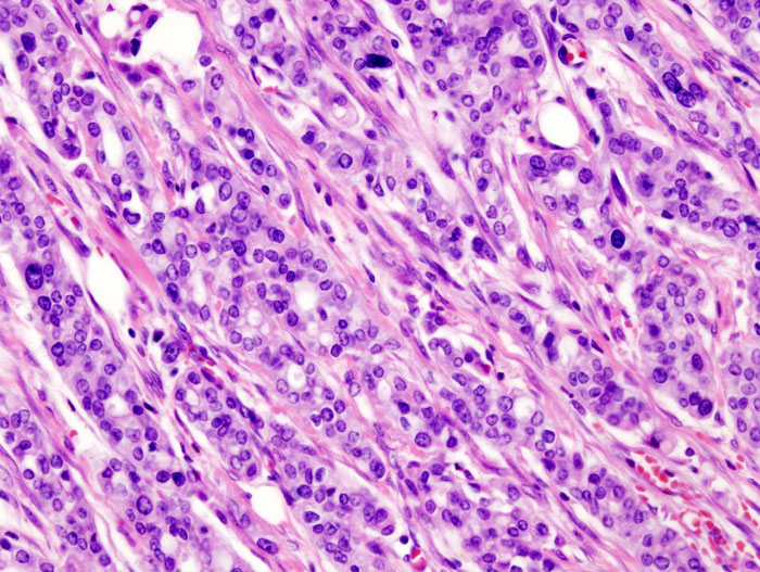 CC BY-SA 3.0, https://commons.wikimedia.org/w/index.php?curid=552900
 -
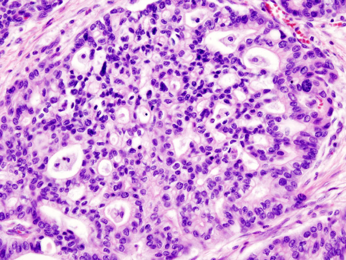 CC BY-SA 3.0, https://commons.wikimedia.org/w/index.php?curid=552901
 -
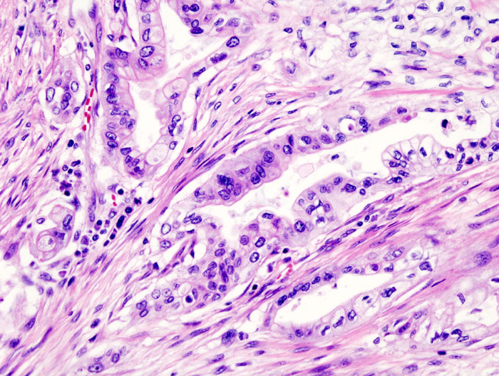 CC BY-SA 3.0, https://commons.wikimedia.org/w/index.php?curid=552904
 -
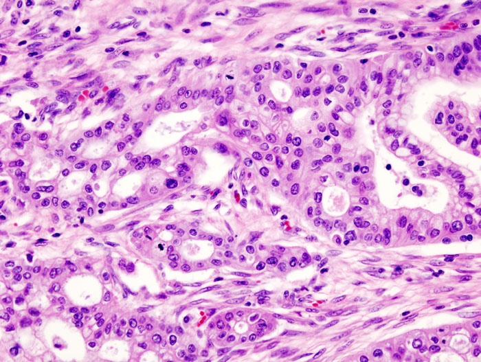 CC BY-SA 3.0, https://commons.wikimedia.org/w/index.php?curid=552903


References
- ↑ 1.0 1.1 Cowgill SM, Muscarella P (2003). “The genetics of pancreatic cancer”. Am. J. Surg. 186 (3): 279–86. PMID 12946833.
- ↑ Hruban RH, Petersen GM, Goggins M, Tersmette AC, Offerhaus GJ, Falatko F, Yeo CJ, Kern SE (1999). “Familial pancreatic cancer”. Ann. Oncol. 10 Suppl 4: 69–73. PMID 10436789.
- ↑ Greer JB, Whitcomb DC, Brand RE (2007). “Genetic predisposition to pancreatic cancer: a brief review”. Am. J. Gastroenterol. 102 (11): 2564–9. doi:10.1111/j.1572-0241.2007.01475.x. PMID 17958761.
- ↑ Soto JL, Barbera VM, Saceda M, Carrato A (2006). “Molecular biology of exocrine pancreatic cancer”. Clin Transl Oncol. 8 (5): 306–12. PMID 16760004.
- ↑ Shi C, Daniels JA, Hruban RH (2008). “Molecular characterization of pancreatic neoplasms”. Adv Anat Pathol. 15 (4): 185–95. doi:10.1097/PAP.0b013e31817bf57d. PMID 18580095.
- ↑ Blansfield JA, Choyke L, Morita SY, Choyke PL, Pingpank JF, Alexander HR, Seidel G, Shutack Y, Yuldasheva N, Eugeni M, Bartlett DL, Glenn GM, Middelton L, Linehan WM, Libutti SK (2007). “Clinical, genetic and radiographic analysis of 108 patients with von Hippel-Lindau disease (VHL) manifested by pancreatic neuroendocrine neoplasms (PNETs)”. Surgery. 142 (6): 814–8, discussion 818.e1–2. doi:10.1016/j.surg.2007.09.012. PMID 18063061.
- ↑ Kouvaraki MA, Shapiro SE, Cote GJ, Lee JE, Yao JC, Waguespack SG, Gagel RF, Evans DB, Perrier ND (2006). “Management of pancreatic endocrine tumors in multiple endocrine neoplasia type 1”. World J Surg. 30 (5): 643–53. doi:10.1007/s00268-006-0360-y. PMID 16680581.
- ↑ 8.0 8.1 Maitra A, Kern SE, Hruban RH (2006). “Molecular pathogenesis of pancreatic cancer”. Best Pract Res Clin Gastroenterol. 20 (2): 211–26. doi:10.1016/j.bpg.2005.10.002. PMID 16549325.
- ↑ Mimeault M, Brand RE, Sasson AA, Batra SK (2005). “Recent advances on the molecular mechanisms involved in pancreatic cancer progression and therapies”. Pancreas. 31 (4): 301–16. PMID 16258363.
- ↑ Talar-Wojnarowska R, Malecka-Panas E (2006). “Molecular pathogenesis of pancreatic adenocarcinoma: potential clinical implications”. Med. Sci. Monit. 12 (9): RA186–93. PMID 16940943.
- ↑ Neuman WL, Wasylyshyn ML, Jacoby R, Erroi F, Angriman I, Montag A, Brasitus T, Michelassi F, Westbrook CA (1991). “Evidence for a common molecular pathogenesis in colorectal, gastric, and pancreatic cancer”. Genes Chromosomes Cancer. 3 (6): 468–73. PMID 1663781.
- ↑ 12.0 12.1 Matthaios D, Zarogoulidis P, Balgouranidou I, Chatzaki E, Kakolyris S (2011). “Molecular pathogenesis of pancreatic cancer and clinical perspectives”. Oncology. 81 (3–4): 259–72. doi:10.1159/000334449. PMID 22116519.
- ↑ Konner J, O’Reilly E (2002). “Pancreatic cancer: epidemiology, genetics, and approaches to screening”. Oncology (Williston Park, N.Y.). 16 (12): 1615–22, 1631–2, discussion 1632–3, 1637–8. PMID 12520639.
- ↑ Hilgers W, Rosty C, Hahn SA (2002). “Molecular pathogenesis of pancreatic cancer”. Hematol. Oncol. Clin. North Am. 16 (1): 17–35, v. PMID 12063826.
- ↑ Hruban RH, Iacobuzio-Donahue C, Wilentz RE, Goggins M, Kern SE (2001). “Molecular pathology of pancreatic cancer”. Cancer J. 7 (4): 251–8. PMID 11561601.
- ↑ Singh S, Chitkara D, Kumar V, Behrman SW, Mahato RI (2013). “miRNA profiling in pancreatic cancer and restoration of chemosensitivity”. Cancer Lett. 334 (2): 211–20. doi:10.1016/j.canlet.2012.10.008. PMID 23073476.
- ↑ Yan L, McFaul C, Howes N, Leslie J, Lancaster G, Wong T, Threadgold J, Evans J, Gilmore I, Smart H, Lombard M, Neoptolemos J, Greenhalf W (2005). “Molecular analysis to detect pancreatic ductal adenocarcinoma in high-risk groups”. Gastroenterology. 128 (7): 2124–30. PMID 15940643.
- ↑ Jones S, Zhang X, Parsons DW, Lin JC, Leary RJ, Angenendt P, Mankoo P, Carter H, Kamiyama H, Jimeno A, Hong SM, Fu B, Lin MT, Calhoun ES, Kamiyama M, Walter K, Nikolskaya T, Nikolsky Y, Hartigan J, Smith DR, Hidalgo M, Leach SD, Klein AP, Jaffee EM, Goggins M, Maitra A, Iacobuzio-Donahue C, Eshleman JR, Kern SE, Hruban RH, Karchin R, Papadopoulos N, Parmigiani G, Vogelstein B, Velculescu VE, Kinzler KW (2008). “Core signaling pathways in human pancreatic cancers revealed by global genomic analyses”. Science. 321 (5897): 1801–6. doi:10.1126/science.1164368. PMC 2848990. PMID 18772397.
- ↑ Li Y, Bhuiyan M, Vaitkevicius VK, Sarkar FH (1998). “Molecular analysis of the p53 gene in pancreatic adenocarcinoma”. Diagn. Mol. Pathol. 7 (1): 4–9. PMID 9646028.
- ↑ Garcea G, Neal CP, Pattenden CJ, Steward WP, Berry DP (2005). “Molecular prognostic markers in pancreatic cancer: a systematic review”. Eur. J. Cancer. 41 (15): 2213–36. doi:10.1016/j.ejca.2005.04.044. PMID 16146690.
- ↑ Schutte M, Hruban RH, Geradts J, Maynard R, Hilgers W, Rabindran SK, Moskaluk CA, Hahn SA, Schwarte-Waldhoff I, Schmiegel W, Baylin SB, Kern SE, Herman JG (1997). “Abrogation of the Rb/p16 tumor-suppressive pathway in virtually all pancreatic carcinomas”. Cancer Res. 57 (15): 3126–30. PMID 9242437.
- ↑ Goggins M, Kern SE, Offerhaus JA, Hruban RH (1999). “Progress in cancer genetics: lessons from pancreatic cancer”. Ann. Oncol. 10 Suppl 4: 4–8. PMID 10436774.
- ↑ 23.0 23.1 Klein AP, Hruban RH, Brune KA, Petersen GM, Goggins M (2001). “Familial pancreatic cancer”. Cancer J. 7 (4): 266–73. PMID 11561603.
- ↑ Mangray S, King TC (1998). “Molecular pathobiology of pancreatic adenocarcinoma”. Front. Biosci. 3: D1148–60. PMID 9820739.
- ↑ Wang C, Lu X (2000). “[Alterations of the p16 gene for the carcinogenesis in pancreas]”. Zhongguo Yi Xue Ke Xue Yuan Xue Bao (in Chinese). 22 (5): 491–3. PMID 12903437.
- ↑ Gerdes B, Bartsch DK, Ramaswamy A, Kersting M, Wild A, Schuermann M, Frey M, Rothmund M (2000). “Multiple primary tumors as an indicator for p16INK4a germline mutations in pancreatic cancer patients?”. Pancreas. 21 (4): 369–75. PMID 11075991.
- ↑ Goggins M, Schutte M, Lu J, Moskaluk CA, Weinstein CL, Petersen GM, Yeo CJ, Jackson CE, Lynch HT, Hruban RH, Kern SE (1996). “Germline BRCA2 gene mutations in patients with apparently sporadic pancreatic carcinomas”. Cancer Res. 56 (23): 5360–4. PMID 8968085.
- ↑ Hahn SA, Greenhalf B, Ellis I, Sina-Frey M, Rieder H, Korte B, Gerdes B, Kress R, Ziegler A, Raeburn JA, Campra D, Grützmann R, Rehder H, Rothmund M, Schmiegel W, Neoptolemos JP, Bartsch DK (2003). “BRCA2 germline mutations in familial pancreatic carcinoma”. J. Natl. Cancer Inst. 95 (3): 214–21. PMID 12569143.
- ↑ Murphy KM, Brune KA, Griffin C, Sollenberger JE, Petersen GM, Bansal R, Hruban RH, Kern SE (2002). “Evaluation of candidate genes MAP2K4, MADH4, ACVR1B, and BRCA2 in familial pancreatic cancer: deleterious BRCA2 mutations in 17%”. Cancer Res. 62 (13): 3789–93. PMID 12097290.
- ↑ Hruban RH, Petersen GM, Ha PK, Kern SE (1998). “Genetics of pancreatic cancer. From genes to families”. Surg. Oncol. Clin. N. Am. 7 (1): 1–23. PMID 9443984.
- ↑ Kojima K, Vickers SM, Adsay NV, Jhala NC, Kim HG, Schoeb TR, Grizzle WE, Klug CA (2007). “Inactivation of Smad4 accelerates Kras(G12D)-mediated pancreatic neoplasia”. Cancer Res. 67 (17): 8121–30. doi:10.1158/0008-5472.CAN-06-4167. PMID 17804724.
- ↑ Almoguera C, Shibata D, Forrester K, Martin J, Arnheim N, Perucho M (1988). “Most human carcinomas of the exocrine pancreas contain mutant c-K-ras genes”. Cell. 53 (4): 549–54. PMID 2453289.
- ↑ Laghi L, Orbetegli O, Bianchi P, Zerbi A, Di Carlo V, Boland CR, Malesci A (2002). “Common occurrence of multiple K-RAS mutations in pancreatic cancers with associated precursor lesions and in biliary cancers”. Oncogene. 21 (27): 4301–6. doi:10.1038/sj.onc.1205533. PMID 12082617.
- ↑ Cheng RF, Wang J, Zhang JY, Sun L, Zhao YR, Qiu ZQ, Sun BC, Sun Y (2016). “MicroRNA-506 is up-regulated in the development of pancreatic ductal adenocarcinoma and is associated with attenuated disease progression”. Chin J Cancer. 35 (1): 64. doi:10.1186/s40880-016-0128-9. PMC 4930606. PMID 27371108.
- ↑ Hayashi N, Egami H, Ogawa M (2002). “[Genetics of pancreatic cancer: recent advances in molecular diagnosis]”. Nihon Geka Gakkai Zasshi (in Japanese). 103 (6): 476–81. PMID 12094699.
- ↑ Howe JR, Conlon KC (1997). “The molecular genetics of pancreatic cancer”. Surg Oncol. 6 (1): 1–18. PMID 9364657.
- ↑ El-Rayes BF, Ali S, Sarkar FH, Philip PA (2004). “Cyclooxygenase-2-dependent and -independent effects of celecoxib in pancreatic cancer cell lines”. Mol. Cancer Ther. 3 (11): 1421–6. PMID 15542781.
- ↑ Hussain T, Gupta S, Adhami VM, Mukhtar H (2005). “Green tea constituent epigallocatechin-3-gallate selectively inhibits COX-2 without affecting COX-1 expression in human prostate carcinoma cells”. Int. J. Cancer. 113 (4): 660–9. doi:10.1002/ijc.20629. PMID 15455372.
- ↑ Wei D, Wang L, He Y, Xiong HQ, Abbruzzese JL, Xie K (2004). “Celecoxib inhibits vascular endothelial growth factor expression in and reduces angiogenesis and metastasis of human pancreatic cancer via suppression of Sp1 transcription factor activity”. Cancer Res. 64 (6): 2030–8. PMID 15026340.
- ↑ Okami J, Yamamoto H, Fujiwara Y, Tsujie M, Kondo M, Noura S, Oshima S, Nagano H, Dono K, Umeshita K, Ishikawa O, Sakon M, Matsuura N, Nakamori S, Monden M (1999). “Overexpression of cyclooxygenase-2 in carcinoma of the pancreas”. Clin. Cancer Res. 5 (8): 2018–24. PMID 10473081.
- ↑ Inoue S, Tezel E, Nakao A (2001). “Molecular diagnosis of pancreatic cancer”. Hepatogastroenterology. 48 (40): 933–8. PMID 11490843.
- ↑ Wang Z, Banerjee S, Li Y, Rahman KM, Zhang Y, Sarkar FH (2006). “Down-regulation of notch-1 inhibits invasion by inactivation of nuclear factor-kappaB, vascular endothelial growth factor, and matrix metalloproteinase-9 in pancreatic cancer cells”. Cancer Res. 66 (5): 2778–84. doi:10.1158/0008-5472.CAN-05-4281. PMID 16510599.
- ↑ Büchler P, Gazdhar A, Schubert M, Giese N, Reber HA, Hines OJ, Giese T, Ceyhan GO, Müller M, Büchler MW, Friess H (2005). “The Notch signaling pathway is related to neurovascular progression of pancreatic cancer”. Ann. Surg. 242 (6): 791–800, discussion 800–1. PMC 1409885. PMID 16327489.
- ↑ Miyamoto Y, Maitra A, Ghosh B, Zechner U, Argani P, Iacobuzio-Donahue CA, Sriuranpong V, Iso T, Meszoely IM, Wolfe MS, Hruban RH, Ball DW, Schmid RM, Leach SD (2003). “Notch mediates TGF alpha-induced changes in epithelial differentiation during pancreatic tumorigenesis”. Cancer Cell. 3 (6): 565–76. PMID 12842085.
- ↑ Wang Z, Zhang Y, Banerjee S, Li Y, Sarkar FH (2006). “Inhibition of nuclear factor kappab activity by genistein is mediated via Notch-1 signaling pathway in pancreatic cancer cells”. Int. J. Cancer. 118 (8): 1930–6. doi:10.1002/ijc.21589. PMID 16284950.
- ↑ Wang Z, Zhang Y, Banerjee S, Li Y, Sarkar FH (2006). “Notch-1 down-regulation by curcumin is associated with the inhibition of cell growth and the induction of apoptosis in pancreatic cancer cells”. Cancer. 106 (11): 2503–13. doi:10.1002/cncr.21904. PMID 16628653.
- ↑ Wang Z, Zhang Y, Li Y, Banerjee S, Liao J, Sarkar FH (2006). “Down-regulation of Notch-1 contributes to cell growth inhibition and apoptosis in pancreatic cancer cells”. Mol. Cancer Ther. 5 (3): 483–93. doi:10.1158/1535-7163.MCT-05-0299. PMID 16546962.
- ↑ Biliran H, Wang Y, Banerjee S, Xu H, Heng H, Thakur A, Bollig A, Sarkar FH, Liao JD (2005). “Overexpression of cyclin D1 promotes tumor cell growth and confers resistance to cisplatin-mediated apoptosis in an elastase-myc transgene-expressing pancreatic tumor cell line”. Clin. Cancer Res. 11 (16): 6075–86. doi:10.1158/1078-0432.CCR-04-2419. PMID 16115953.
- ↑ Kornmann M, Arber N, Korc M (1998). “Inhibition of basal and mitogen-stimulated pancreatic cancer cell growth by cyclin D1 antisense is associated with loss of tumorigenicity and potentiation of cytotoxicity to cisplatinum”. J. Clin. Invest. 101 (2): 344–52. doi:10.1172/JCI1323. PMC 508573. PMID 9435306.
- ↑ Bruns CJ, Harbison MT, Davis DW, Portera CA, Tsan R, McConkey DJ, Evans DB, Abbruzzese JL, Hicklin DJ, Radinsky R (2000). “Epidermal growth factor receptor blockade with C225 plus gemcitabine results in regression of human pancreatic carcinoma growing orthotopically in nude mice by antiangiogenic mechanisms”. Clin. Cancer Res. 6 (5): 1936–48. PMID 10815919.
- ↑ Marshall J (2006). “Clinical implications of the mechanism of epidermal growth factor receptor inhibitors”. Cancer. 107 (6): 1207–18. doi:10.1002/cncr.22133. PMID 16909423.
- ↑ Wang Z, Sengupta R, Banerjee S, Li Y, Zhang Y, Rahman KM, Aboukameel A, Mohammad R, Majumdar AP, Abbruzzese JL, Sarkar FH (2006). “Epidermal growth factor receptor-related protein inhibits cell growth and invasion in pancreatic cancer”. Cancer Res. 66 (15): 7653–60. doi:10.1158/0008-5472.CAN-06-1019. PMID 16885366.
- ↑ Zhang Y, Banerjee S, Wang Z, Xu H, Zhang L, Mohammad R, Aboukameel A, Adsay NV, Che M, Abbruzzese JL, Majumdar AP, Sarkar FH (2006). “Antitumor activity of epidermal growth factor receptor-related protein is mediated by inactivation of ErbB receptors and nuclear factor-kappaB in pancreatic cancer”. Cancer Res. 66 (2): 1025–32. doi:10.1158/0008-5472.CAN-05-2968. PMID 16424038.
- ↑ Zhang Y, Banerjee S, Wang ZW, Marciniak DJ, Majumdar AP, Sarkar FH (2005). “Epidermal growth factor receptor-related protein inhibits cell growth and induces apoptosis of BxPC3 pancreatic cancer cells”. Cancer Res. 65 (9): 3877–82. doi:10.1158/0008-5472.CAN-04-3654. PMID 15867387.
- ↑ Wang W, Abbruzzese JL, Evans DB, Chiao PJ (1999). “Overexpression of urokinase-type plasminogen activator in pancreatic adenocarcinoma is regulated by constitutively activated RelA”. Oncogene. 18 (32): 4554–63. doi:10.1038/sj.onc.1202833. PMID 10467400.
- ↑ Bava SV, Puliappadamba VT, Deepti A, Nair A, Karunagaran D, Anto RJ (2005). “Sensitization of taxol-induced apoptosis by curcumin involves down-regulation of nuclear factor-kappaB and the serine/threonine kinase Akt and is independent of tubulin polymerization”. J. Biol. Chem. 280 (8): 6301–8. doi:10.1074/jbc.M410647200. PMID 15590651.
- ↑ Fahy BN, Schlieman MG, Virudachalam S, Bold RJ (2004). “Inhibition of AKT abrogates chemotherapy-induced NF-kappaB survival mechanisms: implications for therapy in pancreatic cancer”. J. Am. Coll. Surg. 198 (4): 591–9. doi:10.1016/j.jamcollsurg.2003.12.005. PMID 15051014.
- ↑ Fujioka S, Sclabas GM, Schmidt C, Frederick WA, Dong QG, Abbruzzese JL, Evans DB, Baker C, Chiao PJ (2003). “Function of nuclear factor kappaB in pancreatic cancer metastasis”. Clin. Cancer Res. 9 (1): 346–54. PMID 12538487.
- ↑ Karin M (2006). “Nuclear factor-kappaB in cancer development and progression”. Nature. 441 (7092): 431–6. doi:10.1038/nature04870. PMID 16724054.
- ↑ Li L, Aggarwal BB, Shishodia S, Abbruzzese J, Kurzrock R (2004). “Nuclear factor-kappaB and IkappaB kinase are constitutively active in human pancreatic cells, and their down-regulation by curcumin (diferuloylmethane) is associated with the suppression of proliferation and the induction of apoptosis”. Cancer. 101 (10): 2351–62. doi:10.1002/cncr.20605. PMID 15476283.
- ↑ Li Y, Ahmed F, Ali S, Philip PA, Kucuk O, Sarkar FH (2005). “Inactivation of nuclear factor kappaB by soy isoflavone genistein contributes to increased apoptosis induced by chemotherapeutic agents in human cancer cells”. Cancer Res. 65 (15): 6934–42. doi:10.1158/0008-5472.CAN-04-4604. PMID 16061678.
- ↑ Li Y, Chinni SR, Sarkar FH (2005). “Selective growth regulatory and pro-apoptotic effects of DIM is mediated by AKT and NF-kappaB pathways in prostate cancer cells”. Front. Biosci. 10: 236–43. PMID 15574364.
- ↑ Liptay S, Weber CK, Ludwig L, Wagner M, Adler G, Schmid RM (2003). “Mitogenic and antiapoptotic role of constitutive NF-kappaB/Rel activity in pancreatic cancer”. Int. J. Cancer. 105 (6): 735–46. doi:10.1002/ijc.11081. PMID 12767057.
- ↑ {{cite journal |vauthors=Rahman KW, Sarkar FH |title=Inhibition of nuclear translocation of nuclear factor-{kappa}B contributes to 3,3′-diindolylmethane-induced apoptosis in breast cancer cells |journal=Cancer Res. |volume=65 |issue=1 |pages=364–71 |year=2005 |pmid=15665315 |doi= |url=}}
- ↑ Zhang H, Morisaki T, Nakahara C, Matsunaga H, Sato N, Nagumo F, Tadano J, Katano M (2003). “PSK-mediated NF-kappaB inhibition augments docetaxel-induced apoptosis in human pancreatic cancer cells NOR-P1”. Oncogene. 22 (14): 2088–96. doi:10.1038/sj.onc.1206310. PMID 12687011.
- ↑ Nakashima H, Nakamura M, Yamaguchi H, Yamanaka N, Akiyoshi T, Koga K, Yamaguchi K, Tsuneyoshi M, Tanaka M, Katano M (2006). “Nuclear factor-kappaB contributes to hedgehog signaling pathway activation through sonic hedgehog induction in pancreatic cancer”. Cancer Res. 66 (14): 7041–9. doi:10.1158/0008-5472.CAN-05-4588. PMID 16849549.
- ↑ Thayer SP, di Magliano MP, Heiser PW, Nielsen CM, Roberts DJ, Lauwers GY, Qi YP, Gysin S, Fernández-del Castillo C, Yajnik V, Antoniu B, McMahon M, Warshaw AL, Hebrok M (2003). “Hedgehog is an early and late mediator of pancreatic cancer tumorigenesis”. Nature. 425 (6960): 851–6. doi:10.1038/nature02009. PMC 3688051. PMID 14520413.
- ↑ Berman DM, Karhadkar SS, Maitra A, Montes De Oca R, Gerstenblith MR, Briggs K, Parker AR, Shimada Y, Eshleman JR, Watkins DN, Beachy PA (2003). “Widespread requirement for Hedgehog ligand stimulation in growth of digestive tract tumours”. Nature. 425 (6960): 846–51. doi:10.1038/nature01972. PMID 14520411.
- ↑ Mathew E, Zhang Y, Holtz AM, Kane KT, Song JY, Allen BL, Pasca di Magliano M (2014). “Dosage-dependent regulation of pancreatic cancer growth and angiogenesis by hedgehog signaling”. Cell Rep. 9 (2): 484–94. doi:10.1016/j.celrep.2014.09.010. PMC 4362534. PMID 25310976.
- ↑ Castet F, Fabregat-Franco C, Castillo G, et al. Clinical and genomic characterisation of early-onset pancreatic cancer. Eur J Cancer. 2023;194:113338. doi:10.1016/j.ejca.2023.113338
- ↑ Esposito I, Konukiewitz B, Schlitter AM, Klöppel G (2014). “Pathology of pancreatic ductal adenocarcinoma: facts, challenges, and future developments”. World J. Gastroenterol. 20 (38): 13833–41. doi:10.3748/wjg.v20.i38.13833. PMC 4194566. PMID 25320520.
- ↑ Zironda A, Zhang C, Day C, et al. Early vs conventional onset pancreatic ductal adenocarcinoma: analysis of surgical and oncologic outcomes in patients undergoing curative intent resection. HPB (Oxford). 2024;26(1):145-153. doi:10. 1016/j.hpb.2023.09.010
- ↑ Ordonez JE, Hester CA, Zhu H, et al. Clinicopathologic features and outcomes of early-onset pancreatic adenocarcinoma in the United States. Ann Surg Oncol. 2020;27(6):1997- 2006. doi:10.1245/s10434-019-08096-y
- ↑ Rossi ML, Rehman AA, Gondi CS (2014). “Therapeutic options for the management of pancreatic cancer”. World J. Gastroenterol. 20 (32): 11142–59. doi:10.3748/wjg.v20.i32.11142. PMC 4145755. PMID 25170201.
- ↑ Oda Y, Aishima S, Morimatsu K, Shindo K, Fujino M, Mizuuchi Y, Hattori M, Miyazaki T, Tanaka M, Oda Y (2014). “Pancreatic intraepithelial neoplasia in the background of invasive ductal carcinoma of the pancreas as a prognostic factor”. Histopathology. 65 (3): 389–97. doi:10.1111/his.12397. PMID 24931343.
- ↑ Yachida S, Jones S, Bozic I, Antal T, Leary R, Fu B, Kamiyama M, Hruban RH, Eshleman JR, Nowak MA, Velculescu VE, Kinzler KW, Vogelstein B, Iacobuzio-Donahue CA (2010). “Distant metastasis occurs late during the genetic evolution of pancreatic cancer”. Nature. 467 (7319): 1114–7. doi:10.1038/nature09515. PMC 3148940. PMID 20981102.
- ↑ Campbell PJ, Yachida S, Mudie LJ, Stephens PJ, Pleasance ED, Stebbings LA, Morsberger LA, Latimer C, McLaren S, Lin ML, McBride DJ, Varela I, Nik-Zainal SA, Leroy C, Jia M, Menzies A, Butler AP, Teague JW, Griffin CA, Burton J, Swerdlow H, Quail MA, Stratton MR, Iacobuzio-Donahue C, Futreal PA (2010). “The patterns and dynamics of genomic instability in metastatic pancreatic cancer”. Nature. 467 (7319): 1109–13. doi:10.1038/nature09460. PMC 3137369. PMID 20981101.
Editor-In-Chief: C. Michael Gibson, M.S., M.D. [1]; Associate Editor(s)-in-Chief: Sudarshana Datta, MD [2]
Causes
Overview
Pancreatic ductal adenocarcinoma (PDAC) develops from somatic driver mutations that accumulate in steps, on a background of inherited and acquired predisposition. Four genes make up the core molecular signature of PDAC. The first is an activating KRAS mutation, found in about 85–95% of tumors. The other three are tumor suppressor genes that are inactivated: CDKN2A, TP53, and SMAD4. These alterations are acquired one after another as precursor lesions progress to invasive cancer.[1][2][3][4] About 5–10% of unselected patients carry a pathogenic germline variant, most often in DNA damage repair genes. The proportion rises to about 30% in early-onset disease.[5][2][6] Most PDAC is sporadic. Established acquired risk factors include cigarette smoking, obesity, long-standing type 2 diabetes, and chronic pancreatitis.[2][7][8][9] The cause of a patient’s PDAC now shapes management. Universal germline testing is recommended for every patient with PDAC, and tumor profiling finds a minority with targetable alterations.[8][10][11][12]
Causes
Somatic driver mutations
Exome and whole-genome sequencing consistently find four recurrently altered driver genes in PDAC, often called the genomic “mountains.” Alongside them is a long tail of low-frequency “hills.” The average PDAC exome carries only about 40 somatic mutations, which is a low burden for an adenocarcinoma.[1][4][13]
| Gene (locus) | Class | Approximate frequency | Mechanism and timing | Clinical relevance |
|---|---|---|---|---|
| KRAS (12p) | Oncogene | ~85–95% | Activating point mutation, most often at codon 12, less often at codons 13 and 61. It is the earliest, initiating event and is already present in low-grade PanIN. It drives MAPK/ERK signaling. | G12D is the most common variant and is associated with worse survival. G12R is enriched in early-stage disease. G12C is rare but can be targeted with KRAS G12C inhibitors. About 8–13% of tumors are KRAS wild-type, and these are enriched for targetable gene fusions.[1][2][3][7][14] |
| CDKN2A (9p21; p16) | Tumor suppressor | ~60–95% (up to ~95% when deletion and methylation are counted) | Inactivated by homozygous deletion (~40%), loss of heterozygosity plus mutation (~40%), or promoter hypermethylation (~15%). Loss follows KRAS mutation. | When the 9p21 deletion also removes the neighboring interferon gene cluster, the tumor microenvironment becomes immunologically “cold.” Deletion and methylation are missed by mutation-only panels.[4][15][16] |
| TP53 (17p) | Tumor suppressor | ~70–85% | Missense mutation with loss of the second allele. It is a late event, appearing in high-grade PanIN, and drives genomic instability. | Associated with a tendency to metastasize.[3][4][13][15] |
| SMAD4/DPC4 (18q) | Tumor suppressor | ~20–55% | Homozygous deletion, or loss of heterozygosity plus mutation. It is a late event and disrupts TGF-β signaling. | Loss predicts a higher metastatic burden and worse prognosis.[2][3][4][15] |
Several low-frequency drivers are biologically and clinically important:[1][4][13]
- Homologous recombination repair genes: BRCA1, BRCA2, PALB2, and ATM
- Chromatin remodeling genes: ARID1A, KDM6A, PBRM1, and SMARCA4
- TGF-β pathway components: TGFBR2, ACVR1B, and ACVR2A
- GNAS and RNF43: characteristic of PDAC that arises from intraductal papillary mucinous neoplasms (IPMNs)
These drivers fit together into a small number of core signaling pathways. That pathway-level map has replaced older descriptions of nonspecific “deregulation” of signaling molecules.[17][18] Whole-genome analyses also detect large-scale structural events. These include copy-number changes, chromothripsis, and kataegis, which are clusters of substitutions associated with APOBEC enzymes.[13] Prognosis and treatment selection can be refined beyond single-gene status in two further ways. Transcriptomic subtyping separates classical from basal-like tumors. Whole-genome structural classes divide tumors into stable, scattered, and unstable groups.[2][8]
Precursor lesions and multistep progression
About 85–95% of PDACs arise from pancreatic intraepithelial neoplasia (PanIN). About 15–20% arise from macroscopic cystic precursors, most often IPMNs.[14][4][7] The cell of origin is still debated. Genetic and mouse-model data support acinar-to-ductal metaplasia triggered by oncogenic KRAS activation.[14][4] The classic sequence starts with KRAS mutation in low-grade PanIN. CDKN2A inactivation follows, and TP53 and SMAD4 loss occur in high-grade lesions.[2][19][8] Low-grade PanINs are present in 75–85% of adult pancreata, and nearly all of them harbor KRAS mutations, yet very few progress. Progression requires tumor suppressors to be lost one after another over decades. The lesion must also escape immune surveillance and overcome a stroma that does not support tumor growth.[2]
Germline predisposition and hereditary syndromes
About 5–10% of unselected PDAC patients carry a pathogenic germline variant. The figure is about 30% in early-onset disease.[5][2][6] About half of carriers have no family history of the disease.[10]
| Syndrome or pathway | Gene(s) | Key points |
|---|---|---|
| Homologous recombination repair deficiency, including hereditary breast and ovarian cancer syndrome | BRCA2, BRCA1, PALB2, ATM | BRCA2 confers the strongest risk among the common genes (OR ~9), and ATM confers a similar risk (OR ~9). These variants predict sensitivity to platinum chemotherapy and PARP inhibitors. |
| Familial atypical multiple mole melanoma | CDKN2A | Among the highest relative risks. Surveillance is recommended regardless of family history. |
| Peutz-Jeghers syndrome | STK11 | Among the highest relative risks. Surveillance is recommended regardless of family history. |
| Lynch syndrome | MLH1, MSH2, MSH6, PMS2, EPCAM | Produces MSI-high/dMMR tumors that respond to immune checkpoint inhibitors. |
| Li-Fraumeni syndrome | TP53 | Rare germline cause. |
| Hereditary pancreatitis | PRSS1, SPINK1 | Lifetime risk of up to ~40%, driven by recurrent inflammation.[5][8][20][10] |
Acquired and environmental risk factors
Most PDAC is sporadic. The established acquired risk factors are summarized below; see Pancreatic cancer risk factors for more detail.[2][7][8][9]
| Factor | Comments |
|---|---|
| Cigarette smoking | The leading modifiable risk factor. Risk falls after quitting. |
| Obesity (BMI ≥30) | Metabolic syndrome carries an RR of ~1.3. |
| Long-standing type 2 diabetes | Diabetes that starts after age 50 can be an early sign of hidden PDAC. About 0.3–1% of these patients are diagnosed with PDAC within 3 years. |
| Chronic pancreatitis | The association is strongest close to the time of diagnosis. Some of it may reflect pancreatitis caused by a tumor that has not yet been found.[13] |
| Heavy alcohol use (≥3 drinks/day) | The effect is largely mediated through pancreatitis. |
| Other | Non-O ABO blood group, tall height, and diets high in red or processed meat. Associations with certain chronic infections are supported only by modest-quality evidence.[7][9][6] |
Clinical implications of etiology
- Universal germline testing: NCCN and ASCO recommend germline testing for every patient with newly diagnosed PDAC, whether or not there is a family history. Testing should use a multigene panel that includes BRCA1/2, PALB2, ATM, CDKN2A, STK11, and the mismatch repair genes (NCCN category 2A).[8][10][11][12] Testing only patients with a family history misses about half of carriers.[10]
- Somatic tumor profiling: Recommended in locally advanced or metastatic disease to look for actionable alterations. These include BRCA1/2 and PALB2 (platinum or PARP inhibitors), MSI-H/dMMR (immunotherapy), KRAS G12C, and rare NTRK or other fusions. KRAS wild-type tumors should be profiled with particular care.[11][8][18] Even so, matched targeted therapy applies to fewer than 5% of PDACs.[18]
- Risk modification: Counsel patients on quitting smoking. In adults aged 50 or older with new-onset diabetes and other suggestive features, evaluate for an occult malignancy.[2][7]
- Cascade testing and surveillance: Refer high-risk germline carriers for surveillance; see Pancreatic cancer screening.[12][20]
Areas of uncertainty
- Whether PDAC originates from acinar or ductal cells remains unresolved.[14][4]
- It is disputed whether heavy alcohol use is an independent risk factor or acts only through pancreatitis.[8][9]
- Guidelines differ on surveillance eligibility for BRCA1, ATM, and PALB2 carriers who have no family history.[20][12]
- Germline risk profiles vary by ancestry. For example, BRCA1/2, ATM, and CDKN2A variants are less frequent in Asian cohorts, which limits how widely a single panel applies.[21]
References
- ↑ 1.0 1.1 1.2 1.3 Connor AA, Gallinger S (2022). “Pancreatic cancer evolution and heterogeneity: integrating omics and clinical data”. Nat Rev Cancer. 22 (3): 131–142. doi:10.1038/s41568-021-00418-1.
- ↑ 2.00 2.01 2.02 2.03 2.04 2.05 2.06 2.07 2.08 2.09 2.10 Roth S, Apte M, Balachandran VP, Biffi G, Conroy T, Cukierman E; et al. (2026). “Pancreatic cancer”. Nat Rev Dis Primers. 12 (1): 23. doi:10.1038/s41572-026-00699-6.
- ↑ 3.0 3.1 3.2 3.3 Halbrook CJ, Lyssiotis CA, Pasca di Magliano M, Maitra A (2023). “Pancreatic cancer: Advances and challenges”. Cell. 186 (8): 1729–1754. doi:10.1016/j.cell.2023.02.014. PMID 37059070 Check
|pmid=value (help). - ↑ 4.0 4.1 4.2 4.3 4.4 4.5 4.6 4.7 4.8 Luchini C (2026). “Pancreatic ductal adenocarcinoma and its subtypes: clinical relevance of histopathology and molecular characterization, integrating the key updates of the 2026 WHO classification”. Virchows Arch. doi:10.1007/s00428-026-04681-1.
- ↑ 5.0 5.1 5.2 Karloski E, Dudley B, Diergaarde B; et al. (2024). “The role of family history in predicting germline pathogenic variant carriers who develop pancreatic cancer: Results of a multi-center collaboration”. Cancer. 130 (19): 3297–3304. doi:10.1002/cncr.35383.
- ↑ 6.0 6.1 6.2 Jayakrishnan T, Ng K (2025). “Early-onset gastrointestinal cancers: A review”. JAMA. 334 (15): 1373–1385. doi:10.1001/jama.2025.10218.
- ↑ 7.0 7.1 7.2 7.3 7.4 7.5 Stoop TF, Javed AA, Oba A; et al. (2025). “Pancreatic cancer”. Lancet. 405 (10485): 1182–1202. doi:10.1016/S0140-6736(25)00261-2.
- ↑ 8.0 8.1 8.2 8.3 8.4 8.5 8.6 8.7 8.8 Park W, Chawla A, O’Reilly EM (2021). “Pancreatic cancer: A review”. JAMA. 326 (9): 851–862. doi:10.1001/jama.2021.13027.
- ↑ 9.0 9.1 9.2 9.3 Khalaf N, El-Serag HB, Abrams HR, Thrift AP (2021). “Burden of pancreatic cancer: From epidemiology to practice”. Clin Gastroenterol Hepatol. 19 (5): 876–884. doi:10.1016/j.cgh.2020.02.054.
- ↑ 10.0 10.1 10.2 10.3 10.4 Mizrahi JD, Surana R, Valle JW, Shroff RT (2020). “Pancreatic cancer”. Lancet. 395 (10242): 2008–2020. doi:10.1016/S0140-6736(20)30974-0.
- ↑ 11.0 11.1 11.2 Grewal US, Patel RR, Loeffler BT; et al. (2026). “Germline genetic testing among patients with pancreatic adenocarcinoma: A Pancreatic Cancer Action Network patient survey”. Cancer. 132 (10): e70446. doi:10.1002/cncr.70446.
- ↑ 12.0 12.1 12.2 12.3 National Comprehensive Cancer Network. NCCN Clinical Practice Guidelines in Oncology: Genetic/Familial High-Risk Assessment: Breast, Ovarian, Pancreatic, and Prostate. Updated August 31, 2026.
- ↑ 13.0 13.1 13.2 13.3 13.4 Wood LD, Canto MI, Jaffee EM, Simeone DM (2022). “Pancreatic cancer: Pathogenesis, screening, diagnosis, and treatment”. Gastroenterology. 163 (2): 386–402.e1. doi:10.1053/j.gastro.2022.03.056.
- ↑ 14.0 14.1 14.2 14.3 Drizyte-Miller K, Talabi T, Somasundaram A, Cox AD, Der CJ (2025). “KRAS: the Achilles’ heel of pancreas cancer biology”. J Clin Invest. 135 (16): e191939. doi:10.1172/JCI191939.
- ↑ 15.0 15.1 15.2 Wood LD, Yurgelun MB, Goggins MG (2019). “Genetics of familial and sporadic pancreatic cancer”. Gastroenterology. 156 (7): 2041–2055. doi:10.1053/j.gastro.2018.12.039.
- ↑ Ferreira S, Flowers BM, Choi WY; et al. (2026). “Inactivation of CDKN2A ARF promotes p53-independent remodeling of the PDAC tumor microenvironment”. Cancer Res. 86 (13): 3109–3122. doi:10.1158/0008-5472.CAN-25-1969.
- ↑ Hayashi A, Hong J, Iacobuzio-Donahue CA (2021). “The pancreatic cancer genome revisited”. Nat Rev Gastroenterol Hepatol. 18 (7): 469–481. doi:10.1038/s41575-021-00463-z.
- ↑ 18.0 18.1 18.2 Bijlsma MF, Bailey P (2023). “Molecular subtypes and clinical applications”. The Pancreas.
- ↑ Oh G, Dhara S, Simeone D (2023). “Precision cancer medicine”. The Pancreas.
- ↑ 20.0 20.1 20.2 Monahan K, Hampel H, Stoffel E, Kupfer SS (2025). “In our scope of practice: Genetic risk assessment and testing for gastrointestinal cancers and polyposis in gastroenterology”. Gastroenterology. 169 (6): 1147–1165. doi:10.1053/j.gastro.2025.06.001.
- ↑ Yin X, Shen H, Wang H; et al. (2025). “Pathogenic germline variants in Chinese pancreatic adenocarcinoma patients”. Nat Commun. 16 (1): 2214. doi:10.1038/s41467-025-57520-3.
Editor-In-Chief: C. Michael Gibson, M.S., M.D. [1]; Associate Editor(s)-in-Chief: Fahad Hasan, M.D.[2] Aravind Reddy Kothagadi M.B.B.S[3]
Differentiating Pancreatic Cancer from other Disorders

Overview
Pancreatic cancer must be distinguished from other pancreatobiliary pathologies that cause abdominal pain and jaundice such as acute suppurative cholangitis, acute cholecystitis, cholelithiasis, chronic pancreatitis, primary biliary cirrhosis and primary sclerosing cholangitis. Pancreatic cancer must also be differentiated from other pancreatic pathologies such as autoimmune pancreatitis, pancreatic pseudocyst, and neuroendocrine tumors of the pancreas. Pathologies of the bile duct and duodenum such as Choledocholithiasis, gallstones (cholelithiasis), choledochal cysts, Cholangiocarcinoma, Bile duct strictures and ampullary cancer should be differentiated from pancreatic cancer based on imaging and biopsy findings. Metastasis from different sites and vascular causes such as abdominal aortic aneurysms may also mimic pancreatic cancer.
Differentiating Pancreatic Cancer from other Diseases
Pancreatic cancer may also be distinguished from other causes of abdominal pain and jaundice as depicted in the table below:
| ||||||||||||||||||||||||||||||||||||||||||||||||||||||||||||||||||||||||||||||||||||||||||||||||||||||||||||||||||||||||||||||||||||||||||||||||||||||||||||||||||||||||||||||||||||||||||||||||||||||||||||||||||||||||||||||||||||||||||||||||||||||||||||||||||
Differentials based on weight loss, nausea, vomiting, diarrhea, fatigue and abdominal pain
Pancreatic carcinoma should be differentiated from other diseases presenting with weight loss, nausea, vomiting, diarrhea, fatigue and abdominal pain. The differentials include the following:
| Disease | Clinical manifestations | Diagnosis | Comments | |||||||||||||
|---|---|---|---|---|---|---|---|---|---|---|---|---|---|---|---|---|
| Symptoms | Signs | |||||||||||||||
| Abdominal Pain | Fever | Rigors and chills | Nausea or vomiting | Jaundice | Constipation | Diarrhea | Weight loss | GI bleeding | Hypo-
tension |
Guarding | Rebound Tenderness | Bowel sounds | Lab Findings | Imaging | ||
| Chronic pancreatitis | Epigastric | − | − | ± | ± | − | + | + | − | − | − | − | N | CT scan
|
| |
| Pancreatic carcinoma | Epigastric | − | − | + | + | − | + | + | − | − | − | − | N |
Skin manifestations may include: | ||
| Dumping syndrome | Lower and then diffuse | − | − | + | − | − | + | + | − | + | − | − | Hyperactive |
|
|
|
Abbreviations:
RUQ= Right upper quadrant of the abdomen, LUQ= Left upper quadrant, LLQ= Left lower quadrant, RLQ= Right lower quadrant, LFT= Liver function test, SIRS= Systemic inflammatory response syndrome, ERCP= Endoscopic retrograde cholangiopancreatography, IV= Intravenous, N= Normal, AMA= Anti mitochondrial antibodies, LDH= Lactate dehydrogenase, GI= Gastrointestinal, CXR= Chest X ray, IgA= Immunoglobulin A, IgG= Immunoglobulin G, IgM= Immunoglobulin M, CT= Computed tomography, PMN= Polymorphonuclear cells, ESR= Erythrocyte sedimentation rate, CRP= C-reactive protein, TS= Transferrin saturation, SF= Serum Ferritin, SMA= Superior mesenteric artery, SMV= Superior mesenteric vein, ECG= Electrocardiogram
To review a comprehensive differential diagnosis of diseases presenting with abdominal pain, click here.
Differentiating Pancreatic Carcinoma on the basis of involved Organ
The differentials for pancreatic cancer mainly involve pathologies of the pancreas, bile duct, duodenum, lymphovascular tissue or metastasis from other sites. It is difficult to differentiate pancreatic cancer from other neoplasms as they all present with non specific constitutional symptoms. Hence, differentiation is primarily based on anatomic location:[1][2][3][4][5][6][7][8][9][10][11][12][13][14][15][16][17][18][19][20][21][22][23][24][25][26][27][28]
| Origin | Disease | Diagnostic study of choice |
|---|---|---|
| Pancreas |
| |
| Bile duct | ||
| Duodenum |
|
|
| Lymphovascular
tissue |
| |
| Metastasis |
References
- ↑ He XK, Ding Y, Sun LM (2017). “Contrast-enhanced endoscopic ultrasound for differential diagnosis of pancreatic cancer: an updated meta-analysis”. Oncotarget. 8 (39): 66392–66401. doi:10.18632/oncotarget.18915. PMC 5630421. PMID 29029521.
- ↑ Steinman J, Zaheer A, Kluger MD, Remotti H, Hecht EM (2017). “Rare pancreatic tumors”. Abdom Radiol (NY). doi:10.1007/s00261-017-1342-x. PMID 29022085.
- ↑ Ran L, Zhao W, Zhao Y, Bu H (2017). “Value of contrast-enhanced ultrasound in differential diagnosis of solid lesions of pancreas (SLP): A systematic review and a meta-analysis”. Medicine (Baltimore). 96 (28): e7463. doi:10.1097/MD.0000000000007463. PMC 5515757. PMID 28700485.
- ↑ Kołodziejczyk E, Wejnarska K, Oracz G (2016). “Autoimmune pancreatitis in the paediatric population – review of the literature and own experience”. Dev Period Med. 20 (4): 279–286. PMID 28216481.
- ↑ Zhu L, Xue HD, Liu W, Wang X, Sui X, Wang Q, Zhang D, Li P, Jin ZY (2017). “Enhancing pancreatic mass with normal serum CA19-9: key MDCT features to characterize pancreatic neuroendocrine tumours from its mimics”. Radiol Med. 122 (5): 337–344. doi:10.1007/s11547-017-0734-x. PMID 28197876.
- ↑ Sano I, Katanuma A, Yane K, Kin T, Nagai K, Yamazaki H, Koga H, Kitagawa K, Yokoyama K, Ikarashi S, Takahashi K, Maguchi H, Omori Y, Shinohara T (2017). “Pancreatic Metastasis from Rectal Cancer that was Diagnosed by Endoscopic Ultrasonography-guided Fine Needle Aspiration (EUS-FNA)”. Intern. Med. 56 (3): 301–305. doi:10.2169/internalmedicine.56.7213. PMC 5348454. PMID 28154274.
- ↑ Salaria SN, Shi C (2016). “Pancreatic Neuroendocrine Tumors”. Surg Pathol Clin. 9 (4): 595–617. doi:10.1016/j.path.2016.05.006. PMID 27926362.
- ↑ Kawasaki K, Kawaguchi Y, Suzuki Y, Tanaka N (2016). “Renal neuroendocrine tumour and synchronous pancreas metastasis: histopathological diagnosis using prostatic acid phosphatase”. BMJ Case Rep. 2016. doi:10.1136/bcr-2016-214759. PMID 27803081.
- ↑ Nassour I, Choti MA (2016). “Diagnosis and Treatment of Pancreatic Cystic Neoplasms”. JAMA. 316 (12): 1326. doi:10.1001/jama.2016.9130. PMID 27673319.
- ↑ Javadi S, Menias CO, Korivi BR, Shaaban AM, Patnana M, Alhalabi K, Elsayes KM (2017). “Pancreatic Calcifications and Calcified Pancreatic Masses: Pattern Recognition Approach on CT”. AJR Am J Roentgenol. 209 (1): 77–87. doi:10.2214/AJR.17.17862. PMID 28418702.
- ↑ Bergmann F (2016). “[Pancreatic acinar neoplasms : Comparative molecular characterization]”. Pathologe (in German). 37 (Suppl 2): 191–195. doi:10.1007/s00292-016-0235-z. PMID 27807633.
- ↑ Cheng SK, Chuah KL (2016). “Metastatic Renal Cell Carcinoma to the Pancreas: A Review”. Arch. Pathol. Lab. Med. 140 (6): 598–602. doi:10.5858/arpa.2015-0135-RS. PMID 27232353.
- ↑ Haage P, Schwartz CA, Scharwächter C (2016). “[Ductal adenocarcinoma and unusual differential diagnosis]”. Radiologe (in German). 56 (4): 325–37. doi:10.1007/s00117-016-0090-1. PMID 27000276.
- ↑ Scialpi M, Reginelli A, D’Andrea A, Gravante S, Falcone G, Baccari P, Manganaro L, Palumbo B, Cappabianca S (2016). “Pancreatic tumors imaging: An update”. Int J Surg. 28 Suppl 1: S142–55. doi:10.1016/j.ijsu.2015.12.053. PMID 26777740.
- ↑ Senosiain Lalastra C, Foruny Olcina JR (2015). “[Autoimmune pancreatitis]”. Gastroenterol Hepatol (in Spanish; Castilian). 38 (9): 549–55. doi:10.1016/j.gastrohep.2015.01.006. PMID 25799073.
- ↑ Barbier L, Delpero JR (2014). “[Pancreatic tumours]”. Rev Prat (in French). 64 (9): 1307–12. PMID 25638877.
- ↑ Díte P, Uvírová M, Bojková M, Novotný I, Dvorácková J, Kianicka B, Nechutová H, Dovrtelová L, Floreánová K, Martínek A (2014). “Differentiating autoimmune pancreatitis from pancreatic cancer”. Minerva Gastroenterol Dietol. 60 (4): 247–53. PMID 25288201.
- ↑ Lalwani N, Mannelli L, Ganeshan DM, Shanbhogue AK, Dighe MK, Tiwari HA, Maximin S, Monti S, Ragucci M, Prasad SR (2015). “Uncommon pancreatic tumors and pseudotumors”. Abdom Imaging. 40 (1): 167–80. doi:10.1007/s00261-014-0189-7. PMID 25063236.
- ↑ Dite P, Nechutova H, Uvirova M, Dvorackova J, Kianicka B, Martinek A (2014). “Autoimmune pancreatitis”. Biomed Pap Med Fac Univ Palacky Olomouc Czech Repub. 158 (1): 17–22. doi:10.5507/bp.2013.094. PMID 24572485.
- ↑ Mastoraki A, Tzortzopoulou A, Tsela S, Danias N, Sakorafas G, Smyrniotis V, Arkadopoulos N (2014). “Hereditary pancreatitis: dilemmas in differential diagnosis and therapeutic approach”. J Gastrointest Cancer. 45 (1): 22–6. doi:10.1007/s12029-013-9559-6. PMID 24242859.
- ↑ Beyer G, Menzel J, Krüger PC, Ribback S, Lerch MM, Mayerle J (2013). “[Autoimmune pancreatitis]”. Dtsch. Med. Wochenschr. (in German). 138 (46): 2359–70, quiz 2371–4. doi:10.1055/s-0033-1349475. PMID 24193862.
- ↑ Al-Hawary MM, Kaza RK, Azar SF, Ruma JA, Francis IR (2013). “Mimics of pancreatic ductal adenocarcinoma”. Cancer Imaging. 13 (3): 342–9. doi:10.1102/1470-7330.2013.9012. PMC 3800430. PMID 24060833.
- ↑ Bednar F, Scheiman JM, McKenna BJ, Simeone DM (2013). “Breast cancer metastases to the pancreas”. J. Gastrointest. Surg. 17 (10): 1826–31. doi:10.1007/s11605-013-2291-5. PMID 23918083.
- ↑ DiMagno MJ, DiMagno EP (2013). “Chronic pancreatitis”. Curr. Opin. Gastroenterol. 29 (5): 531–6. doi:10.1097/MOG.0b013e3283639370. PMC 4387887. PMID 23852141.
- ↑ Berger AW, Seufferlein T, Kleger A (2017). “[Cystic pancreatic tumors: diagnostics and new biomarkers]”. Chirurg (in German). 88 (11): 905–912. doi:10.1007/s00104-017-0493-1. PMID 28831506.
- ↑ Nougaret S, Mannelli L, Pierredon MA, Schembri V, Guiu B (2016). “Cystic pancreatic lesions: From increased diagnosis rate to new dilemmas”. Diagn Interv Imaging. 97 (12): 1275–1285. doi:10.1016/j.diii.2016.08.017. PMID 27840080.
- ↑ Xu MM, Yin S, Siddiqui AA, Salem RR, Schrope B, Sethi A, Poneros JM, Gress FG, Genkinger JM, Do C, Brooks CA, Chabot JA, Kluger MD, Kowalski T, Loren DE, Aslanian H, Farrell JJ, Gonda TA (2017). “Comparison of the diagnostic accuracy of three current guidelines for the evaluation of asymptomatic pancreatic cystic neoplasms”. Medicine (Baltimore). 96 (35): e7900. doi:10.1097/MD.0000000000007900. PMC 5585501. PMID 28858107.
- ↑ Dong J, Cong L, Zhang TP, Zhao YP (2016). “Pancreatic metastasis of renal cell carcinoma”. HBPD INT. 15 (1): 30–8. PMID 26818541.
Editor-In-Chief: C. Michael Gibson, M.S., M.D. [1]; Associate Editor(s)-in-Chief: Sudarshana Datta, MD [2]
Epidemiology and Demographics
Overview
Pancreatic cancer is uncommon compared with other solid tumors but is extremely lethal. It is currently the third leading cause of cancer death in the United States and is projected to become the second by about 2030.[1][2] About 90% of cases are pancreatic ductal adenocarcinoma (PDAC), so population statistics for pancreatic cancer largely reflect PDAC.[2] In 2021, U.S. age-adjusted incidence was 18.7 per 100,000 and mortality was 15.8 per 100,000. These two rates are nearly the same, which reflects how poor the prognosis is.[3] Five-year relative survival has risen from about 4% in 2000 to about 13%, but this is still among the lowest of any major cancer.[4]
Incidence rises steeply after age 50 and is higher in men and in non-Hispanic Black individuals. Rates are highest in very-high Human Development Index (HDI) regions. Early-onset disease (diagnosis before age 50) is increasing in the U.S., mainly among women.[1][5][6]
Epidemiology and Demographics
Burden in the United States
| Measure | Estimate |
|---|---|
| Age-adjusted incidence (2021) | 18.7 per 100,000; 55,643 new diagnoses[3] |
| Age-adjusted mortality (2021) | 15.8 per 100,000; 47,906 deaths[3] |
| Annual estimates (American Cancer Society–based) | About 62,000 new cases and 48,000–49,000 deaths[1] |
| Lifetime risk | About 1.7% by age 75[1] |
| Incidence trend | Rising about 0.5% per year since 2010[1] |
| Rank among causes of cancer death | 3rd; projected to be 2nd (after lung cancer) by about 2030[1][2] |
Histologic Distribution
- About 90% of cases are PDAC and about 5% are pancreatic neuroendocrine tumors. The rest are acinar cell carcinoma, pancreatoblastoma, and other rare types.[2]
Survival and Stage at Diagnosis
- Overall 5-year relative survival has improved from about 4% (2000) to about 13%.[4]
- Only 15–20% of patients have surgically resectable disease when diagnosed. This late presentation is the main reason overall survival is poor.[4][1]
- Stage I disease can reach about 80% 5-year survival, so the stage at detection is the strongest population-level survival lever.[7][1]
| SEER summary stage | Proportion at diagnosis | 5-year relative survival |
|---|---|---|
| Localized | About 17% | About 37–44% |
| Regional | About 26% | About 12% |
| Distant | About 46% | About 3% |
| Unstaged | About 10% | — |
Sources: Roth et al., 2026; Stoffel et al., 2023; Aslanian et al., 2020.[4][1][7]
Age
- Age is the strongest demographic risk factor. Age-specific incidence climbs steeply after age 50 and peaks at 70–79 years.[4]
- Mean age at diagnosis is about 65–71 years. Only about 20% of cases are diagnosed before age 60.[1][8]
Sex
- Incidence and mortality are consistently higher in men. The global male-to-female incidence ratio is about 1.3–1.4:1, and this excess is not fully explained.[5][9]
- Worldwide age-standardized rates in 2022:[4]
- Incidence: 5.5 per 100,000 in men vs. 4.0 per 100,000 in women
- Mortality: 5.0 per 100,000 in men vs. 3.5 per 100,000 in women
Race and Ethnicity (United States)
| Group | Incidence (per 100,000) |
|---|---|
| Non-Hispanic Black | 15.9 |
| Non-Hispanic White | 13.4 |
| Hispanic | 12.2 |
| Asian/Pacific Islander | 10.3 |
- Non-Hispanic Black individuals have the highest incidence and mortality of all major U.S. racial/ethnic groups.[1]
- In SEER data, median overall survival is 5 months for Black patients and 6 months for White patients. Black race remains linked to worse overall survival after propensity-score matching (HR about 1.16).[10]
- Lower rates of surgery (about 14% vs. 17%) and longer time to treatment explain only part of this gap. About 56% of the disparity remains unexplained after accounting for surgery and income.[10] Black patients also have worse survival than White patients within the same income level.[11]
- Five-year relative survival improved in all groups between 2002–2006 and 2015–2019. For example, it rose from 6.3% to 12.5% in non-Hispanic White patients and from 5.0% to 10.5% in non-Hispanic Black patients. The Black–White gap has not closed.[12]
Global Burden and Geographic Variation
- GLOBOCAN 2022 estimated 510,992 new cases and 467,409 deaths worldwide. Pancreatic cancer ranks about 12th in incidence but 6th–7th in cancer mortality because most cases are fatal.[13][4][14]
- Incidence and mortality are about 5-fold higher in very-high-HDI/sociodemographic index (SDI) countries than in low-resource settings. Western Europe (about 8.5 per 100,000) and North America (about 8.0 per 100,000) have the highest rates, compared with about 1.3 per 100,000 in Southeast Asia.[1][5]
- Global lifetime risk is about 0.89%. It ranges from about 0.15% in Middle Africa to about 2.06% in Western Europe.[14]
- Between 1990 and 2021, global incident cases more than doubled, reaching about 509,000 in 2021. This rise is mainly driven by population aging and growth. Over the same period, the age-standardized incidence rate rose only slightly, from 5.47 to 5.96 per 100,000.[15][16]
Early-Onset Pancreatic Cancer
- Early-onset pancreatic cancer (EOPC) is usually defined as diagnosis before age 50, although some studies use cutoffs of 55 or 60 years.[6]
- U.S. EOPC incidence rose from 1.1 to 1.4 per 100,000 between 2013 and 2022 (average annual percent change [AAPC] 2.4%). In 2022, SEER recorded 1,324 EOPC cases, about 5.2% of all pancreatic cancers.[6][17]
- Sex: The increase is mostly in women, whose rate rose from 1.0 to 1.4 per 100,000 (AAPC 2.8%). In men, the rate rose from 1.3 to 1.4 per 100,000 (AAPC 1.0%).[6][17]
- Race/ethnicity: EOPC incidence is highest in non-Hispanic Black individuals (about 1.7 per 100,000). The largest relative increases have been in Hispanic individuals (AAPC 4.6%) and non-Hispanic White individuals.[6][17]
Population-Attributable Burden
- The rising burden reflects population aging plus a growing prevalence of modifiable risk factors. Details are covered in the risk factors microchapter.
- Cigarette smoking is the leading modifiable risk factor and accounts for about 21% of pancreatic cancer deaths.[14]
- Excess body weight accounts for about 17% of U.S. cases (population-attributable fraction).[18]
Secular Trends and Projections
- In the U.S. and other high-SDI settings, age-standardized rates rose sharply after 2001 and leveled off after about 2016. Some analyses show slight recent declines. Absolute numbers of cases and deaths keep rising as the population ages.[9]
- Globally, GLOBOCAN-based models project that pancreatic cancer incidence will rise about 95% by 2050. This is the largest relative increase among gastrointestinal cancers, with the fastest growth in low- and medium-HDI countries.<ref name=”Danpanichkul2026″>Danpanichkul P, et al. Projected global burden of gastrointestinal cancers to 2050. Cancer. 2026.
Areas of Uncertainty
- The cause of the higher rates in men is still unexplained. Hormonal, behavioral, and biological hypotheses have not been proven.[5]
- EOPC trends differ by data source. SEER/registry data show a rising U.S. incidence, while some Global Burden of Disease (GBD) analyses show stable or declining global age-standardized early-onset rates. This difference is partly a method artifact from grouping PDAC with neuroendocrine tumors.
- Projection models disagree. Some forecast continued rises in age-standardized incidence and mortality through 2040–2050. Some GBD-based Bayesian models project declining age-standardized rates even as absolute counts rise.[16][19]
- It is not yet known how much of the racial survival disparity comes from tumor biology versus access to care.[10]
References
- ↑ 1.00 1.01 1.02 1.03 1.04 1.05 1.06 1.07 1.08 1.09 1.10 1.11 1.12 Stoffel EM, Brand RE, Goggins M (2023). “Pancreatic cancer: changing epidemiology and new approaches to risk assessment, early detection, and prevention”. Gastroenterology. 164 (5): 752–765. doi:10.1053/j.gastro.2023.02.012. PMID 36804602 Check
|pmid=value (help). - ↑ 2.0 2.1 2.2 2.3 Park W, Chawla A, O’Reilly EM (2021). “Pancreatic cancer: a review”. JAMA. 326 (9): 851–862. doi:10.1001/jama.2021.13027. PMID 34547082 Check
|pmid=value (help). - ↑ 3.0 3.1 3.2 Peery AF, Murphy CC, Anderson C; et al. (2025). “Burden and cost of gastrointestinal, liver, and pancreatic diseases in the United States: update 2024”. Gastroenterology. 168 (5): 1000–1024. doi:10.1053/j.gastro.2024.12.029.
- ↑ 4.0 4.1 4.2 4.3 4.4 4.5 4.6 Roth S, Apte M, Balachandran VP; et al. (2026). “Pancreatic cancer”. Nat Rev Dis Primers.
- ↑ 5.0 5.1 5.2 5.3 Huang J, Lok V, Ngai CH; et al. (2021). “Worldwide burden of, risk factors for, and trends in pancreatic cancer”. Gastroenterology. 160 (3): 744–754. doi:10.1053/j.gastro.2020.10.007. PMID 33058868 Check
|pmid=value (help). - ↑ 6.0 6.1 6.2 6.3 6.4 Jayakrishnan T, Ng K (2025). “Early-onset gastrointestinal cancers: a review”. JAMA. doi:10.1001/jama.2025.10218.
- ↑ 7.0 7.1 Aslanian HR, Lee JH, Canto MI (2020). “AGA clinical practice update on pancreas cancer screening in high-risk individuals: expert review”. Gastroenterology. 159 (1): 358–362. doi:10.1053/j.gastro.2020.03.088. PMID 32416142 Check
|pmid=value (help). - ↑ Mizrahi JD, Surana R, Valle JW, Shroff RT (2020). “Pancreatic cancer”. Lancet. 395 (10242): 2008–2020. doi:10.1016/S0140-6736(20)30974-0. PMID 32593337 Check
|pmid=value (help). - ↑ 9.0 9.1 Shouse et al. Pancreatic cancer epidemiologic trends. Pancreas. 2026.
- ↑ 10.0 10.1 10.2 Dhali et al. Racial disparities in pancreatic cancer survival (SEER analysis). Am J Clin Oncol. 2026.
- ↑ Wu et al. Racial and socioeconomic disparities in pancreatic cancer survival. Am J Surg. 2026.
- ↑ Chen et al. Trends in 5-year cancer survival disparities by race and ethnicity in the US between 2002–2006 and 2015–2019. Sci Rep. 2024.
- ↑ Bray F, Laversanne M, Sung H; et al. (2024). “Global cancer statistics 2022: GLOBOCAN estimates of incidence and mortality worldwide for 36 cancers in 185 countries”. CA Cancer J Clin. 74 (3): 229–263. doi:10.3322/caac.21834. PMID 38572751 Check
|pmid=value (help). - ↑ 14.0 14.1 14.2 Stoop TF, Javed AA, Oba A; et al. (2025). “Pancreatic cancer”. Lancet. 405 (10485): 1182–1202. doi:10.1016/S0140-6736(25)00261-2. PMID 40187844 Check
|pmid=value (help). - ↑ Yu W, Zhou D, Meng F; et al. (2025). “The global, regional burden of pancreatic cancer and its attributable risk factors from 1990 to 2021”. BMC Cancer. 25: 186. doi:10.1186/s12885-025-13471-y.
- ↑ 16.0 16.1 Li T, Lin C, Wang W (2025). “Global, regional, and national burden of pancreatic cancer from 1990 to 2021, its attributable risk factors, and projections to 2050: a systematic analysis of the global burden of disease study 2021”. BMC Cancer. 25 (1). doi:10.1186/s12885-025-13597-z.
- ↑ 17.0 17.1 17.2 Surveillance, Epidemiology, and End Results Program. SEER*Explorer: an interactive website for SEER cancer statistics. Surveillance Research Program, National Cancer Institute; April 16, 2025. Accessed May 19, 2025. SEER*Explorer
- ↑ Islami et al. Population-attributable fractions of cancer due to excess body weight. Nat Rev Clin Oncol. 2020.
- ↑ Invalid
<ref>tag; no text was provided for refs namedDanpanichkul2026
Editor-In-Chief: C. Michael Gibson, M.S., M.D. [1]; Associate Editor(s)-in-Chief: Fahad Hasan, M.D.[2]
Risk Factors
Overview
Pancreatic ductal adenocarcinoma (PDAC) accounts for approximately 90% of pancreatic neoplasms.[1] Its risk factors fall into three groups: modifiable lifestyle and metabolic factors, non-modifiable demographic factors, and inherited (germline) susceptibility. Most individual factors carry only a modest relative risk. The factors with the most clinical leverage are:
- Cigarette smoking, the dominant modifiable cause (~20% of cases).
- New-onset diabetes mellitus in older adults, which often reflects occult PDAC rather than causing it.
- High-penetrance hereditary syndromes.[2][1]
Approximately 5–10% of unselected patients with PDAC carry a pathogenic germline variant. At least half of these carriers have no suggestive family history, which is the rationale for universal germline testing of all patients with PDAC.[3][4]
Risk Factors
Lifestyle and Metabolic Risk Factors
| Risk factor | Key clinical points |
|---|---|
| Cigarette smoking | Leading modifiable cause, attributable to ~20% (range 11–32%) of PDAC. Clear dose–response. Risk falls after cessation and approaches baseline ~20 years after quitting.[2][1] |
| Heavy alcohol use (≥3 drinks/day, ~30 g/day) | Higher intake carries higher risk. The effect is partly mediated through pancreatitis and acetaldehyde-related DNA damage. Independent causal evidence remains modest because of confounding by smoking.[1][4][5] |
| Obesity (BMI ≥30 kg/m²) | Independent of diabetes. Central adiposity, insulin resistance, and chronic inflammation are the proposed mechanisms.[4][6] |
| Metabolic syndrome | Risk rises with the number of components. Excess risk attenuates after recovery from metabolic syndrome.[2] |
| Long-standing type 2 diabetes (>3 years) | A modest causal risk factor. Distinct from new-onset diabetes (see below).[3][2] |
Diabetes Mellitus and New-Onset Diabetes
- Diabetes and PDAC are related in both directions. Long-standing type 2 diabetes is a modest causal factor. In contrast, new-onset diabetes (NOD) within ~3 years before diagnosis is increasingly viewed as a paraneoplastic manifestation of occult PDAC.[2][6]
- Up to ~50% of patients have diabetes at the time of PDAC diagnosis. PDAC incidence is elevated for years before diagnosis, and the association is strongest within 1 year of diabetes onset (adjusted HR ~9).[7]
- Absolute 3-year PDAC risk after NOD in adults ≥50 years is below 1%. This is too low to justify imaging every patient with NOD, but high enough to justify phenotype-based enrichment.[6]
- Features that raise risk within NOD:[6]
- Older age
- Unintentional weight loss
- Rapidly escalating glycemia
- The ENDPAC (Enriching New-Onset Diabetes for Pancreatic Cancer) model uses age, weight change, and glucose change. A score ≥3 identified a subgroup with ~1.2% annual PDAC incidence.[6]
- Post-pancreatitis diabetes mellitus carries higher PDAC risk than type 2 diabetes alone (~6.9-fold vs type 2 diabetes).[8]
Pancreatitis
Non-Modifiable Demographic Risk Factors
- Age is the dominant non-modifiable factor. Incidence rises steeply after age 50 and peaks at 70–79 years.[4]
- Male sex carries modestly higher incidence than female sex.[4]
- Race/ethnicity: In the United States, incidence is highest among non-Hispanic Black individuals, followed by White, Hispanic, and Asian–Pacific Islander populations.[3][4]
- Ashkenazi Jewish ancestry:
- Elevated risk reflects largely a higher prevalence of founder BRCA1/BRCA2 variants.[10]
- In a prospective cohort of 49,410 men, Ashkenazi Jewish heritage alone showed only a trend toward higher risk (HR 1.29; 95% CI 0.94–1.77).
- Risk was markedly elevated among Ashkenazi Jewish men with a family history of breast cancer (HR 2.61) or colorectal cancer (HR 1.92).[11]
- Non-O ABO blood group is a reproducible, modest risk factor (OR ~1.3–1.7 vs group O; adjusted OR 1.37 in a population-based case–control study). The association is stronger in people aged ≤70 years.[12][13]
Inherited (Germline) Susceptibility
- Approximately 5–10% of unselected patients with PDAC carry a pathogenic or likely pathogenic germline variant, most often in DNA damage-repair or homologous recombination genes.[3][4]
- At least half of carriers report no family history and do not meet classic syndrome criteria.[3][14]
| Gene (syndrome) | Clinical notes |
|---|---|
| STK11 (Peutz-Jeghers syndrome) | Highest-penetrance PDAC gene |
| PRSS1 (hereditary pancreatitis) | Risk mediated by recurrent pancreatic inflammation. Lifetime risk estimates have been revised downward (see below). |
| CDKN2A (familial atypical multiple mole melanoma syndrome, FAMMM) | Concurrent melanoma risk |
| BRCA2 | Most common identifiable germline variant in PDAC. Relevant to PARP inhibitor and platinum therapy. |
| PALB2 | Homologous recombination repair gene |
| BRCA1 | Lower risk than BRCA2 |
| ATM | Moderate penetrance |
| Lynch syndrome (MLH1, MSH2, MSH6, PMS2, EPCAM) | Microsatellite instability-high/mismatch repair-deficient tumors; sensitive to checkpoint inhibitors |
| TP53 (Li-Fraumeni syndrome) | Multi-organ cancer predisposition |
| APC (familial adenomatous polyposis, including the Gardner variant) | Weak association |
Estimates are compiled from guideline and review sources.[15][16][17][3]
- Hereditary pancreatitis lifetime risk: Older teaching cited lifetime PDAC risk as high as ~40–70%. More recent evidence suggests a lifetime risk below 10%, although relative risk remains very high.[3]
- Familial pancreatic cancer (FPC) is defined as ≥2 affected first-degree relatives without an identified syndrome. It accounts for ~10% of cases.[15][4]
- In FPC, relative risk rises with the number and age of affected relatives:
- In a prospective cohort, a family history of pancreatic cancer in a first-degree relative conferred an HR of 2.79 (95% CI 1.28–6.07).[11]
Early-Onset Pancreatic Cancer
- Early-onset GI cancer is typically defined as diagnosis before age 50.[19] Some pancreatic cancer consortia instead define early-onset as before age 60 and very-early-onset as before age 45.[20]
- Established risk factors are more strongly associated with earlier-onset disease than with later-onset disease. These include smoking, obesity, diabetes, tall height, and non-O blood group.
- Carrying 3–5 of these factors conferred an HR of 9.24 in people aged ≤60 years, versus 1.46 in those aged >70 years.[13]
- Alcohol intake >26 g/day (≈2 drinks) was associated with early-onset PDAC (OR 1.49), especially before age 45 (OR 2.18).[20]
- Approximately 15–30% of early-onset GI cancers carry pathogenic germline variants, for example in BRCA1/2 or mismatch repair genes. All patients with early-onset GI cancer should therefore undergo genetic testing.[19]
Factors With Uncertain or Disputed Association
- Helicobacter pylori — evidence is inconsistent:
- A case–control study linked CagA-negative H. pylori seropositivity with increased risk (OR 1.68).[12]
- A nested analysis of 5 prospective US cohorts found no association, regardless of CagA status (OR 0.83; 95% CI 0.65–1.06).[21]
- A 2025 meta-analysis found no significant overall association (OR 1.15; 95% CI 0.93–1.41).[22]
- Not currently an actionable risk factor.
- Alcohol as an independent cause — causal evidence remains incomplete because of confounding by smoking and pancreatitis.[5]
- Moderate-penetrance carriers without a family history — guidelines disagree on whether BRCA1, ATM, or PALB2 carriers without a family history of PDAC should be considered high risk for surveillance purposes.[17][10]
Clinical Implications of Risk Stratification
- Universal germline testing: Offer multigene panel germline testing to all patients diagnosed with PDAC, regardless of age or family history.[10][15]
- Relatives: Refer first-degree relatives for genetic counseling. Use cascade testing when a pathogenic variant is identified.[10]
- Screening: Screening unselected patients with diabetes or chronic pancreatitis is not supported, because absolute risk is too low. Enrichment by genotype, family history, or NOD phenotype is required.[3][6]
- NOD evaluation: Consider occult PDAC in adults ≥50 years with new diabetes plus unexplained weight loss or rapidly worsening glycemia.[6]
- Risk reduction: Counseling should prioritize smoking cessation, which carries the largest attributable benefit. Weight control, alcohol moderation, and metabolic optimization are also recommended.[4]
- Gene-specific surveillance eligibility and start ages are covered in Pancreatic cancer screening.
References
- ↑ 1.0 1.1 1.2 1.3 1.4 Park W, Chawla A, O’Reilly EM (2021). “Pancreatic Cancer: A Review”. JAMA. 326 (9): 851–862. doi:10.1001/jama.2021.13027. PMID 34547082 Check
|pmid=value (help). - ↑ 2.0 2.1 2.2 2.3 2.4 Stoop TF, Javed AA, Oba A, Groot Koerkamp B, Seufferlein T; et al. (2025). “Pancreatic cancer”. Lancet. doi:10.1016/S0140-6736(25)00261-2.
- ↑ 3.00 3.01 3.02 3.03 3.04 3.05 3.06 3.07 3.08 3.09 Stoffel EM, Brand RE, Goggins M (2023). “Pancreatic Cancer: Changing Epidemiology and New Approaches to Risk Assessment, Early Detection, and Prevention”. Gastroenterology. 164 (5): 752–765. doi:10.1053/j.gastro.2023.02.012. PMID 36804602 Check
|pmid=value (help). - ↑ 4.00 4.01 4.02 4.03 4.04 4.05 4.06 4.07 4.08 4.09 4.10 Roth S, Apte M, Balachandran VP, Biffi G, Conroy T; et al. (2026). “Pancreatic cancer”. Nat Rev Dis Primers. 12 (1): 23. doi:10.1038/s41572-026-00699-6.
- ↑ 5.0 5.1 Stolzenberg-Solomon R (2026). “Is Heavy Alcohol Use Associated With Young-Onset Pancreatic Cancer?”. J Clin Oncol. 44: 730–732.
- ↑ 6.0 6.1 6.2 6.3 6.4 6.5 6.6 Wood LD, Canto MI, Jaffee EM, Simeone DM (2022). “Pancreatic Cancer: Pathogenesis, Screening, Diagnosis, and Treatment”. Gastroenterology. 163 (2): 386–402.e1. doi:10.1053/j.gastro.2022.03.056.
- ↑ Hee NKY, Lim QH, Khunti K, Ooi YG, Yen IW, Saad M, Ekinci EI, Li HY, Vethakkan SR, Chan JCN, Lim LL (2026). “Secondary causes of diabetes: a crossroad of endocrinology and oncology”. Lancet Diabetes Endocrinol. doi:10.1016/S2213-8587(26)00027-6.
- ↑ Cho J, Scragg R, Petrov MS (2020). “Postpancreatitis Diabetes Confers Higher Risk for Pancreatic Cancer Than Type 2 Diabetes: Results From a Nationwide Cancer Registry”. Diabetes Care. 43: 2106–2112. doi:10.2337/dc20-0207.
- ↑ Thierens NDE, Verdonk RC, Löhr JM, van Santvoort HC, Bouwense SAW, van Hooft JE (2024). “Chronic pancreatitis”. Lancet. 404 (10471): 2605–2618. PMID 39647500 Check
|pmid=value (help). - ↑ 10.0 10.1 10.2 10.3 National Comprehensive Cancer Network (2026). “NCCN Clinical Practice Guidelines in Oncology: Genetic/Familial High-Risk Assessment: Breast, Ovarian, Pancreatic, and Prostate. Version 2.2026”.
- ↑ 11.0 11.1 Hamada T, Yuan C, Yurgelun MB; et al. (2019). “Family history of cancer, Ashkenazi Jewish ancestry, and pancreatic cancer risk”. Br J Cancer. doi:10.1038/s41416-019-0426-5.
- ↑ 12.0 12.1 Risch HA, Yu H, Lu L, Kidd MS (2010). “ABO blood group, Helicobacter pylori seropositivity, and risk of pancreatic cancer: a case-control study”. J Natl Cancer Inst. 102 (7): 502–505. doi:10.1093/jnci/djq007. PMID 20181960.
- ↑ 13.0 13.1 Yuan C, Kim J, Wang QL; et al. (2022). “The age-dependent association of risk factors with pancreatic cancer”. Ann Oncol. 33 (7): 693–701. doi:10.1016/j.annonc.2022.03.276. PMID 35398288 Check
|pmid=value (help). - ↑ Grewal US, Patel RR, Loeffler BT; et al. (2026). “Germline genetic testing among patients with pancreatic adenocarcinoma: A Pancreatic Cancer Action Network patient survey”. Cancer. 132 (10): e70446.
- ↑ 15.0 15.1 15.2 Stoffel EM, McKernin SE, Brand R; et al. (2019). “Evaluating Susceptibility to Pancreatic Cancer: ASCO Provisional Clinical Opinion”. J Clin Oncol. 37 (2): 153–164. doi:10.1200/JCO.18.01489.
- ↑ Mizrahi JD, Surana R, Valle JW, Shroff RT (2020). “Pancreatic cancer”. Lancet. 395 (10242): 2008–2020. doi:10.1016/S0140-6736(20)30974-0.
- ↑ 17.0 17.1 Monahan K, Hampel H, Stoffel E, Kupfer SS (2025). “In Our Scope of Practice: Genetic Risk Assessment and Testing for Gastrointestinal Cancers and Polyposis in Gastroenterology”. Gastroenterology. 169 (6): 1147–1165. PMID 40484140 Check
|pmid=value (help). - ↑ Vincent A, Herman J, Schulick R, Hruban RH, Goggins M (2011). “Pancreatic cancer”. Lancet. 378: 607–620.
- ↑ 19.0 19.1 Jayakrishnan T, Ng K (2025). “Early-Onset Gastrointestinal Cancers: A Review”. JAMA. doi:10.1001/jama.2025.10218. PMID 40674064 Check
|pmid=value (help). - ↑ 20.0 20.1 McWilliams RR, Maisonneuve P, Bamlet WR; et al. (2016). “Risk Factors for Early-Onset and Very-Early-Onset Pancreatic Adenocarcinoma: A Pancreatic Cancer Case-Control Consortium (PanC4) Analysis”. Pancreas. 45 (2): 311–316. doi:10.1097/MPA.0000000000000392.
- ↑ Lee AA, Wang QL, Kim J, Babic A, Zhang X; et al. (2023). “Helicobacter pylori Seropositivity, ABO Blood Type, and Pancreatic Cancer Risk From 5 Prospective Cohorts”. Clin Transl Gastroenterol. 14 (5): e00573. doi:10.14309/ctg.0000000000000573.
- ↑ Qin C; et al. (2025). “A study of the association between Helicobacter pylori infection type and pancreatic cancer risk: A systematic review and meta-analysis”. Oncol Lett. doi:10.3892/ol.2025.14920.
Editor-In-Chief: C. Michael Gibson, M.S., M.D. [1]; Associate Editor(s)-in-Chief: Fahad Hasan, M.D.[2] Aravind Reddy Kothagadi M.B.B.S[3]
Screening
Overview
The U.S. Preventive Services Task Force (USPSTF) recommends against screening for pancreatic cancer in asymptomatic, average-risk adults (Grade D). There is no evidence that screening, or treatment of screen-detected disease, reduces morbidity or mortality, and the harms are at least moderate.[1]
Targeted surveillance is recommended for high-risk individuals. This group includes carriers of a pathogenic or likely pathogenic (P/LP) germline variant in a pancreatic cancer susceptibility gene and people with a qualifying family history, generally a lifetime risk above 5%. NCCN, ASGE, AGA, and the International Cancer of the Pancreas Screening (CAPS) Consortium recommend annual contrast-enhanced magnetic resonance imaging (MRI) with magnetic resonance cholangiopancreatography (MRCP) and/or endoscopic ultrasound (EUS), performed at experienced high-volume centers.[2][3][4]
The case for surveillance in enriched populations rests on stage. Pancreatic ductal adenocarcinoma (PDAC) has a lifetime risk of only about 1.3%–1.7% in the general population. It is still the third-leading cause of U.S. cancer death, with about 13% overall 5-year survival. Most cases present at regional or distant stage, where 5-year survival is 3%–16%, whereas survival for stage IA disease approaches about 80%.[5][6] About 10% of PDAC is attributable to hereditary predisposition, and current surveillance targets only this fraction of the disease burden.[7][8]
Screening
Average-risk individuals
- Screening is not recommended (USPSTF Grade D, 2019 reaffirmation of the 2004 recommendation). In a low-prevalence population, false-positive findings drive downstream testing and surgery with no demonstrated benefit.[1]
- The Grade D recommendation applies to asymptomatic adults with new-onset diabetes, smoking, obesity, or chronic pancreatitis. It does not apply to people with inherited cancer syndromes or familial pancreatic cancer.[1]
- New-onset diabetes mellitus (NOD) carries a standardized incidence ratio for PDAC of only about 1.5. Prediction models are not yet validated for routine use, and there is no demonstrated survival benefit, so NOD alone does not warrant imaging-based screening. It remains an active research focus.[9][10]
- Chronic pancreatitis raises relative risk about 7-fold, but the absolute risk is too low to justify screening.[11]
- CA 19-9 is not a validated screening biomarker, and no accurate liquid biomarker is currently available.[9][12]
High-risk individuals
High-risk individuals fall into two groups:
- Carriers of P/LP germline variants in PDAC susceptibility genes.
- People with a qualifying family history, including familial pancreatic cancer (FPC) kindreds.[2][6]
NCCN criteria (category 2A) are shown below. For every row, surveillance may start earlier: at 10 years younger than the youngest affected relative.[2][3]
| Group | Family history requirement (NCCN) | Starting age (NCCN) | Comments |
|---|---|---|---|
| STK11 (Peutz-Jeghers syndrome) | None; screen regardless of family history | 30–35 years | Highest relative risk (RR ~132). AGA and ASGE start at age 35.[2][10][4] |
| CDKN2A (familial atypical multiple mole melanoma [FAMMM]) | None; screen regardless of family history | 40 years | [2][9][13] |
| ATM, BRCA2 | ≥1 first- or second-degree relative with exocrine PDAC on the same side of the family | 50 years | ASGE/AGA may offer surveillance without family history (see disagreements below).[2][8] |
| BRCA1, PALB2, TP53, MLH1, MSH2, MSH6, EPCAM (3′ deletions affecting MSH2; Lynch syndrome) | ≥1 first- or second-degree relative with exocrine PDAC on the same side as the variant | 50 years | TP53 carriers usually have whole-body MRI surveillance, which images the pancreas, so separate pancreatic imaging is often unnecessary.[2][8] |
| Familial pancreatic cancer without an identified variant | ≥2 first-degree relatives (FDRs); or 1 FDR plus ≥1 second-degree relative; or ≥3 affected relatives (≥1 an FDR) with exocrine PDAC | 50–55 years | CAPS experts were split between age 50 and 55.[2][6][3] |
| Hereditary pancreatitis (PRSS1 variant plus clinical phenotype) | None required | 20 years after pancreatitis onset or age 40, whichever is earlier | ASGE/AGA use age 40. CAPS reached no consensus, and ACR advises against screening (see below).[2][14][11] |
- Not PDAC surveillance indications: familial adenomatous polyposis, Von Hippel-Lindau disease, and multiple endocrine neoplasia type 1. The pancreatic tumors relevant to VHL and MEN1 are neuroendocrine.[2][4]
- Multisociety guidance does not recommend PDAC screening for chronic pancreatitis associated with SPINK1, CFTR, CTRC, CPA1, or CEL.[14]
- A single FDR with PDAC and no other risk factor confers about a 2-fold risk (about 3% lifetime risk). This does not justify surveillance on its own, but it does justify germline testing.[2][15]
Germline genetic testing
- Offer germline genetic testing, preferably with a multigene panel, to:
Screening modalities, interval, and setting
- Preferred tests: contrast-enhanced MRI/MRCP and/or EUS, performed annually when no concerning lesion is present.[2][3][15]
- The two modalities complement each other. EUS better visualizes small solid lesions, and MRCP better detects small cysts. Their accuracy for high-grade dysplasia and early PDAC appears similar, and the added value of MRI on top of EUS is debated.[15][9][4]
- ASGE recommends choosing EUS, MRI, or alternating between them based on patient factors:[4]
- EUS is preferred for very-high-risk carriers (Peutz-Jeghers syndrome, FAMMM), when it can be combined with upper endoscopy or colonoscopy, or when MRI is contraindicated.
- MRI is preferred to avoid invasive testing or anesthesia.
- Escalation: in an established high-risk individual, a concerning lesion or new-onset diabetes should prompt further diagnostic studies or a shorter surveillance interval.[10][18]
- Setting: surveillance should take place at high-volume centers, ideally within a longitudinal research protocol. In-depth counseling should come first and cover cost, the frequency of benign or indeterminate findings, and the uncertain benefit.[2][3][19]
- Stopping surveillance: consider stopping when the patient is more likely to die of non-PDAC causes, or has comorbidities that would preclude resection. There is no firm consensus on this.[20]
Guideline disagreements and areas of uncertainty
- Moderate-risk genes without family history: NCCN and ACG require an affected FDR or second-degree relative for BRCA1, ATM, PALB2, and the Lynch genes. ASGE and AGA offer surveillance to BRCA2, BRCA1, and PALB2 carriers based on a lifetime-risk threshold above 5%, even without family history.[15][19][8]
- Family history requirement for variant carriers: CAPS considers carriers with only one affected FDR eligible for surveillance.[3][8]
- Starting age for FPC: CAPS experts were split between age 50 and 55.[3]
- Hereditary pancreatitis: CAPS reached no consensus. ACR advises against imaging surveillance in chronic or hereditary pancreatitis because inflammation and calcification confound interpretation. This conflicts with NCCN and ASGE recommendations to screen PRSS1 carriers.[3][12][11]
- Modality strategy: whether to alternate modalities, and the incremental value of combining MRI with EUS, remain unresolved.[9][3]
Common pitfalls
- Applying average-risk logic (“do not screen”) to true high-risk carriers. The opposite error is screening average-risk patients because of NOD or a single affected FDR.[9][15]
- Performing surveillance outside high-volume centers, which leads to overinvestigation of benign cysts.[2]
- Treating FAP, VHL, MEN1, or CFTR-associated chronic pancreatitis as PDAC screening indications.[2][14]
- Missing new-onset diabetes in an established high-risk individual, which should trigger escalation.[10][18]
References
- ↑ 1.0 1.1 1.2 US Preventive Services Task Force, Owens DK, Davidson KW; et al. (2019). “Screening for Pancreatic Cancer: US Preventive Services Task Force Reaffirmation Recommendation Statement”. JAMA. 322 (5): 438–444. doi:10.1001/jama.2019.10232. PMID 31386141.
- ↑ 2.00 2.01 2.02 2.03 2.04 2.05 2.06 2.07 2.08 2.09 2.10 2.11 2.12 2.13 2.14 2.15 National Comprehensive Cancer Network. NCCN Clinical Practice Guidelines in Oncology: Genetic/Familial High-Risk Assessment: Breast, Ovarian, Pancreatic, and Prostate. Version 1.2027. Updated August 31, 2026. https://www.nccn.org/guidelines
- ↑ 3.0 3.1 3.2 3.3 3.4 3.5 3.6 3.7 3.8 Goggins M, Overbeek KA, Brand R; et al. (2020). “Management of patients with increased risk for familial pancreatic cancer: updated recommendations from the International Cancer of the Pancreas Screening (CAPS) Consortium”. Gut. 69 (1): 7–17. doi:10.1136/gutjnl-2019-319352. PMID 31672839.
- ↑ 4.0 4.1 4.2 4.3 4.4 Sawhney MS, Calderwood AH, Thosani NC; et al. (2022). “ASGE guideline on screening for pancreatic cancer in individuals with genetic susceptibility: summary and recommendations”. Gastrointest Endosc. 95 (5): 817–826. doi:10.1016/j.gie.2021.12.001. PMID 35183358 Check
|pmid=value (help). - ↑ Genkinger JM, Klein AP (2025). “Why is earlier detection, the key to reducing pancreatic cancer, so challenging despite technological advances?”. J Clin Oncol. 43 (26): 2843–2846. doi:10.1200/JCO-25-00796.
- ↑ 6.0 6.1 6.2 Ahmed TM, Kawamoto S, Lopez-Ramirez F; et al. (2024). “Early detection of pancreatic cancer in the era of precision medicine”. Abdom Radiol (NY). 49 (10): 3559–3573. doi:10.1007/s00261-024-04358-w.
- ↑ Roth S, Apte M, Balachandran VP; et al. (2026). “Pancreatic cancer”. Nat Rev Dis Primers. 12 (1): 23. doi:10.1038/s41572-026-00699-6.
- ↑ 8.0 8.1 8.2 8.3 8.4 Karloski E, Dudley B, Diergaarde B; et al. (2024). “The role of family history in predicting germline pathogenic variant carriers who develop pancreatic cancer: results of a multi-center collaboration”. Cancer. 130 (19): 3297–3304. doi:10.1002/cncr.35383.
- ↑ 9.0 9.1 9.2 9.3 9.4 9.5 Stoop TF, Javed AA, Oba A; et al. (2025). “Pancreatic cancer”. Lancet. 405 (10485): 1182–1202. doi:10.1016/S0140-6736(25)00261-2.
- ↑ 10.0 10.1 10.2 10.3 Aslanian HR, Lee JH, Canto MI (2020). “AGA Clinical Practice Update on Pancreas Cancer Screening in High-Risk Individuals: Expert Review”. Gastroenterology. 159 (1): 358–362. doi:10.1053/j.gastro.2020.03.088.
- ↑ 11.0 11.1 11.2 Thierens N, Verdonk RC, Löhr JM; et al. (2024). “Chronic pancreatitis”. Lancet. 404 (10471): 2605–2618. doi:10.1016/S0140-6736(24)02187-1.
- ↑ 12.0 12.1 Expert Panel on Gastrointestinal Imaging, Fung A, Zaheer A; et al. (2025). “ACR Appropriateness Criteria® Screening, Locoregional Assessment, and Surveillance of Pancreatic Ductal Adenocarcinoma: 2025 Update”. J Am Coll Radiol. 22 (11S): S610–S624. doi:10.1016/j.jacr.2025.08.037.
- ↑ Jayakrishnan T, Ng K (2025). “Early-onset gastrointestinal cancers: a review”. JAMA. 334 (15): 1373–1385. doi:10.1001/jama.2025.10218.
- ↑ 14.0 14.1 14.2 Calderwood AH, Sawhney MS, Thosani NC; et al. (2022). “American Society for Gastrointestinal Endoscopy guideline on screening for pancreatic cancer in individuals with genetic susceptibility: methodology and review of evidence”. Gastrointest Endosc. 95 (5): 827–854.e3. doi:10.1016/j.gie.2021.12.002.
- ↑ 15.0 15.1 15.2 15.3 15.4 Stoffel EM, Brand RE, Goggins M (2023). “Pancreatic cancer: changing epidemiology and new approaches to risk assessment, early detection, and prevention”. Gastroenterology. 164 (5): 752–765. doi:10.1053/j.gastro.2023.02.012.
- ↑ Park W, Chawla A, O’Reilly EM (2021). “Pancreatic cancer: a review”. JAMA. 326 (9): 851–862. doi:10.1001/jama.2021.13027.
- ↑ Rainone M, Singh I, Salo-Mullen EE, Stadler ZK, O’Reilly EM (2020). “An emerging paradigm for germline testing in pancreatic ductal adenocarcinoma and immediate implications for clinical practice: a review”. JAMA Oncol. 6 (5): 764–771. doi:10.1001/jamaoncol.2019.5963.
- ↑ 18.0 18.1 Wood LD, Canto MI, Jaffee EM, Simeone DM (2022). “Pancreatic cancer: pathogenesis, screening, diagnosis, and treatment”. Gastroenterology. 163 (2): 386–402.e1. doi:10.1053/j.gastro.2022.03.056.
- ↑ 19.0 19.1 Monahan K, Hampel H, Stoffel E, Kupfer SS (2025). “In our scope of practice: genetic risk assessment and testing for gastrointestinal cancers and polyposis in gastroenterology”. Gastroenterology. 169 (6): 1147–1165. doi:10.1053/j.gastro.2025.06.001.
- ↑ Shen MJ, Jamali A, Katona BW (2025). “Challenges of early detection of pancreatic cancer”. J Clin Invest. 135 (20): e191942. doi:10.1172/JCI191942.
Editor-In-Chief: C. Michael Gibson, M.S., M.D. [1] ; Associate Editor(s)-in-Chief: Fahad Hasan, M.D.[2] Aravind Reddy Kothagadi M.B.B.S[3]
Natural History, Complications and Prognosis
Overview
Pancreatic ductal adenocarcinoma (PDAC) accounts for most pancreatic malignancies. It arises from precursor lesions through stepwise accumulation of KRAS, CDKN2A, TP53, and SMAD4 alterations.[1] At the molecular level the disease develops over many years, but it progresses rapidly once clinically apparent.[2] The major complications are:
- Biliary obstruction
- Gastric outlet/duodenal obstruction
- Cancer-related pain
- Exocrine pancreatic insufficiency
- Cachexia
- Venous thromboembolism (VTE)
Overall 5-year relative survival is about 12%. It is about 46% for localized disease and about 3% for distant disease.[3]
Natural History
Precursor lesions and molecular progression
- PDAC arises from two main precursor types:[1][4]
- Pancreatic intraepithelial neoplasia (PanIN), which is microscopic and not visible on imaging.
- Cystic precursors, such as intraductal papillary mucinous neoplasm (IPMN) and mucinous cystic neoplasm (MCN).
- The canonical genetic sequence begins with activating KRAS mutations. It is followed by progressive inactivation of the tumor suppressors CDKN2A, TP53, and SMAD4.[1]
- Fewer than 10% of cystic and PanIN lesions progress to cancer.[4]
Timeline of progression
- Genomic modeling estimates the following intervals:[2]
- About 10–12 years from the initiating mutation to the founder non-metastatic clone.
- About 6–7 further years to the metastatic clone.
- About 2–3 years from dissemination to death.
- A microsimulation model gave these estimates for lesions that became malignant:[4]
- Mean time from onset to cancer was about 9.0 years for PanIN and 8.8 years for cysts.
- Progression from high-grade dysplasia to cancer took about 4 years.
- This long latency is a theoretical window for early detection.
Pattern of spread and clinical course
- The liver is the most common site of initial metastasis, far more common than the lung.[5]
- Tumor location shapes how the disease presents:[6]
- Head tumors tend to present earlier, with obstructive jaundice.
- Body/tail tumors present later, with abdominal or back pain, and are usually advanced at diagnosis.
- Early disease is asymptomatic or causes nonspecific symptoms. Treatment is stratified by resectability category: resectable, borderline resectable, locally advanced, or metastatic.[6]
- At diagnosis in the United States (SEER):[3]
- About 13% of cases are localized.
- About 29% are regional.
- About 51% are distant (metastatic).
- New-onset diabetes mellitus can precede the diagnosis.[6]
Complications
Biliary and gastric outlet obstruction
- Endoscopic stent placement is used to relieve biliary obstruction.[7]
- Duodenal obstruction is managed with endoscopic enteral stenting or gastrojejunostomy.[7]
- Stents give faster symptom relief.
- Gastrojejunostomy is more durable.
- The choice depends on expected survival.[6]
Pain
- Abdominal or back pain is reported in up to 62% of patients before diagnosis.[8]
- Causes include perineural invasion, visceral ingrowth, pancreatic duct obstruction, and metastases.
- Management steps up from opioids to interventional options, including EUS-guided celiac plexus neurolysis.[8]
- The effect of neurolysis usually lasts about 1–3 months, so it is best suited to patients with limited life expectancy.
Exocrine pancreatic insufficiency
- Exocrine pancreatic insufficiency (EPI) affects about 72% (95% CI 55–86%) of patients with advanced PDAC.[9]
- It is significantly more common with pancreatic head tumors (RR 3.36, 95% CI 1.07–10.54).
- Pancreatic enzyme replacement therapy (PERT) was associated with a 3.8-month survival benefit (95% CI 1.37–6.19).[9]
Cancer cachexia
- Cancer cachexia is present in about 63% of patients at diagnosis.[8]
- It reduces tolerance of treatment and worsens survival.
- No pharmacotherapy is approved.
- Investigational: the GDF-15 antibody ponsegromab increased body weight in a phase 2 trial in cancer cachexia.[10]
Venous thromboembolism
- PDAC carries one of the highest VTE risks of any solid tumor. About 1 in 5 newly diagnosed patients develop VTE.[11]
- In the prospective BACAP cohort:[12]
- Cumulative VTE incidence was 8.1% at 3 months and 19.2% at 12 months.
- About half of events were incidental (asymptomatic).
- Locally advanced or metastatic stage was an independent risk factor.
- VTE independently predicted shorter progression-free and overall survival.
- Prophylaxis: ITAC recommends primary prophylaxis for ambulatory patients with locally advanced or metastatic pancreatic cancer who are receiving systemic anticancer therapy and have low bleeding risk.[13]
- Low molecular weight heparin (LMWH): grade 1A.
- Rivaroxaban or apixaban: grade 1B.
- Treatment of established VTE: LMWH or a direct oral anticoagulant (DOAC) is recommended. DOACs carry a higher bleeding risk in luminal gastrointestinal cancers.[14]
Other complications
- Malignant ascites is managed with intermittent paracentesis, spironolactone, or an indwelling drainage catheter.[7]
- Acute pancreatitis from duct obstruction can be the presenting feature.[6]
- Postoperative complications of pancreaticoduodenectomy are covered in the surgery microchapter.
Areas of uncertainty
- Scope of ambulatory thromboprophylaxis:
- PERT and survival: the survival benefit comes from meta-analysis of predominantly non-randomized data.[9]
- Incidental splanchnic vein thrombosis: the benefit of anticoagulation is uncertain and should be decided case by case.[7]
Prognosis
Overall outlook
Over the last decade, overall 5-year survival rose from 6% to 12%.[3]
- The gain is concentrated in localized disease, where survival rose from 24% to 46%.
- Survival for distant disease rose only from 2% to 3%.
Stage-specific survival
Staging uses the AJCC 8th edition TNM system:[15]
- T category is defined by tumor size: T1 ≤2 cm, T2 >2–4 cm, T3 >4 cm.
- N category is defined by the number of positive nodes: N1 = 1–3, N2 ≥4.
| Summary stage | Proportion at diagnosis | 5-year relative survival |
|---|---|---|
| Localized | ~13% | ~46% |
| Regional | ~29% | ~15% |
| Distant (metastatic) | ~51% | ~3% |
Actual (observed, not actuarial) 5-year survival by AJCC stage in a real-world SEER cohort was:[16]
- Stage IA: 31.7%
- Stage IB: 11.8%
- Stage IIA: 9.0%
- Stage IIB: 8.7%
- Stage III: 1.9%
- Stage IV: 0.5%
Prognostic factors
| Setting | Independent prognostic factors |
|---|---|
| Resected disease | Nodal status, T stage, and tumor grade. Resection margin is also associated with survival where it was recorded.[17] |
| After neoadjuvant therapy and resection | Margin status, nodal disease, AJCC T stage, and normalization of CA 19-9. Age, sex, perineural invasion, baseline tumor size, and baseline CA 19-9 are not independently prognostic.[18] |
| Metastatic disease | Initial lung metastasis is associated with lower risk of death than initial liver metastasis (HR 0.61).[5] VTE independently predicts shorter survival.[12] |
- The weight of prognostic factors changes over time.[19]
- Aggressive tumor biology drives early mortality.
- After neoadjuvant therapy, frailty and comorbidity become more important to later survival.
References
- ↑ 1.0 1.1 1.2 Ryan DP, Hong TS, Bardeesy N (2014). “Pancreatic adenocarcinoma”. N Engl J Med. 371 (11): 1039–49. doi:10.1056/NEJMra1404198. PMID 25207767.
- ↑ 2.0 2.1 Connor AA, Gallinger S (2022). “Pancreatic cancer evolution and heterogeneity: integrating omics and clinical data”. Nat Rev Cancer. 22 (3): 131–142. doi:10.1038/s41568-021-00418-1. PMID 34789870 Check
|pmid=value (help). - ↑ 3.0 3.1 3.2 3.3 Rahib L, Coffin T, Kenner B (2025). “Factors Driving Pancreatic Cancer Survival Rates”. Pancreas. 54 (6): e530–e536. PMC 12175802 Check
|pmc=value (help). - ↑ 4.0 4.1 4.2 Koopmann B, Dunnewind N, van Duuren LA, Lansdorp-Vogelaar I, Naber SK, Cahen DL, Bruno MJ, de Kok I (2023). “The Natural Disease Course of Pancreatic Cyst-Associated Neoplasia, Dysplasia, and Ductal Adenocarcinoma: Results of a Microsimulation Model”. Gastroenterology. 165 (6): 1522–1532. doi:10.1053/j.gastro.2023.08.027. PMID 37633497 Check
|pmid=value (help). Vancouver style error: initials (help) - ↑ 5.0 5.1 Mancheski E, Baek D, Bowne W, Lavu H, Yeo CJ, Nevler A (2026). “Initial Site of Metastasis Influences Prognosis in Pancreatic Ductal Adenocarcinoma”. Cancer Med. 15 (4): e71760. doi:10.1002/cam4.71760. PMID 41889072 Check
|pmid=value (help). - ↑ 6.0 6.1 6.2 6.3 6.4 Park W, Chawla A, O’Reilly EM (2021). “Pancreatic Cancer: A Review”. JAMA. 326 (9): 851–862. doi:10.1001/jama.2021.13027. PMID 34547082 Check
|pmid=value (help). - ↑ 7.0 7.1 7.2 7.3 Sohal D, Kennedy EB, Cinar P, Conroy T, Copur MS, Crane CH, Garrido-Laguna I, Lau MW, Johnson T, Krishnamurthi S, Moravek C, O’Reilly EM, Philip PA, Pant S, Shah MA, Sahai V, Uronis HE, Zaidi N, Laheru DA (2020). “Metastatic Pancreatic Cancer: ASCO Guideline Update”. J Clin Oncol. 38 (27): 3217–3230. doi:10.1200/JCO.20.01364. PMID 32755482 Check
|pmid=value (help). Vancouver style error: initials (help) - ↑ 8.0 8.1 8.2 Stoop TF, Javed AA, Oba A, Groot Koerkamp B, Seufferlein T, Wilmink JW, Besselink MG (2025). “Pancreatic cancer”. Lancet. 405 (10485): 1182–1202. doi:10.1016/S0140-6736(25)00261-2. PMID 40187844 Check
|pmid=value (help). - ↑ 9.0 9.1 9.2 de la Iglesia D, Avci B, Kiriukova M, Panic N, Bozhychko M, Sandru V, de-Madaria E, Capurso G (2020). “Pancreatic exocrine insufficiency and pancreatic enzyme replacement therapy in patients with advanced pancreatic cancer: A systematic review and meta-analysis”. United European Gastroenterol J. 8 (9): 1115–1125. doi:10.1177/2050640620938987. PMID 32631175 Check
|pmid=value (help). - ↑ Groarke JD, Crawford J, Collins SM, Lubaczewski S, Roeland EJ, Naito T, Hendifar AE, Fallon M, Takayama K, Asmis T, Dunne RF, Karahanoglu I, Northcott CA, Harrington MA, Rossulek M, Qiu R, Saxena AR (2024). “Ponsegromab for the Treatment of Cancer Cachexia”. N Engl J Med. 391 (24): 2291–2303. doi:10.1056/NEJMoa2409515. PMID 39282907 Check
|pmid=value (help). - ↑ Willems R, Michiels N, Lanting VR, Bouwense S, van den Broek B, Graus M, Klok FA, Groot Koerkamp B, de Laat B, Roest M, Wilmink JW, van Es N, Mieog J, ten Cate H, de Vos-Geelen J (2023). “Venous Thromboembolism and Primary Thromboprophylaxis in Perioperative Pancreatic Cancer Care”. Cancers (Basel). 15 (14): 3546. doi:10.3390/cancers15143546. PMID 37509209 Check
|pmid=value (help). Vancouver style error: initials (help) - ↑ 12.0 12.1 Frere C, Bournet B, Gourgou S, Fraisse J, Canivet C, Connors JM, Buscail L, Farge D (2020). “Incidence of Venous Thromboembolism in Patients With Newly Diagnosed Pancreatic Cancer and Factors Associated With Outcomes”. Gastroenterology. 158 (5): 1346–1358.e4. doi:10.1053/j.gastro.2019.12.009. PMID 31843588.
- ↑ 13.0 13.1 Farge D, Frere C, Connors JM, Khorana AA, Kakkar A, Ay C, Muñoz A, Brenner B, Prata PH, Brilhante D, Antic D, Casais P, Guillermo Esposito MC, Ikezoe T, Abutalib SA, Meillon-García LA, Bounameaux H, Pabinger I, Douketis J (2022). “2022 international clinical practice guidelines for the treatment and prophylaxis of venous thromboembolism in patients with cancer, including patients with COVID-19”. Lancet Oncol. 23 (7): e334–e347. doi:10.1016/S1470-2045(22)00160-7. PMID 35772465 Check
|pmid=value (help). - ↑ 14.0 14.1 Key NS, Khorana AA, Kuderer NM, Bohlke K, Lee A, Arcelus JI, Wong SL, Balaban EP, Flowers CR, Gates LE, Kakkar AK, Tempero MA, Gupta S, Lyman GH, Falanga A (2023). “Venous Thromboembolism Prophylaxis and Treatment in Patients With Cancer: ASCO Guideline Update”. J Clin Oncol. 41 (16): 3063–3071. doi:10.1200/JCO.23.00294. PMID 37075273 Check
|pmid=value (help). Vancouver style error: initials (help) - ↑ Amin MB, Edge SB, Greene FL, Byrd DR, Brookland RK, Washington MK, Gershenwald JE, Compton CC, Hess KR, Sullivan DC, Jessup JM, Brierley JD, Gaspar LE, Schilsky RL, Balch CM, Winchester DP, Asare EA, Madera M, Gress DM, Meyer LR (2017). AJCC Cancer Staging Manual (8th ed.). New York: Springer. ISBN 978-3-319-40617-6.
- ↑ Bengtsson A, Andersson R, Ansari D (2020). “The actual 5-year survivors of pancreatic ductal adenocarcinoma based on real-world data”. Sci Rep. 10 (1): 16425. doi:10.1038/s41598-020-73525-y. PMID 33009477 Check
|pmid=value (help). - ↑ Huang L, Balavarca Y, van der Geest L, Lemmens V, Van Eycken L, De Schutter H, Johannesen TB, Zadnik V, Primic-Žakelj M, Mägi M, Grützmann R, Besselink MG, Schrotz-King P, Brenner H, Jansen L (2019). “Development and validation of a prognostic model to predict the prognosis of patients who underwent chemotherapy and resection of pancreatic adenocarcinoma: a large international population-based cohort study”. BMC Med. 17 (1): 66. doi:10.1186/s12916-019-1304-y. PMID 30905320.
- ↑ Javed AA, Habib A, Mahmud O, Fatimi AS, Grewal M, Mughal N, He J, Wolfgang CL, Daamen L, Besselink MG (2025). “Prognostic factors in localized pancreatic ductal adenocarcinoma after neoadjuvant therapy and resection: a systematic review and meta-analysis”. J Natl Cancer Inst. 117 (5): 840–867. doi:10.1093/jnci/djae294. PMID 39563429 Check
|pmid=value (help). - ↑ Javed AA, Fatimi AS, Rompen IF, Mahmud O, van Goor I, Habib JR, Andel P, Campbell BA, Schouten TJ, Bagante F, Mughal NA, Stoop TF, Lafaro KJ, Burkhart RA, Burns WR, Hewitt DB, Sacks GD, van Santvoort HC, den Dulk M, Daams F, Mieog J, Stommel M, Patijn GA, de Hingh I, Festen S, Nijkamp MW, Klaase JM, Lips DJ, Wijsman JH, van der Harst E, Manusama ER, van Eijck C, Groot Koerkamp B, Kazemier G, Busch OR, Molenaar IQ, Daamen LA, Besselink MG, He J, Wolfgang CL (2025). “Time-varying impact of established prognostic factors in resected pancreatic ductal adenocarcinoma”. J Natl Cancer Inst. 117 (12): 2526–2534. doi:10.1093/jnci/djaf196. PMID 40900668 Check
|pmid=value (help). Vancouver style error: initials (help)
Editor-In-Chief: C. Michael Gibson, M.S., M.D. [1]; Associate Editor(s)-in-Chief: Fahad Hasan, M.D.[2] Aravind Reddy Kothagadi M.B.B.S[3] Sudarshana Datta, MD [4]
Diagnosis
Diagnosis
Staging | History and Symptoms | Physical Examination | Laboratory Findings | Chest X Ray | CT | MRI | Ultrasound | Other Imaging Findings | Other Diagnostic Studies
Treatment
Treatment
Medical Therapy | Surgery | Treatment by Stage | Primary Prevention | Secondary Prevention | Cost-Effectiveness of Therapy | Future or Investigational Therapies
Editor-In-Chief: For patient information click here C. Michael Gibson, M.S., M.D. [1]; Associate Editor(s)-in-Chief: Sudarshana Datta, MD [2] Fahad Hasan, M.D.[3] Aravind Reddy Kothagadi M.B.B.S[4]