
Cirrhosis
For patient information click here
Editor-In-Chief: C. Michael Gibson, M.S., M.D. [1]; Associate Editor(s)-in-Chief: Sudarshana Datta, MD [2]
Synonyms and keywords: Cirrhosis of liver; hepatic fibrosis; hepatic sclerosis
Overview
Editor-In-Chief: C. Michael Gibson, M.S., M.D. [1] Associate Editor(s)-in-Chief: Sudarshana Datta, MD [2]
Overview
Cirrhosis is a consequence of chronic liver disease characterized by the replacement of liver tissue by fibrotic scar tissue as well as regenerative nodules, leading to progressive loss of liver function. The first description of the clinical picture and pathologic appearance of cirrhosis, was published in 1761. René Laennec coined the term “cirrhosis” in 1819. The word “cirrhosis” was derived from the Greek term kirrhos, meaning “tawny” (to describe the orange-yellow or tan color of the diseased liver). The pathological hallmark of cirrhosis is the development of scar tissue which leads to replacement of normal liver parenchyma, leading to blockade of portal blood flow and disturbance of normal liver function. When fibrosis of the liver reaches an advanced stage where distortion of the hepatic vasculature also occurs, it is termed as cirrhosis of the liver. The pathogenesis of cirrhosis involves inflammation, hepatic stellate cell activation, angiogenesis and fibrogenesis. Fibrosis eventually leads to formation of septae that grossly distort the liver architecture which includes both the liver parenchyma and the vasculature, accompanied by regenerative nodule formation.The most common cause of cirrhosis in the United States is Hepatitis C and chronic, heavy alcohol use, while the most common cause of cirrhosis worldwide and in Asian countries is the hepatitis virus. The devastating complications include complete liver failure or the development of hepatocellular carcinoma. Other complications include portal hypertension, ascites, jaundice, itching, esophageal varices, spontaneous bacterial peritonitis, hepatic encephalopathy, hepatorenal syndrome, hepatopulmonary syndrome and cardiomyopathy. Prognosis depends on the causes, existing complications and a variety of factors which make the prediction of life expectancy questionable. Liver cirrhosis may present with non specific constitutional symptoms such as fever, anorexia, fatigue, nausea, vomiting, menstrual irregularities and testicular atrophy. Patients with decompensated cirrhosis may develop complications and present with jaundice, increase in abdominal girth due to ascites, pruritus, hematochezia, melena and confusion due to hepatic encephalopathy. Patients with cirrhosis usually present with signs of jaundice, palmar erythema, spider angiomata, gynaecomastia and alteration of mental status arising due to complications of cirrhosis. Abdominal examination may show signs of abdominal distension, caput medusae, splenomegaly and flank dullness on percussion. On ultrasonography, changes in the liver contour and increased nodularity may be evident. The echo texture may appear coarse along with an increase in echogenecity from focal fatty changes and irregular appearing areas. USG may also be used to screen for hepatocellular carcinoma, portal hypertension and Budd-Chiari syndrome. The gold standard diagnostic test for cirrhosis in developing countries is liver biopsy, although it is rarely necessary for diagnosis or treatment. In developed countries, Fibroscan is fast replacing liver biopsy as the gold standard diagnostic modality. The change that cirrhosis causes to the liver is irreversible, therefore treatment is mostly centered on ameliorating the complications of cirrhosis such as ascites, hepatic encephalopathy, spontaneous bacterial peritonitis, esophageal varices and hepatorenal syndrome. Chronic constitutional symptoms such as pruritus, hypogonadism, osteoporosis and anorexia must be treated in patients. Maintenance of adequate nutrition (especially protein intake) is extremely important in cirrhosis patients. The underlying cause of cirrhosis needs to be managed and the treatment varies depending upon the cause of cirrhosis. Patients with decompensated liver cirrhosis are usually candidates for liver transplantation. Liver transplantation may be carried out based on MELD score and assessment of the patient’s quality of life, absence of contraindications and disease severity. In cases where transplantation is contraindicated, a patient may undergo the TIPS procedure. A transjugular intrahepatic portosystemic shunt, also TIPS, is an artificial channel in the liver from the portal vein to a hepatic vein, created via the jugular vein. The main purpose of the TIPS procedure is to decompress the portal vein which would in turn help to prevent rebleeding from varices, and also prevent ascites formation. TIPS is used to treat portal hypertension which is often due to cirrhosis.
Historical Perspective
The first description of the clinical picture and pathologic appearance of cirrhosis, was published in 1761. René Laennec coined the term “cirrhosis” in 1819. The word “cirrhosis” was derived from the Greek term kirrhos, meaning “tawny” (to describe the orange-yellow or tan color of the diseased liver). In 1930, the first theory explaining the pathogenesis of cirrhosis was advanced by Roessle and terms such as parenchymal degeneration, regeneration and scarring were mentioned.
Classification
Cirrhosis of the liver may be classified using two methods: classification based on etiology, and classification based on morphology. Currently, classifying cirrhosis based on morphology is not used, as it requires an invasive procedure to examine the gross appearance of the liver, and it provides little diagnostic value. Classifying cirrhosis according to etiology is a more acceptable form of classification, as it may be attained through non-invasive laboratory testing, and has a higher diagnostic value.
Pathophysiology
Cirrhosis occurs due to long term liver injury which causes an imbalance between matrix production and degradation. The pathological hallmark of cirrhosis is the development of scar tissue which leads to replacement of normal liver parenchyma, leading to blockade of portal blood flow and disturbance of normal liver function. When fibrosis of the liver reaches an advanced stage where distortion of the hepatic vasculature also occurs, it is termed as cirrhosis of the liver. The pathogenesis of cirrhosis involves inflammation, hepatic stellate cell activation, angiogenesis and fibrogenesis. Kupffer cells are hepatic macrophages responsible for hepatic stellate cell activation during injury. Hepatic stellate cells (HSC) which are located in the subendothelial space of Disse, become activated in areas of liver injury and secrete TGF-β1, which leads to a fibrotic response and proliferation of connective tissue. Cirrhosis may also lead to hepatic microvascular changes including the formation of intra hepatic shunts (due to angiogenesis and loss of parenchymal cells) and endothelial dysfunction. Fibrosis eventually leads to formation of septae that grossly distort the liver architecture which includes both the liver parenchyma and the vasculature, accompanied by regenerative nodule formation.
Causes
There are a wide range of causes for cirrhosis, including alcohol abuse, genetic diseases, cardiac causes, toxins, viruses, and malnutrition. The consequence to the liver is the same in all cases with the functional liver tissue being replaced by fibrous scar tissue and regenerative nodules.
Differentiating Cirrhosis from other Diseases
Cirrhosis may present in a similar way to some other diseases. History, physical examination, and diagnostic testing may help to differentiate cirrhosis from other diseases such as malignancy, constrictive pericarditis, Budd-Chiari syndrome, portal vein thrombosis and splenic vein thrombosis.
Epidemiology and Demographics
The most common cause of cirrhosis in the United States is Hepatitis C and chronic, heavy alcohol use, while the most common cause of cirrhosis worldwide and in Asian countries is the hepatitis virus. The gender that is most commonly affected by cirrhosis varies, depending on the underlying etiology. The incidence of cirrhosis increases with age; the median age of diagnosis of cirrhosis due to alcoholic liver disease is 52 years. The median age of diagnosis of cryptogenic/NAFLD/NASH cirrhosis is 60 years.
Risk Factors
Lifestyle and genetic risk factors play an important role in the development of cirrhosis in patients. Chronic alcohol use, chronic hepatitis B and C infection and genetic diseases such as cystic fibrosis are also significant risk factors for the development of cirrhosis.
Screening
Screening for HCC must be done in every cirrhotic patient. It is performed at a frequency of 6-12 months. Screening endoscopy for detection of esophageal varices must be performed at regular intervals in all patients.
Natural History, Prognosis and Complications
Cirrhosis is an irreversible process, the course of which is highly variable in patients. The natural history progresses in such a way that there is a lengthy stage of compensation, followed by the development of complications and sequelae as a result of the cirrhosis. The devastating complications include complete liver failure or the development of hepatocellular carcinoma. Other complications include portal hypertension, ascites, jaundice, itching, esophageal varices, spontaneous bacterial peritonitis, hepatic encephalopathy, hepatorenal syndrome, hepatopulmonary syndrome and cardiomyopathy. Prognosis depends on the causes, existing complications and a variety of factors which make the prediction of life expectancy questionable. There are scores which classify disease severity and to determine suitability for liver transplantation in patients.
Diagnosis
Diagnostic Study of Choice
Liver biopsy is the gold standard test for the diagnosis of cirrhosis. The presence of bridging fibrous septa, parenchymal nodules bearing a mixture of replicating and sensecent hepatocytes and involvement of most or all of the liver are confirmatory of cirrhosis. Liver biopsy helps in confirmation of the diagnosis, determination of prognosis, underlying etiology, management of rejection subsequent to liver transplantation and evaluation of abnormal hepatic investigations. Sample of the liver may be obtained by Percutaneous, transjugular and laparoscopic radiographically- guided fine-needle approach. However, percutaneous liver biopsy is considered as the cornerstone of diagnosis.
History and Symptoms
Liver cirrhosis may present with non specific constitutional symptoms such as fever, anorexia, fatigue, nausea, vomiting, menstrual irregularities and testicular atrophy. Patients with decompensated cirrhosis may develop complications and present with jaundice, increase in abdominal girth due to ascites, pruritus, hematochezia, melena and confusion due to hepatic encephalopathy. Symptoms may also vary depending upon the underlying cause of cirrhosis. A detailed history of alcohol use, blood transfusions, history of viral hepatitis and family history of liver disease must be taken in all patients.
Physical Examination
Patients with cirrhosis usually present with signs of jaundice, palmar erythema, spider angiomata, gynaecomastia and alteration of mental status arising due to complications of cirrhosis. Abdominal examination may show signs of abdominal distension, caput medusae, splenomegaly and flank dullness on percussion. Other findings on examination include nail changes, presence of Clubbing, dupuytren’s contracture(flexion deformities of the fingers) and Asterixis in cases with hepatic encephalopathy.
Laboratory Findings
A range of laboratory values may be obtained in the evaluation of cirrhosis, in order to determine disease severity and causation. Liver function tests, complete blood count, basic metabolic panel and coagulation factors are standard in the evaluation of cirrhosis. More specific testing for markers and serum enzymes may be performed when certain etiologies are suspected.
Imaging Findings
Electrocardiogram
In a few patients with cirrhosis, circulating toxins may cause the ECG to show prolongation of the QT interval. Low voltage complexes in the precordial leads may also be noticed in patients with fluid overload.
Chest X Ray
Chest x ray has a limited role in the diagnosis and management of cirrhosis, but can be helpful in identifying certain complications that can occur as a result of cirrhosis.
CT
Although CT scans are not routinely used in evaluation and diagnosis of cirrhosis, it may show the presence of lobar atrophic and hypertrophic changes in the liver, as well as ascites and varices in advanced disease. CT can also visualize the presence of tumors and blocked bile ducts, as well as evaluate the size of the liver.
MRI
The use of magnetic resonance imaging (MRI) as a diagnostic test for cirrhosis is uncertain. MRI differentiates regenerating or dysplastic nodules and hepatocellular carcinoma. MRI is best used as a follow-up study to determine whether lesions have changed in appearance and size. MRI may accurately diagnose cirrhosis and determines disease severity. Decreased signal intensity on MRI may also reveal an iron overload and provides an estimate of the hepatic iron concentration. MR angiography is more sensitive than ultrasonography in diagnosing complications of cirrhosis such as portal vein thrombosis.
Echocardiography or Ultrasound
On ultrasonography, changes in the liver contour and increased nodularity may be evident. The echo texture may appear coarse along with an increase in echogenecity from focal fatty changes and irregular appearing areas. USG may also be used to screen for hepatocellular carcinoma, portal hypertension and Budd-Chiari syndrome.
Other Imaging Findings
Tc-99m labeled sulfur may be used in nuclear imaging to provide some indication of hepatic function in cirrhotic patients. In candidates for liver transplant, CTA is used to asses the drainage of the liver. All patients with cirrhosis must undergo a diagnostic endoscopy for the evaluation of varices. Gastric endoscopy is an option if gastric varices are suspected, and endoscopic ultrasound may also help in the visualization of varices. ERCP is performed if a biliary pathology such as primary sclerosing cholangitis is suspected as the underlying cause of cirrhosis.
Other Diagnostic Studies
The gold standard diagnostic test for cirrhosis in developing countries is liver biopsy, although it is rarely necessary for diagnosis or treatment. In developed countries, Fibroscan is fast replacing liver biopsy as the gold standard diagnostic modality. NT-proBNP may be used to evaluate the complications of cirrhosis.
Treatment
Medical Therapy
The change that cirrhosis causes to the liver is irreversible, therefore treatment is mostly centered on ameliorating the complications of cirrhosis such as ascites, hepatic encephalopathy, spontaneous bacterial peritonitis, esophageal varices and hepatorenal syndrome. Chronic constitutional symptoms such as pruritus, hypogonadism, osteoporosis and anorexia must be treated in patients. Maintenance of adequate nutrition (especially protein intake) is extremely important in cirrhosis patients. The underlying cause of cirrhosis needs to be managed and the treatment varies depending upon the cause of cirrhosis.
Surgery
Patients with decompensated liver cirrhosis are usually candidates for liver transplantation. Liver transplantation may be carried out based on MELD score and assessment of the patient’s quality of life, absence of contraindications and disease severity. In cases where transplantation is contraindicated, a patient may undergo the TIPS procedure. A transjugular intrahepatic portosystemic shunt, also TIPS, is an artificial channel in the liver from the portal vein to a hepatic vein, created via the jugular vein. The main purpose of the TIPS procedure is to decompress the portal vein which would in turn help to prevent rebleeding from varices, and also prevent ascites formation. TIPS is used to treat portal hypertension which is often due to cirrhosis.
Prevention
Primary prevention of cirrhosis includes avoidance of causative agents such as alcohol, high doses of certain supplements (vitamin A, copper and iron) and vaccination against hepatitis. Adequate caloric intake, physical activity, prevention of high risk behaviors, screening of blood products and vaccination for Hepatitis B play an important role in primary prevention. Secondary prevention in patients with cirrhosis is aimed at preventing further damage to the liver. Avoidance of alcohol and other hepatotoxins, treatment of underlying chronic liver disease and immunization against viral hepatitis for susceptible patients are key measures of secondary prevention of cirrhosis.
References
Historical Perspective
Editor-In-Chief: C. Michael Gibson, M.S., M.D. [1] Associate Editor(s)-in-Chief: Sudarshana Datta, MD [2]
Overview
The first description of the clinical picture and pathologic appearance of cirrhosis, was published in 1761. René Laennec coined the term “cirrhosis” in 1819. The word “cirrhosis” was derived from the Greek term kirrhos, meaning “tawny” (to describe the orange-yellow or tan color of the diseased liver). In 1930, the first theory explaining the pathogenesis of cirrhosis was presented by Roessle and terms such as parenchymal degeneration, regeneration and scarring were mentioned.
Historical Perspective
Discovery
- In 1761, the transformation of the liver in cirrhotic patients was identified by the first anatomic pathologist, Gianbattista Morgagni after conducting 500 autopsies.[1]
- In 1819, René Laennec coined the term “cirrhosis“.[2]
- The word “cirrhosis” was derived from the Greek term kirrhos, meaning “tawny” (to describe the orange-yellow or tan color of the diseased liver).[1]
- In 1851, primary biliary cirrhosis was described by Addison and Gull at Guy’s Hospital Report, the title of the study being, “On a certain affectation of skin-vitiligoiedea-alpha plana-beta tuberosa”.
Landmark Events
- In1892, Hanot reported a similar study on primary biliary cirrhosis which was titled “La cirrhosis hypertrophique avec ictere et chroniqe”.
- In 1930, the first theory explaining the pathogenesis of this disorder was presented by Roessle and terms such as parenchymal degeneration, regeneration and scarring were mentioned.
- In 1950, Ahrens et al. coined the term primary biliary cirrhosis.[3]
References
- ↑ 1.0 1.1 “Liver Cirrhosis”.
- ↑ Roguin A. Rene Theophile Hyacinthe Laennec (1781-1826): the man behind the stethoscope. Clin Med Res 2006;4:230-5. PMID 17048358.
- ↑ Heathcote EJ (2003). “Primary biliary cirrhosis: historical perspective”. Clin Liver Dis. 7 (4): 735–40. PMID 14594128.
Classification
Editor-In-Chief: C. Michael Gibson, M.S., M.D. [1] Associate Editor(s)-in-Chief: Charmaine Patel, M.D. [2]Sudarshana Datta, MD [3]
Overview
Cirrhosis of the liver may be classified using two classification methods based on etiology and morphology. Currently, classifying cirrhosis based on morphology is not recommended, as it requires an invasive procedure to examine the gross appearance of the liver, and provides little diagnostic value. Classifying cirrhosis according to etiology is a more acceptable form of classification, as it may be attained through non-invasive laboratory testing, and has a higher diagnostic value.
Classification Based On Etiology
Cirrhosis may be classified on the basis of etiology. This is the most widely accepted method of classification.
(a) Alcoholic cirrhosis
- Most common cause of cirrhosis
- Caused by continuous and prolonged alcohol abuse
- According to American Academy of Family Physicians (AAFP), approximately 60-70 percent of all cases of cirrhosis are due to alcohol abuse
(b) Post-necrotic cirrhosis
- Occurs after a massive event causes liver cell death
- Viral hepatitis is the most common cause
- Agents that are toxic to the liver may also be a cause
(c) Biliary cirrhosis
- Results from any disease that leads to biliary obstruction
- May be due to a blockage in the bile duct and inflammation
- Excess bile in the liver may cause tissue destruction, resulting in jaundice
(d) Cardiac cirrhosis
- Caused by congestive heart failure leading to poor circulation of oxygenated blood to the liver
- Poor circulation may result in liver cell death, and subsequent replacement of dead cells by fibrous tissue.
(e) Cirrhosis due to genetic disorders
- Caused by genetic disorders such as hemochromatosis, Wilson’s disease, or alpha-1 antitrypsin deficiency.
(f) Cirrhosis due to malnutrition
- Cirrhosis caused by various forms of malnutrition, particularly chronic starvation.
Classification Based On Morphology
Cirrhosis has historically been classified based upon the nodular morphology that is seen on upon the gross appearance of the liver. Accurate assessment of the liver morphology can only be obtained through surgery, biopsy, or autopsy, therefore more recently, more non-invasive means of classifying and determining the causes of cirrhosis are used.
| Micronodular | Macronodular | Mixed |
|---|---|---|
| Micronodular cirrhosis is characterized by nodules that are less than 3mm in diameter | Macronodular cirrhosis is characterized by nodules that are more than 3mm in diameter | Micronodular cirrhosis can often progress into macronodular cirrhosis. During this transformation, a mixed form of cirrhosis may be seen.[1] |
Causes:
|
Causes:
|
Mixed nodular cirrhosis is also seen in Indian childhood cirrhosis. [2] |
Classification Based On Severity
- Child-Pugh scoring system is used for predicting the risk of complications and severity of cirrhosis.
- The Child-Pugh score employs five clinical measures of liver disease. Each measure is scored 1-3, with 3 indicating most severe derangement.
| Measure | 1 point | 2 points | 3 points | units |
| Bilirubin (total) | <34.2 (<2) | 34.2-51.3 (2-3) | >51.3 (>3) | μmol/l (mg/dL) |
| Serum albumin | >35 | 28-35 | <28 | g/L |
| INR | <1.7 | 1.71-2.3 | > 2.3 | no unit |
| Ascites | None | Suppressed with medication | Refractory | no unit |
| Hepatic encephalopathy | None | Grade I-II (or suppressed with medication) | Grade III-IV (or refractory) | no unit |
- It should be noted that different textbooks and publications use different measures. Some older reference works substitute PT prolongation for INR.
- If the PT is <4 seconds than control, it is assigned 1 point.
- If the PT is 4-6 seconds over control, then it scores 2 points and if PT is >6 seconds over control, it scores 3 points.
- In primary sclerosing cholangitis (PSC) and primary biliary cirrhosis (PBC), the bilirubin references are changed to reflect the fact that these diseases feature high conjugated bilirubin levels:
- The upper limit for 1 point is 68 μmol/l (4 mg/dL) and the upper limit for 2 points is 170 μmol/l (10 mg/dL).
Interpretation
- Chronic liver disease is classified into Child-Pugh class A to C:
| Points | Class | One year survival | Two year survival |
| 5-6 | A (Compensated cirrhosis) | 100% | 85% |
| 7-9 | B (Failing) | 80% | 60% |
| 10-15 | C (Decompensated cirrhosis) | 45% | 35% |
- Child-Pugh scores may be used to predict development of complications of cirrhosis.
- A Child-Pugh class C indicates higher chance of developing bleeding varices than those with class A.[3]
References
- ↑ Fauerholdt L, Schlichting P, Christensen E, Poulsen H, Tygstrup N, Juhl E (1983). “Conversion of micronodular cirrhosis into macronodular cirrhosis”. Hepatology. 3 (6): 928–31. PMID 6629323.
- ↑ Nayak NC, Ramalingaswami V (1975). “Indian childhood cirrhosis”. Clin Gastroenterol. 4 (2): 333–49. PMID 47794.
- ↑ de Franchis R, Primignani M (1992). “Why do varices bleed?”. Gastroenterology Clinics of North America. 21 (1): 85–101. PMID 1568779.
|access-date=requires|url=(help)
Pathophysiology
Editor-In-Chief: C. Michael Gibson, M.S., M.D. [1];Associate Editor(s)-in-Chief: Sudarshana Datta, MD [2] Alberto Castro Molina, M.D.
Overview
Cirrhosis occurs due to long term liver injury which causes an imbalance between matrix production and degradation. The pathological hallmark of cirrhosis is the development of scar tissue which leads to replacement of normal liver parenchyma, leading to blockade of portal blood flow and disturbance of normal liver function. When fibrosis of the liver reaches an advanced stage where distortion of the hepatic vasculature also occurs, it is termed as cirrhosis of the liver. The pathogenesis of cirrhosis involves inflammation, hepatic stellate cell activation, angiogenesis, and fibrogenesis. Kupffer cells are hepatic macrophages responsible for hepatic stellate cell activation during injury. Hepatic stellate cells (HSC) which are located in the subendothelial space of Disse, become activated in areas of liver injury and secrete transforming growth factor-beta 1 (TGF-β1), which leads to a fibrotic response and proliferation of connective tissue. Cirrhosis may also lead to hepatic microvascular changes including the formation of intra-hepatic shunts (due to angiogenesis and loss of parenchymal cells) and endothelial dysfunction. Fibrosis eventually leads to formation of septae that grossly distort the liver architecture which includes both the liver parenchyma and the vasculature, accompanied by regenerative nodule formation. HAYOP
Pathophysiology
The pathogenesis of cirrhosis is as follows:[1][2][3][4][5][6]
- When an injured tissue is replaced by a collagenous scar, it is termed as fibrosis. The development of fibrosis requires several months, or even years of ongoing injury.
- The pathological hallmark of cirrhosis is the development of scar tissue that leads to replacement of normal liver parenchyma, leading to blockade of portal blood flow and disturbance of normal liver function.
- When fibrosis of the liver reaches a point where distortion of the hepatic vasculature also occurs, it is termed as cirrhosis of the liver. If the damage progresses, panlobular cirrhosis may result.
- The cellular mechanisms responsible for cirrhosis are similar regardless of the type of initial insult and site of injury within the liver lobule.
- Viral hepatitis involves the periportal region, whereas involvement in alcoholic liver disease is largely pericentral.
- Cirrhosis involves the following steps:[7]
Hepatic stellate cell activation
The role of hepatic stellate cells in the pathogenesis of cirrhosis is described below:
- Kupffer cells are hepatic macrophages responsible for hepatic stellate cell activation during injury.
- The stellate cell, (also known as the perisinusoidal cell or Ito cell) is a type of cell that normally stores vitamin A and plays a pivotal role in the development of cirrhosis.
- Hepatic stellate cells (HSC) are usually located in the subendothelial space of Disse and become activated to a myofibroblast-like cell in areas of liver injury. This contractile cell (known as a myofibroblast) obstructs blood flow in the circulation.
- The stellate cell secretes transforming growth factor-beta 1 (TGF-β1), which leads to a fibrotic response and proliferation of connective tissue.
- Connective tissue proliferation leads to the formation of extracellular matrix around hepatocytes that is composed of collagens (especially type I, III, IV), glycoprotein and proteoglycans.
- Collagen and non-collagenous matrix proteins responsible for fibrosis are produced by the activated hepatic stellate cells (HSC).
- Hepatocyte damage causes the release of lipid peroxidases from injured cell membranes leading to necrosis of parenchymal cells.
- Activated HSC induce the production of numerous cytokines and their receptors, such as platelet-derived growth factor (PDGF) and TGF-f31, which are responsible for fibrogenesis.
- The matrix formed due to HSC activation is deposited in the space of Disse and leads to loss of fenestrations of endothelial cells, through a process called capillarization.
- Stellate cell activation leads to disturbance of the balance between matrix metalloproteinases and the naturally occurring inhibitors (TIMP 1 and TIMP2). This is followed by matrix breakdown and replacement by connective tissue-secreted matrix.[8]
- Matrix metalloproteinase (MMP) are calcium dependent enzymes that specifically degrade collagen and non collagenous substrate.
- MMP-2 and stromyelysin-1 are produced by stellate cells.
- MMP-2 degrades collagen and stromelysin-1 degrades proteoglycan and glycoprotein.
Microvascular changes
Cirrhosis leads to hepatic microvascular changes characterised by:[9]
- Formation of intra hepatic shunts (due to angiogenesis and loss of parenchymal cells)
- Hepatic endothelial dysfunction
- Sinusoidal endothelial cells are also important contributors of early fibrosis. Endothelial cells from a normal liver produces collagen, laminin and fibronectin.[10][11]
- The endothelial dysfunction is characterised by:[12]
- Insufficient release of vasodilators, such as nitric oxide due to oxidative stress
- Increased production of vasoconstrictors (mainly adrenergic stimulation and activation of endothelins and RAAS)
- The liver responds to injury with new blood vessel formation. Mediators involved in angiogenesis include:
Angiogenesis
- Angiogenesis in cirrhosis results in the production of immature and permeable vascular endothelial growth factor (VEGF) induced neo-vessels that further exacerbate liver injury.[13][14]
Fibrosis
The role of fibrosis in the pathogenesis of cirrhosis is described below:
- Fibrosis eventually leads to formation of septae that grossly distort the liver architecture which includes both the liver parenchyma and the vasculature.
- A cirrhotic liver compromises hepatic sinusoidal exchange by shunting arterial and portal blood directly into the central veins (hepatic outflow).
- Vascularized fibrous septa connect central veins with portal tracts leading to islands of hepatocytes surrounded by fibrous bands without central veins.[15][16][17]
- These mechanisms simultaneously occurring in the liver lead to fibrous tissue band (septa) and regenerative hepatocyte nodule formation, which eventually replace the entire liver architecture, leading to decreased blood flow throughout.
- The formation of fibrotic bands is accompanied by regenerative nodule formation in the hepatic parenchyma.
- Advancement of cirrhosis may lead to parenchymal dysfunction and development of portal hypertension.
- The pathological hallmark of cirrhosis is the development of scar tissue that replaces normal parenchyma, leading to blockade of portal blood flow and disturbance of normal liver function.
- Due to portal hypertension, the spleen becomes congested, which leads to hypersplenism and increased platelet sequestration.
Pathogenesis of cirrhosis according to cause
Pathogenesis of cirrhosis based upon the underlying cause is as follows:
- Alcoholic liver disease: Alcohol seems to injure the liver by blocking the normal metabolism of protein, fats, and carbohydrates. Patients may also have concurrent alcoholic hepatitis with fever, hepatomegaly, jaundice, and anorexia. Liver damage due to alcoholic hepatitis may progress to cirrhosis.
- Chronic hepatitis C: Infection with the hepatitis C virus causes inflammation and low grade damage to the liver that may eventually lead to cirrhosis after decades.
- Non-alcoholic steatohepatitis (NASH): In NASH, fat builds up in the liver and eventually causes scar tissue. This type of hepatitis appears to be associated with diabetes, protein malnutrition, obesity, coronary artery disease, and treatment with corticosteroid medications.
- Primary sclerosing cholangitis (PSC): PSC is a progressive cholestatic disorder presenting with pruritus, steatorrhea, fat soluble vitamin deficiencies, and metabolic bone disease.
- There is a strong association with inflammatory bowel disease (IBD), especially ulcerative colitis.
- Autoimmune hepatitis: Immunologic damage to the liver leads to inflammation, scarring and cirrhosis.
- Portal hypertension may result from a combination of the following:
- Structural disturbances associated with advanced liver disease account for 70% of total hepatic vascular resistance.
- Functional abnormalities such as endothelial dysfunction and increased hepatic vascular tone account for 30% of total hepatic vascular resistance.
Pathophysiology of Cirrhosis due to Alcohol
Mechanisms of alcohol-induced liver damage include:[18][19][20][21]
- Impairment of:
- Ethanol intake leads to elevated accumulation of intracellular triglycerides by:[22][23][24]
- Lipoprotein secretion
- Decreased fatty acid oxidation
- Increased fatty acid uptake
- Alcohol is converted by alcohol dehydrogenase to acetaldehyde.
- Due to the high reactivity of acetaldehyde, it forms acetaldehyde–protein adducts which cause damage to cells by:
- Trafficking of hepatic proteins
- Interrupting microtubule formation
- Interfering with enzyme activities
- Reactive oxygen species begin to form as a result of hepatocyte damage that activate Kupffer cells.[6]
- Kupffer cell activation leads to the production of profibrogenic cytokines which in turn, stimulates stellate cells.
- Stellate cell activation leads to connective tissue formation due to deposition extracellular matrix and collagen.
- Portal triads develop connections with central veins due to connective tissue formation in pericentral and periportal zones, leading to the formation of regenerative nodules.
- Shrinkage of the liver occurs over years due to repeated insults that lead to:
- Loss of hepatocytes
- Increased production and deposition of collagen and regenerative nodule formation on a background of fibrosis
Pathophysiology of Portal Hypertension due to Cirrhosis
Increased resistance
- Portal hypertension is related to elevation of resistance in the portal vasculature.
- Increased resistance in portal system may be due to both intra-hepatic and also portosystemic collateral resistance.
- Intra-hepatic resistance
- The main factor responsible for intra-hepatic resistance is hepatic vascular compliance, which is greatly decreased in liver fibrosis or cirrhosis.
- Portal hypertension occurs when compliance is decreased and blood flow is increased in liver.[25]
- Pre-hepatic and post-hepatic portal hypertension arise due to some secondary obstruction before or after liver vasculature, respectively.[26]
- Alcoholic hepatitis causes both sinusoidal and post-sinusoidal pathologies.[27][28]
- Hepatic vascular endothelium synthesizes and secretes both vasodilators (e.g., nitric oxide, prostacyclins) and vasoconstrictors (e.g., endothelin and prostanoids).[29][30]
- Increased resistance due to the elevation of vascular tone may be caused by excess of vasoconstrictors or lack of vasodilators.
- It is postulated that in cirrhotic liver the nitric oxide level is lower and the response to endothelin in myofibrils is stronger than in normal liver.[31]
- Portosystemic collateral resistance
- Collateral blood circulation develops as a consequence of portal hypertension which is the main contributor to the esophageal and gastric varices
- The main purpose of the collaterals is to decompress and bypass portal blood flow.
- However, portosystemic collaterals may not lead to a complete decompression.
- Portosystemic circulation occurs between the short gastric, left gastric vein, and the esophageal, azygos and the intercostal veins; the superior, the middle, and the inferior hemorrhoidal veins; the paraumbilical venous plexus, the venous system of abdominal organs juxtaposed with the retroperitoneum and abdominal wall; the left renal vein, the splanchnic, the adrenal, and the spermatic veins.[32]
- Intra-hepatic resistance
Hyperdynamic circulation in portal hypertension
- Peripheral vasodilatation is the basis for decreased systemic vascular resistance and mean arterial pressure, plasma volume expansion, elevated splanchnic blood flow, and elevated cardiac index.[33]
- Systemic vasodilation
- Three main mechanisms which contribute to the peripheral vasodilation are as follows:
- Increased vasodilators production in systemic circulation[34]
- Increased vasodilators production in local endothelium[35]
- Decreased vascular response to local vasoconstrictors[36]
- Three main mechanisms which contribute to the peripheral vasodilation are as follows:
- Plasma volume
- There are several events which contribute to the hyperdynamic circulation such as:
- Initial vasodilatation, induced by systemic and local endothelial factors
- Subsequent plasma volume expansion[37]
- There are several events which contribute to the hyperdynamic circulation such as:
Acute kidney injury in cirrhosis
Acute kidney injury (AKI) is a common complication of decompensated cirrhosis and is strongly associated with short term morbidity and mortality.[38][39]
Definitions and classification
- The International Club of Ascites (ICA) defines AKI in cirrhosis as an increase in serum creatinine of at least 0.3 mg per deciliter within 48 hours, or an increase of at least 50 percent from baseline within 7 days.[40]
- ICA AKI staging is commonly used to risk stratify patients (stage 1, 2, and 3), and progression in stage is associated with worse outcomes.[40]
- The major etiologies of AKI in cirrhosis include hypovolemia related (including overdiuresis or gastrointestinal bleeding), infection associated AKI, Acute tubular necrosis (ATN), and Hepatorenal syndrome AKI (HRS AKI); postrenal obstruction is uncommon but should be excluded when clinically suspected.[38][41]
Hepatorenal syndrome AKI
- HRS reflects functional kidney failure related to advanced cirrhosis and severe circulatory dysfunction, traditionally explained by marked splanchnic vasodilation with neurohormonal activation and renal vasoconstriction.[42][43]
- ICA diagnostic criteria for HRS AKI include cirrhosis with ascites and AKI, no response after 2 consecutive days of diuretic withdrawal plus plasma volume expansion with albumin (1 g per kg per day, up to 100 g per day), absence of shock, no current or recent nephrotoxic drugs, and no evidence of structural kidney disease (for example, significant proteinuria, marked hematuria, or abnormal kidney ultrasonography).[40][43]
- AKI in cirrhosis frequently has mixed mechanisms, and careful reassessment for infection, hypovolemia, and ATN is essential even when HRS AKI is suspected.[38]
Common precipitants and triggers
Common precipitants include bacterial infections (including spontaneous bacterial peritonitis), large volume paracentesis without adequate albumin replacement, gastrointestinal bleeding, excessive diuresis, diarrhea or poor oral intake, and exposure to nephrotoxic agents such as NSAIDs or iodinated contrast.[38][44]
Diagnostic approach
- Initial evaluation focuses on identifying reversible causes, assessing intravascular volume, ruling out shock, and promptly diagnosing and treating infection.[38][40]
- Suggested tests include serum chemistries, urinalysis and urine sediment microscopy, urine protein quantification when indicated, and kidney ultrasonography if obstruction or intrinsic renal disease is a concern.[38][45]
- Traditional urine indices such as urine sodium and fractional excretion of sodium may have limited discriminatory value in cirrhosis, particularly with diuretic exposure; urine sediment and clinical context remain important.[38]
- Urinary biomarkers (for example NGAL) may help distinguish ATN from functional causes of AKI and provide prognostic information, but availability and standardized thresholds vary across settings.[46][47][48]
Management
- Management begins with rapid identification and treatment of precipitants (especially infection), stopping nephrotoxins, holding or reducing diuretics when appropriate, and assessing volume status.[38][40]
- Plasma volume expansion with intravenous albumin is commonly used early (including for suspected hypovolemia and as part of the diagnostic algorithm for HRS AKI). A common approach is albumin 1 g per kg per day (up to 100 g per day) for 2 days, with reassessment of creatinine and hemodynamics.[40][43]
- If AKI persists or progresses and HRS AKI is diagnosed, vasoconstrictor therapy plus albumin is recommended to improve kidney function and bridge eligible patients to liver transplantation.[38][43]
Vasoconstrictor therapy for HRS AKI
- Terlipressin plus albumin is a commonly recommended first line regimen where available; randomized trials have shown higher rates of HRS reversal compared with placebo, though careful monitoring is required due to adverse events including ischemic complications and respiratory failure in higher risk patients.[49][50]
- Norepinephrine plus albumin is an alternative option, typically used in an ICU setting, with trials and meta analyses suggesting similar efficacy to terlipressin in some settings.[51][52]
- Midodrine plus octreotide with albumin has been used in some centers, particularly outside the ICU, but may be less effective than terlipressin or norepinephrine based on comparative studies.[53]
Kidney replacement therapy and transplantation
- Dialysis may be needed for standard indications (refractory hyperkalemia, severe acidosis, volume overload, uremic complications) and is often used as a bridge to transplantation in selected patients.[38][45]
- Liver transplantation is the definitive treatment for HRS AKI; the likelihood of renal recovery after transplant depends on the duration and severity of kidney dysfunction and the presence of structural kidney injury such as ATN.[38][54]
- Guidance documents provide criteria for considering simultaneous liver kidney transplantation in carefully selected patients with sustained and severe renal dysfunction.[55]
Prevention
- Prevention strategies include avoiding nephrotoxins (especially NSAIDs), careful titration of diuretics, prompt treatment of infections, and albumin administration after large volume paracentesis to reduce circulatory dysfunction and AKI risk.[38][56]
- In spontaneous bacterial peritonitis, adjunctive albumin reduces the risk of renal failure and improves survival in selected patients, and prophylactic antibiotics are recommended for high risk populations per guideline based approaches.[57][55]
- Patients with cirrhosis and AKI benefit from early nephrology and hepatology involvement, and early transplant evaluation should be considered when appropriate.[38]
Genetics
- Certain TERT (Telomerase reverese transcriptase) gene variants resulting in reduced telomerase activity have been found to be a risk factor for sporadic cirrhosis[58]
- An uncharacterized nucleolar protein, NOL11, has a role in the pathogenesis of North American Indian childhood cirrhosis[59]
- Loss of interaction between the C-terminus of a protein called Utp4/cirhin and other SSU processome proteins may cause cirrhosis in children[60]
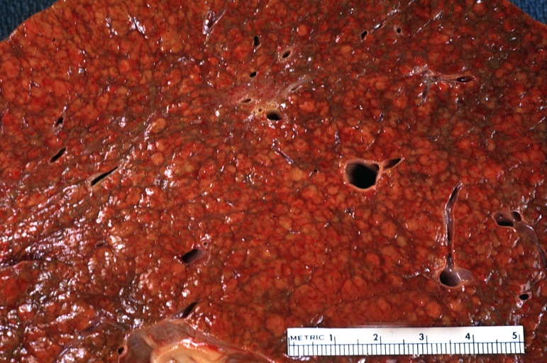
Gross Pathology
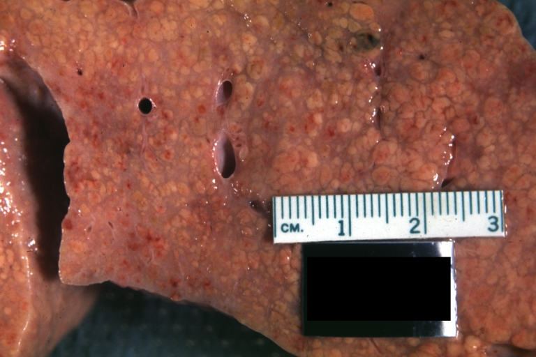
On gross examination, the liver may initially be enlarged, but with progression of the disease, it becomes smaller. Its surface is irregular, the consistency is firm, and the color is often yellow (if associates steatosis). Depending on the size of the nodules there are three macroscopic types: micronodular, macronodular and mixed cirrhosis.
- In the micronodular form (Laennec‘s cirrhosis or portal cirrhosis) regenerating nodules are under 3 mm.
- In macronodular cirrhosis (post-necrotic cirrhosis), the nodules are larger than 3 mm.
- The mixed cirrhosis consists of a variety of nodules with different sizes.
On gross pathology, cirrhotic liver, splenomegaly, and esophageal varices are characteristic findings in portal hypertension.
CirrhosisOn gross pathology there are two types of cirrhosis:
|
 |
 |
SplenomegalyOn gross pathology, diffuse enlargement and congestion of the spleen are characteristic findings of splenomegaly. |
 | |
Esophageal VaricesOn gross pathology, prominent, congested, and tortoise veins in the lower parts of esophagus are characteristic findings of esophageal varices. |
 | |
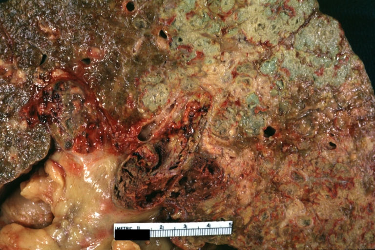
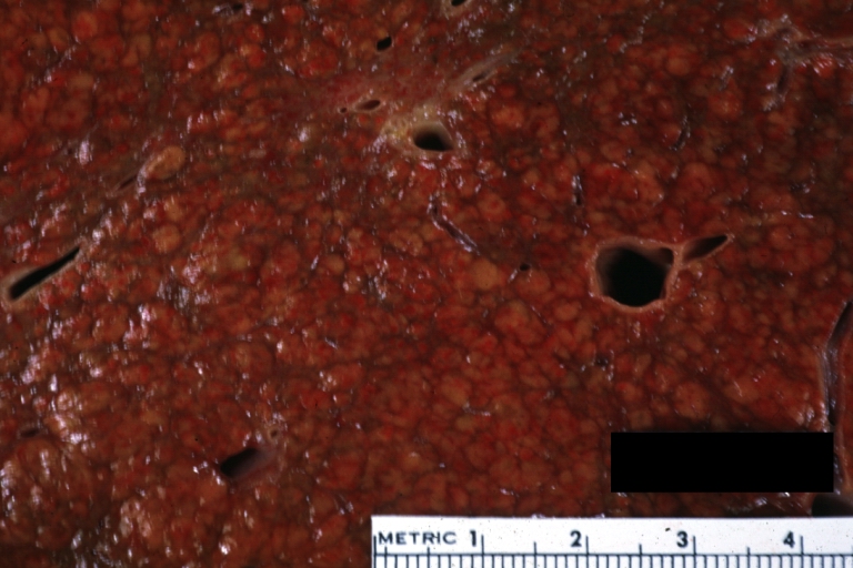
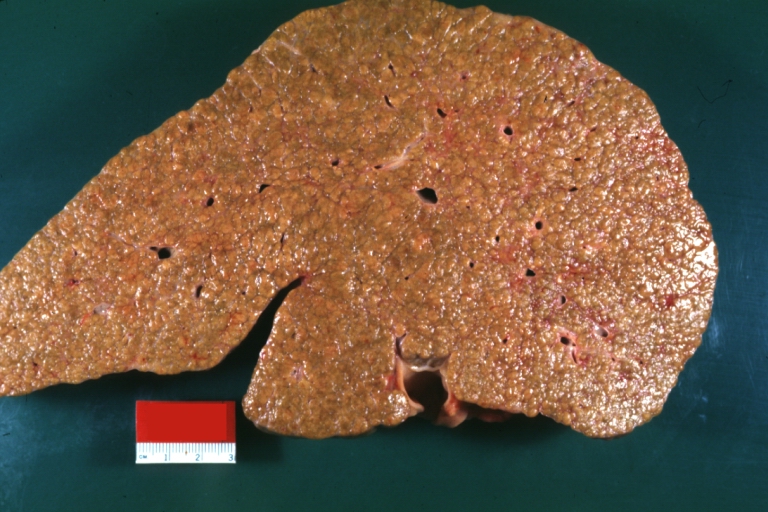
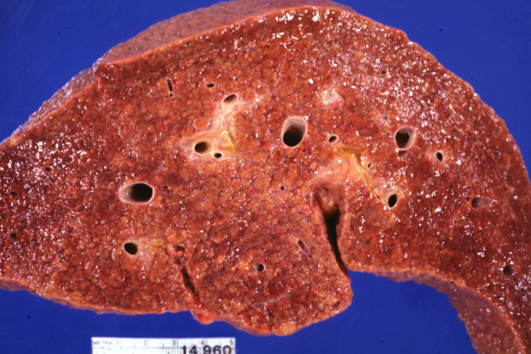
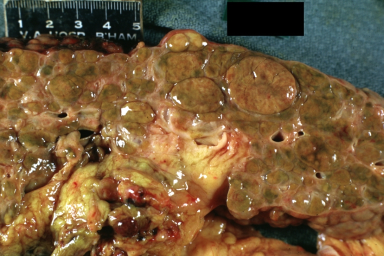
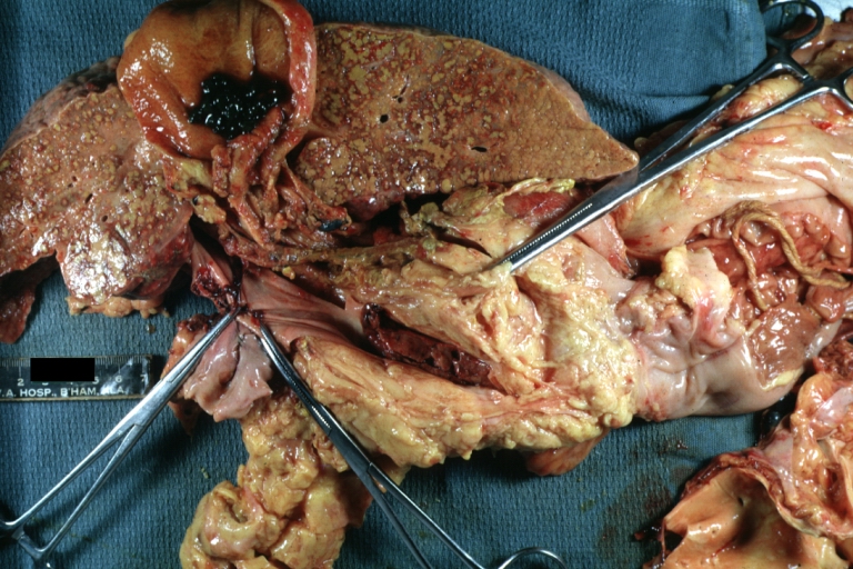
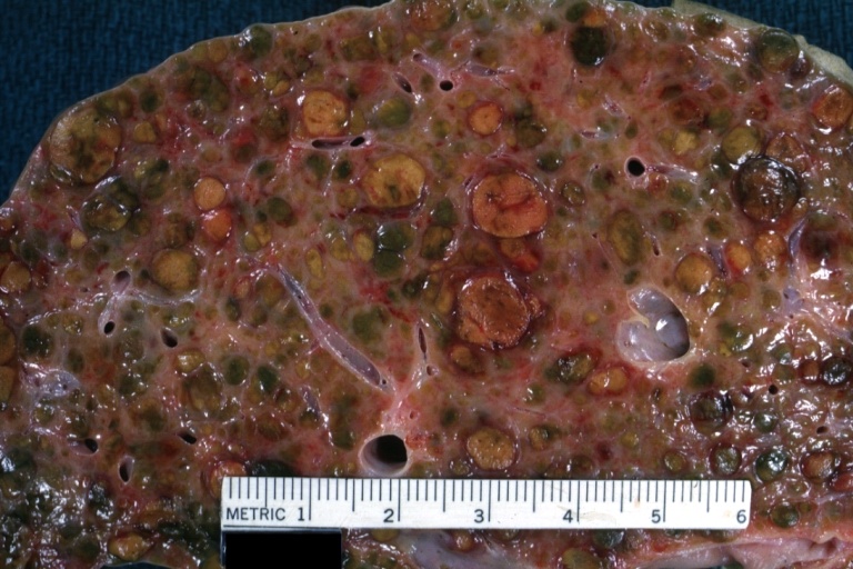
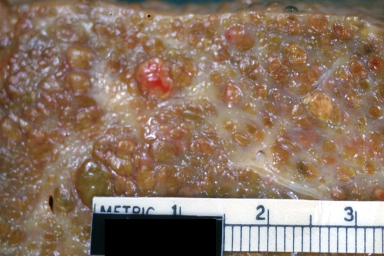
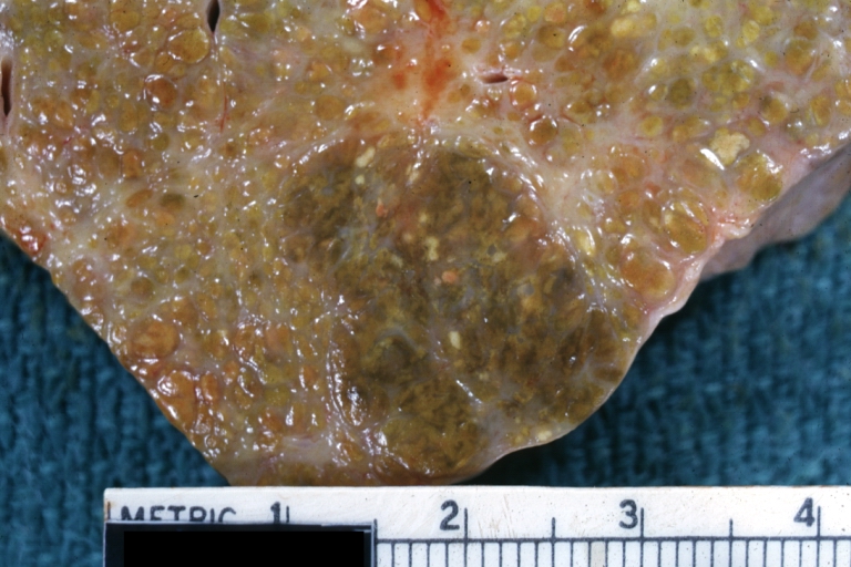
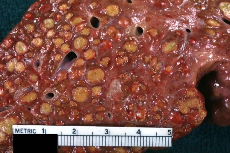
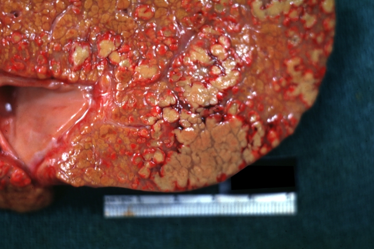
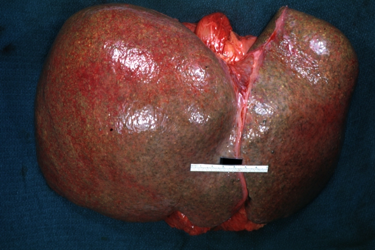
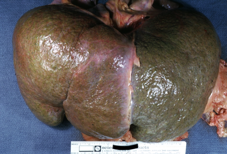
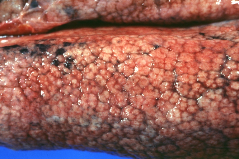
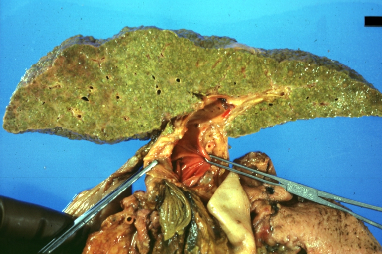
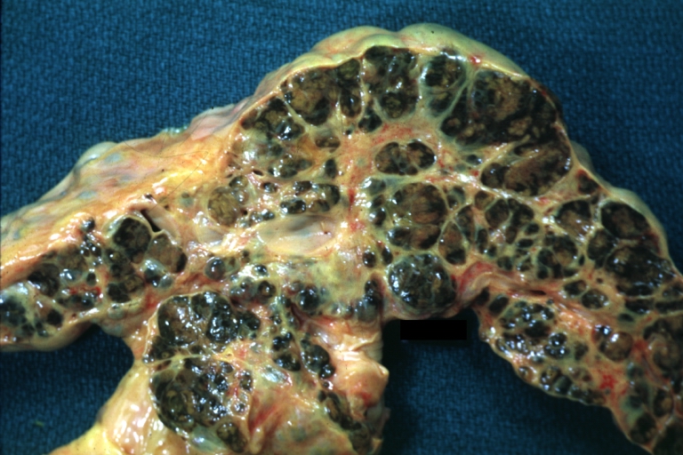
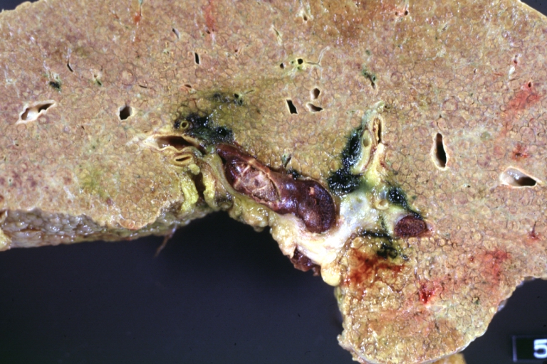
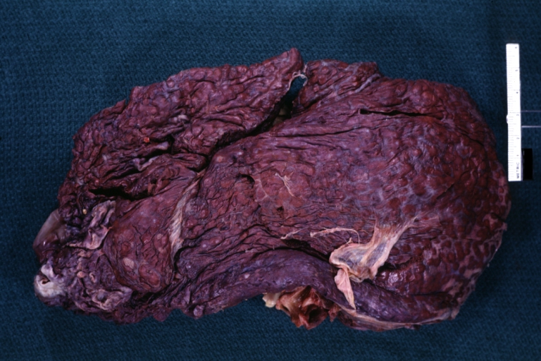
Images of gross pathology of cirrhosis
-
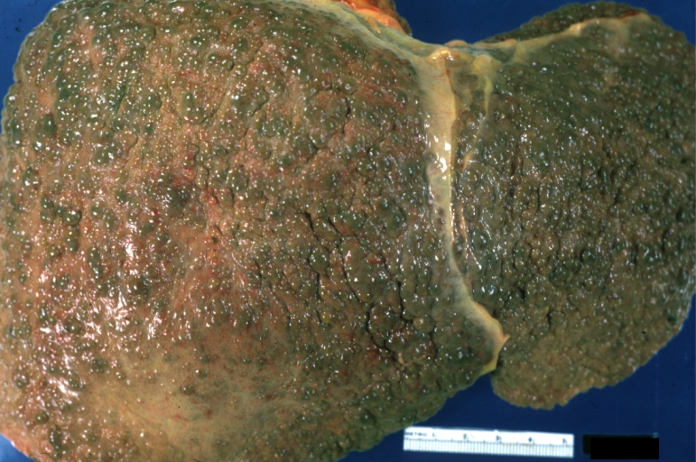 Cirrhosis: Gross, external view of micronodular cirrhosis
 -
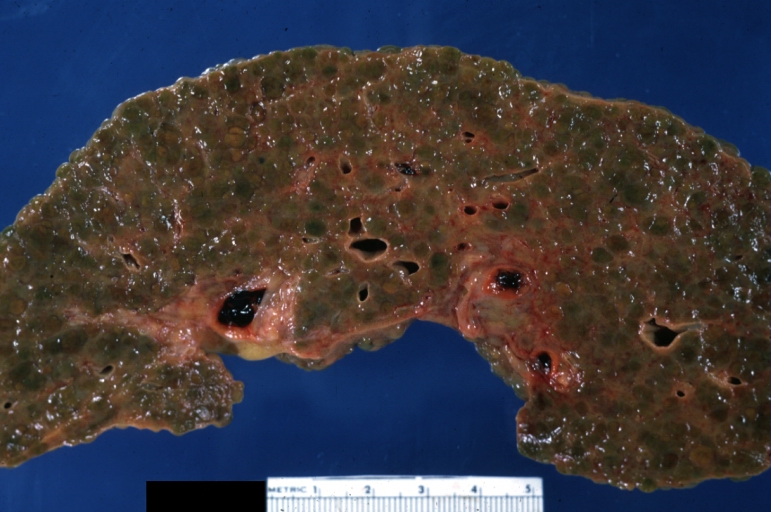 Cirrhosis: Gross, cut section of previous one (an excellent example)
 -
 Cirrhosis: Gross, close-up image
 -
 Macronodular cirrhosis and hepatoma


-
 Cirrhosis: Gross, close-up, natural color (an excellent example)
 -
 Cirrhosis: Gross, close-up (an excellent example)
 -
 Cirrhosis: Gross, close-up view
 -
 Micronodular cirrhosis: Gross, external view (an excellent example)


-
 Micronodular cirrhosis: Gross, close-up image
 -
 Micronodular cirrhosis: Gross (an excellent example)
 -
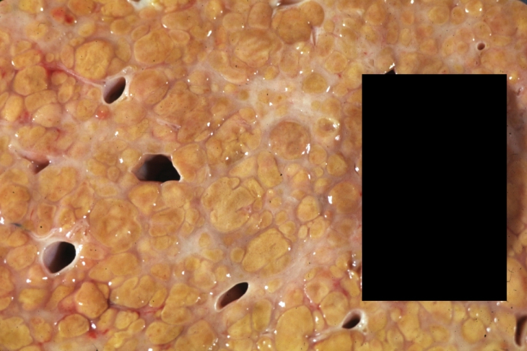
 Macronodular cirrhosis: Gross, natural color (perfect color for cirrhosis), close-up, an excellent example
 -
 Cirrhosis with portocaval shunt: Gross, severe cirrhosis with extensive liver necrosis due to thrombosis of portocaval shunt (well shown)


-
 Endstage cirrhosis: Gross, natural color, close-up (an excellent example)
 -
 Endstage cirrhosis: Gross, natural color, close-up view is an excellent example for nodules of yellow-orange liver tissue and broad irregular bands of fibrosis
 -
 Endstage cirrhosis: Gross, natural color, close-up cut surface, very well shown nodules of yellow and necrotic opaque liver tissue with broad and irregular bands of fibrosis (an excellent example)
 -
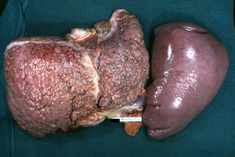 Macronodular cirrhosis: Gross, natural color, external view of liver and very enlarged spleen (liver has variable size nodules up to about 2 cm)


-
 Macronodular cirrhosis: Gross, natural color, cut surface, large irregular bands of fibrosis with variable size liver cell nodules up to about 8 mm and all necrotic appears to be an end stage liver disease.
 -
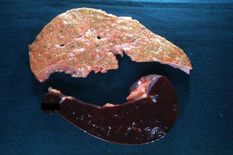 Macronodular cirrhosis: Gross, natural color view of frontal sections of liver and spleen showing a contracted macronodular liver and an enlarged spleen as large as the liver
 -
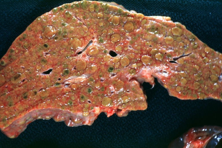 Macronodular cirrhosis: Gross, natural color slab of liver
 -
 Fatty change and early cirrhosis: Gross, natural color, rather close-up image showing typical fatty color, and in lighting at lower right of micrography micronodularity is evident (quite good example)


-
 Cirrhosis with portal vein thrombosis: Gross, natural color, sectioned liver with portal vein exposed and filled with red thrombus. A good example of end stage cirrhosis.
 -
 Endstage cirrhosis with lobular necrosis: Gross, natural color, very close-up view (an excellent example of alcoholic cirrhosis)
 -
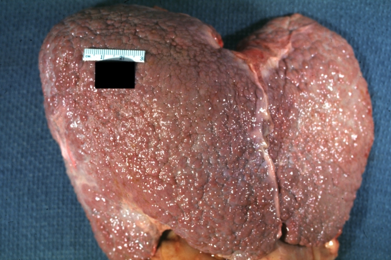
 Micronodular cirrhosis: Gross, natural color view of whole liver through capsule with obvious cirrhosis (note to quite large liver)
 -
 Micronodular cirrhosis: Gross, natural color, view of whole liver showing external surface typical cirrhotic liver (history of alcoholism)


-
 Lung: Idiopathic Interstitial Fibrosis: Gross, natural color, an excellent photo of lung cirrhosis (close-up view)
 -
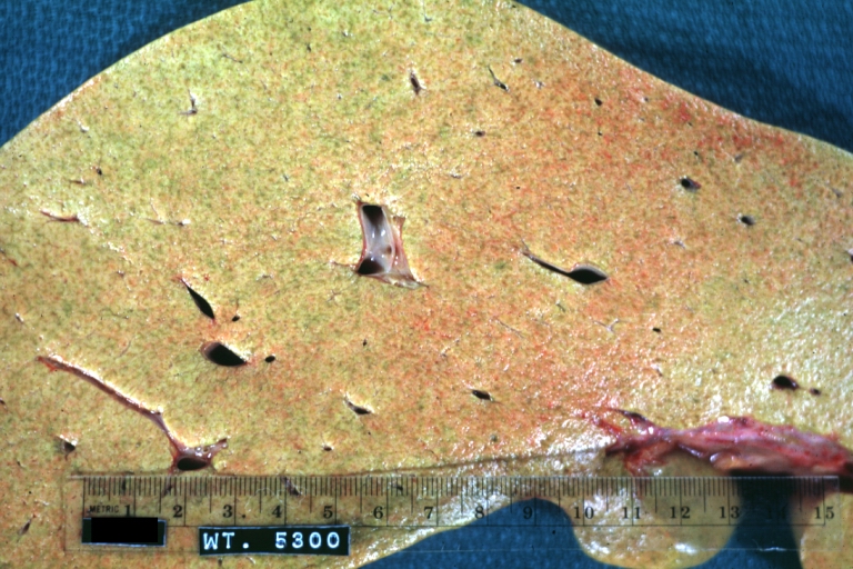
 Endstage cirrhosis: Gross, natural color, slice of liver. Portal vein is opened to show size and patency.
 -
 Endstage cirrhosis: Gross, natural color, severe cirrhosis with bile stasis
 -
 Portal Vein Thrombosis with cirrhosis: Gross, close-up, micronodular cirrhosis with portal vein thrombosis


-
 Lung: Hematite: Gross, natural color, external view of “pulmonary cirrhosis” with typical hematite color
 -
 Gross, natural color of liver and stomach view from external surfaces, micronodular cirrhosis and hemorrhagic gastritis (as the surgeon would see these in natural color)


Microscopic Pathology
- Microscopic pathology reveals the four stages of cirrhosis as it progresses:
- Chronic nonsuppurative destructive cholangitis: inflammation and necrosis of portal tracts with lymphocyte infiltration leads to the destruction of the bile ducts
- Development of biliary stasis and fibrosis
- Periportal fibrosis progresses to bridging fibrosis
- Increased proliferation of smaller bile ductules leads to regenerative nodule formation
- Microscopically, cirrhosis is characterized by regeneration nodules surrounded by fibrous septa.
- In these nodules, regenerating hepatocytes are present.
- Portal tracts, central veins and the radial pattern of hepatocytes are absent.
- Fibrous septa are present and inflammatory infiltrate composed of lymphocytes and macrophages) are also visible.
- If the underlying cause is secondary biliary cirrhosis, biliary ducts are damaged, proliferated or distended leading to bile stasis.
- Dilated ducts contain inspissated bile which appears as bile casts or bile thrombi (brown-green, amorphous).
- Bile retention may be found also in the parenchyma and are referred to as “bile lakes”.[65]
Microscopic pathology
The main microscopic histopathological findings in portal hypertension are related to cirrhosis, esophageal varices, hepatic amyloidosis, and congestive hepatopathy due to heart failure or Budd-Chiari syndrome.
CirrhosisRobbins definition of microscopic histopathological findings in cirrhosis includes (all three is needed for diagnosis):[66] |
 |
Esophageal varicesThe main microscopic histopathological findings in esophageal varices are:
|
 |
Hepatic amyloidosisThe main microscopic histopathological findings in hepatic amyloidosis is amorphous extracellular pink stuff on H&E staining. |
 |
Congestive hepatopathyThe main microscopic histopathological findings in congestive hepatopathy (due to heart failure or Budd-Chiari syndrome) are:
|
 |
Videos
{{#ev:youtube|CzKGvWZrUpU}}
{{#ev:youtube|CV8OYeIUXko}}
{{#ev:youtube|Jj8ozr_IttM}}
References
- ↑ Arthur MJ, Iredale JP (1994). “Hepatic lipocytes, TIMP-1 and liver fibrosis”. J R Coll Physicians Lond. 28 (3): 200–8. PMID 7932316.
- ↑ Friedman SL (1993). “Seminars in medicine of the Beth Israel Hospital, Boston. The cellular basis of hepatic fibrosis. Mechanisms and treatment strategies”. N. Engl. J. Med. 328 (25): 1828–35. doi:10.1056/NEJM199306243282508. PMID 8502273.
- ↑ Iredale JP (1996). “Matrix turnover in fibrogenesis”. Hepatogastroenterology. 43 (7): 56–71. PMID 8682489.
- ↑ Gressner AM (1994). “Perisinusoidal lipocytes and fibrogenesis”. Gut. 35 (10): 1331–3. PMC 1374996. PMID 7959178.
- ↑ Iredale JP (2007). “Models of liver fibrosis: exploring the dynamic nature of inflammation and repair in a solid organ”. J. Clin. Invest. 117 (3): 539–48. doi:10.1172/JCI30542. PMC 1804370. PMID 17332881.
- ↑ 6.0 6.1 Arthur MJ (2002). “Reversibility of liver fibrosis and cirrhosis following treatment for hepatitis C”. Gastroenterology. 122 (5): 1525–8. PMID 11984538.
- ↑ Wanless IR, Wong F, Blendis LM, Greig P, Heathcote EJ, Levy G (1995). “Hepatic and portal vein thrombosis in cirrhosis: possible role in development of parenchymal extinction and portal hypertension”. Hepatology. 21 (5): 1238–47. PMID 7737629.
- ↑ Iredale JP. Cirrhosis: new research provides a basis for rational and targeted treatments. BMJ 2003;327:143-7.Fulltext. PMID 12869458.
- ↑ Fernández M, Semela D, Bruix J, Colle I, Pinzani M, Bosch J (2009). “Angiogenesis in liver disease”. J. Hepatol. 50 (3): 604–20. doi:10.1016/j.jhep.2008.12.011. PMID 19157625.
- ↑ Maher JJ, McGuire RF (1990). “Extracellular matrix gene expression increases preferentially in rat lipocytes and sinusoidal endothelial cells during hepatic fibrosis in vivo”. J. Clin. Invest. 86 (5): 1641–8. doi:10.1172/JCI114886. PMC 296914. PMID 2243137. Unknown parameter
|month=ignored (help) - ↑ Herbst H, Frey A, Heinrichs O; et al. (1997). “Heterogeneity of liver cells expressing procollagen types I and IV in vivo”. Histochem. Cell Biol. 107 (5): 399–409. PMID 9208331. Unknown parameter
|month=ignored (help) - ↑ García-Pagán JC, Gracia-Sancho J, Bosch J (2012). “Functional aspects on the pathophysiology of portal hypertension in cirrhosis”. J. Hepatol. 57 (2): 458–61. doi:10.1016/j.jhep.2012.03.007. PMID 22504334.
- ↑ Lee JS, Semela D, Iredale J, Shah VH (2007). “Sinusoidal remodeling and angiogenesis: a new function for the liver-specific pericyte?”. Hepatology. 45 (3): 817–25. doi:10.1002/hep.21564. PMID 17326208. Unknown parameter
|month=ignored (help) - ↑ Rosmorduc O, Housset C (2010). “Hypoxia: a link between fibrogenesis, angiogenesis, and carcinogenesis in liver disease”. Semin. Liver Dis. 30 (3): 258–70. doi:10.1055/s-0030-1255355. PMID 20665378. Unknown parameter
|month=ignored (help) - ↑ Schuppan D, Afdhal NH (2008). “Liver cirrhosis”. Lancet. 371 (9615): 838–51. doi:10.1016/S0140-6736(08)60383-9. PMC 2271178. PMID 18328931.
- ↑ Desmet VJ, Roskams T (2004). “Cirrhosis reversal: a duel between dogma and myth”. J. Hepatol. 40 (5): 860–7. doi:10.1016/j.jhep.2004.03.007. PMID 15094237.
- ↑ Wanless IR, Nakashima E, Sherman M (2000). “Regression of human cirrhosis. Morphologic features and the genesis of incomplete septal cirrhosis”. Arch. Pathol. Lab. Med. 124 (11): 1599–607. doi:10.1043/0003-9985(2000)124<1599:ROHC>2.0.CO;2. PMID 11079009.
- ↑ Ceni E, Mello T, Galli A (2014). “Pathogenesis of alcoholic liver disease: role of oxidative metabolism”. World J. Gastroenterol. 20 (47): 17756–72. doi:10.3748/wjg.v20.i47.17756. PMC 4273126. PMID 25548474.
- ↑ You M, Crabb DW (2004). “Recent advances in alcoholic liver disease II. Minireview: molecular mechanisms of alcoholic fatty liver”. Am. J. Physiol. Gastrointest. Liver Physiol. 287 (1): G1–6. doi:10.1152/ajpgi.00056.2004. PMID 15194557.
- ↑ Freeman TL, Tuma DJ, Thiele GM, Klassen LW, Worrall S, Niemelä O, Parkkila S, Emery PW, Preedy VR (2005). “Recent advances in alcohol-induced adduct formation”. Alcohol. Clin. Exp. Res. 29 (7): 1310–6. PMID 16088993.
- ↑ Niemelä O (2007). “Acetaldehyde adducts in circulation”. Novartis Found. Symp. 285: 183–92, discussion 193–7. PMID 17590995.
- ↑ Fischer M, You M, Matsumoto M, Crabb DW (2003). “Peroxisome proliferator-activated receptor alpha (PPARalpha) agonist treatment reverses PPARalpha dysfunction and abnormalities in hepatic lipid metabolism in ethanol-fed mice”. J. Biol. Chem. 278 (30): 27997–8004. doi:10.1074/jbc.M302140200. PMID 12791698.
- ↑ You M, Matsumoto M, Pacold CM, Cho WK, Crabb DW (2004). “The role of AMP-activated protein kinase in the action of ethanol in the liver”. Gastroenterology. 127 (6): 1798–808. PMID 15578517.
- ↑ Ji C, Chan C, Kaplowitz N (2006). “Predominant role of sterol response element binding proteins (SREBP) lipogenic pathways in hepatic steatosis in the murine intragastric ethanol feeding model”. J. Hepatol. 45 (5): 717–24. doi:10.1016/j.jhep.2006.05.009. PMID 16879892.
- ↑ Greenway CV, Stark RD (1971). “Hepatic vascular bed”. Physiol. Rev. 51 (1): 23–65. PMID 5543903.
- ↑ Schiff, Eugene (2012). Schiff’s diseases of the liver. Chichester, West Sussex, UK: John Wiley & Sons. ISBN 9780470654682.
- ↑ SCHAFFNER F, POPER H (1963). “Capillarization of hepatic sinusoids in man”. Gastroenterology. 44: 239–42. PMID 13976646.
- ↑ Reynolds TB, Hidemura R, Michel H, Peters R (1969). “Portal hypertension without cirrhosis in alcoholic liver disease”. Ann. Intern. Med. 70 (3): 497–506. PMID 5775031.
- ↑ Rubanyi GM (1991). “Endothelium-derived relaxing and contracting factors”. J. Cell. Biochem. 46 (1): 27–36. doi:10.1002/jcb.240460106. PMID 1874796.
- ↑ Epstein, Franklin H.; Vane, John R.; Änggård, Erik E.; Botting, Regina M. (1990). “Regulatory Functions of the Vascular Endothelium”. New England Journal of Medicine. 323 (1): 27–36. doi:10.1056/NEJM199007053230106. ISSN 0028-4793.
- ↑ Rockey DC, Weisiger RA (1996). “Endothelin induced contractility of stellate cells from normal and cirrhotic rat liver: implications for regulation of portal pressure and resistance”. Hepatology. 24 (1): 233–40. doi:10.1002/hep.510240137. PMID 8707268.
- ↑ Mosca P, Lee FY, Kaumann AJ, Groszmann RJ (1992). “Pharmacology of portal-systemic collaterals in portal hypertensive rats: role of endothelium”. Am. J. Physiol. 263 (4 Pt 1): G544–50. PMID 1415713.
- ↑ Colombato LA, Albillos A, Groszmann RJ (1992). “Temporal relationship of peripheral vasodilatation, plasma volume expansion and the hyperdynamic circulatory state in portal-hypertensive rats”. Hepatology. 15 (2): 323–8. PMID 1735537.
- ↑ Genecin P, Polio J, Colombato LA, Ferraioli G, Reuben A, Groszmann RJ (1990). “Bile acids do not mediate the hyperdynamic circulation in portal hypertensive rats”. Am. J. Physiol. 259 (1 Pt 1): G21–5. PMID 2372062.
- ↑ Casadevall, María; Panés, Julián; Piqué, Josep M.; Marroni, Norma; Bosch, Jaume; Whittle, Brendan J. R. (1993). “Involvement of nitric oxide and prostaglandins in gastric mucosal hyperemia of portal-hypertensive anesthetized rats”. Hepatology. 18 (3): 628–634. doi:10.1002/hep.1840180323. ISSN 0270-9139.
- ↑ Sieber CC, Groszmann RJ (1992). “In vitro hyporeactivity to methoxamine in portal hypertensive rats: reversal by nitric oxide blockade”. Am. J. Physiol. 262 (6 Pt 1): G996–1001. PMID 1616049.
- ↑ Albillos A, Colombato LA, Lee FY, Groszmann RJ (1993). “Octreotide ameliorates vasodilatation and Na+ retention in portal hypertensive rats”. Gastroenterology. 104 (2): 575–9. PMID 8425700.
- ↑ 38.00 38.01 38.02 38.03 38.04 38.05 38.06 38.07 38.08 38.09 38.10 38.11 38.12 Nadim MK, Garcia-Tsao G (2023). “Acute Kidney Injury in Cirrhosis”. N Engl J Med. 388: 733–745. doi:10.1056/NEJMra2215289.
- ↑ Belcher JM, Garcia-Tsao G, Sanyal AJ, et al. (2013). “Association of AKI with mortality and complications in hospitalized patients with cirrhosis”. Hepatology. 57: 753–762.
- ↑ 40.0 40.1 40.2 40.3 40.4 40.5 Angeli P, Ginès P, Wong F, et al. (2015). “Diagnosis and management of acute kidney injury in patients with cirrhosis: revised consensus recommendations of the International Club of Ascites”. J Hepatol. 62: 968–974.
- ↑ Piano S, Rosi S, Maresio G, et al. (2013). “Evaluation of the Acute Kidney Injury Network criteria in hospitalized patients with cirrhosis and ascites”. J Hepatol. 59: 482–489.
- ↑ Schrier RW, Arroyo V, Bernardi M, Epstein M, Henriksen JH, Rodés J (1988). “Peripheral arterial vasodilation hypothesis: a proposal for the initiation of renal sodium and water retention in cirrhosis”. Hepatology. 8: 1151–1157.
- ↑ 43.0 43.1 43.2 43.3 Angeli P, Gines P, Wong F, et al. (2019). “Hepatorenal syndrome in cirrhosis: updated definition and management recommendations”. J Hepatol. 71: 811–822.
- ↑ Salerno F, Gerbes A, Ginès P, Wong F, Arroyo V (2007). “Diagnosis, prevention and treatment of hepatorenal syndrome in cirrhosis”. Gut. 56: 1310–1318.
- ↑ 45.0 45.1 Francoz C, Durand F, Kahn JA, Genyk YS, Nadim MK (2019). “Hepatorenal syndrome”. Clin J Am Soc Nephrol. 14: 774–781.
- ↑ Belcher JM, Sanyal AJ, Peixoto AJ, et al. (2014). “Kidney biomarkers and differential diagnosis of patients with cirrhosis and acute kidney injury”. Hepatology. 60: 622–632.
- ↑ Allegretti AS, Parada XV, Endres P, et al. (2021). “Urinary NGAL as a diagnostic and prognostic marker for acute kidney injury in cirrhosis: a prospective study”. Clin Transl Gastroenterol. 12 (5).
- ↑ Francoz C, Nadim MK, Durand F (2016). “Kidney biomarkers in cirrhosis”. J Hepatol. 65: 809–824.
- ↑ Wong F, Pappas SC, Curry MP, et al. (2021). “Terlipressin plus albumin for the treatment of hepatorenal syndrome”. N Engl J Med. 384: 818–828.
- ↑ Solà E, et al. (2022). “Factors associated with respiratory failure during terlipressin therapy in hepatorenal syndrome”. Hepatology.
- ↑ Sharma P, et al. (2008). “Norepinephrine vs terlipressin in hepatorenal syndrome: randomized study”. J Hepatol.
- ↑ Nassar Junior AP, Farias AQ, d’ Albuquerque LA, Carrilho FJ, Malbouisson LM (2014). “Terlipressin versus norepinephrine in the treatment of hepatorenal syndrome: a systematic review and meta-analysis”. PLoS One. 9 (9).
- ↑ Cavallin M, Kamath PS, Merli M, et al. (2015). “Terlipressin plus albumin versus midodrine and octreotide plus albumin in the treatment of hepatorenal syndrome: a randomized trial”. Hepatology. 62: 567–574.
- ↑ Nadim MK, Sung RS, Davis CL, et al. (2012). “Impact of the etiology of acute kidney injury on outcomes following liver transplantation: acute tubular necrosis vs hepatorenal syndrome”. Liver Transpl. 18: 539–548.
- ↑ 55.0 55.1 Biggins SW, Angeli P, Garcia-Tsao G, et al. (2021). “Diagnosis, evaluation, and management of ascites, spontaneous bacterial peritonitis, and hepatorenal syndrome”. Hepatology. 74: 1014–1048.
- ↑ Bernardi M, Caraceni P, Navickis RJ, Wilkes MM (2012). “Albumin infusion in patients undergoing large-volume paracentesis: a meta-analysis of randomized trials”. Hepatology. 55: 1172–1181.
- ↑ Sort P, Navasa M, Arroyo V, et al. (1999). “Effect of intravenous albumin on renal impairment and mortality in patients with cirrhosis and spontaneous bacterial peritonitis”. N Engl J Med. 341: 403–409.
- ↑ Calado RT, Brudno J, Mehta P; et al. (2011). “Constitutional telomerase mutations are genetic risk factors for cirrhosis”. Hepatology. 53 (5): 1600–7. doi:10.1002/hep.24173. PMC 3082730. PMID 21520173. Unknown parameter
|month=ignored (help) - ↑ Freed EF, Prieto JL, McCann KL, McStay B, Baserga SJ (2012). “NOL11, Implicated in the Pathogenesis of North American Indian Childhood Cirrhosis, Is Required for Pre-rRNA Transcription and Processing”. PLoS Genet. 8 (8): e1002892. doi:10.1371/journal.pgen.1002892. PMC 3420923. PMID 22916032. Unknown parameter
|month=ignored (help) - ↑ Freed EF, Baserga SJ (2010). “The C-terminus of Utp4, mutated in childhood cirrhosis, is essential for ribosome biogenesis”. Nucleic Acids Res. 38 (14): 4798–806. doi:10.1093/nar/gkq185. PMC 2919705. PMID 20385600. Unknown parameter
|month=ignored (help) - ↑ <CC BY-SA 4.0 (https://creativecommons.org/licenses/by-sa/4.0)>
- ↑ “www.meddean.luc.edu”.
- ↑ Amadalvarez – Own work, <“https://creativecommons.org/licenses/by-sa/4.0” title=”Creative Commons Attribution-Share Alike 4.0″>CC BY-SA 4.0, <“https://commons.wikimedia.org/w/index.php?curid=49669333“>Link
- ↑ <http://wellcomeimages.org/indexplus/obf_images/29/b4/13f38971164f946a97f9d32ddd93.jpg>Gallery: <“http://wellcomeimages.org/indexplus/image/L0074357.html“><“http://creativecommons.org/licenses/by/4.0> CC BY 4.0, <“https://commons.wikimedia.org/w/index.php?curid=36297209“>
- ↑ Pathology atlas, “cirrhosis”.
- ↑ Mitchell, Richard (2012). Pocket companion to Robbins and Cotran pathologic basis of disease. Philadelphia, PA: Elsevier Saunders. ISBN 978-1416054542.
- ↑ “File:Cirrhosis high mag.jpg – Libre Pathology”.
- ↑ “Esophageal varices – Libre Pathology”.
- ↑ “File:Hepatic amyloidosis – high mag.jpg – Libre Pathology”.
- ↑ “File:2 CEN NEC 1 680x512px.tif – Libre Pathology”.
Causes
Editor-In-Chief: C. Michael Gibson, M.S., M.D. [1] Associate Editor(s)-in-Chief: Aditya Govindavarjhulla, M.B.B.S. [2] Sudarshana Datta, MD [3]
Overview
There are a wide range of causes for cirrhosis, including alcohol abuse, genetic diseases, cardiac causes, toxins, viruses, and malnutrition. The consequence is the same in all cases with the functional liver tissue being replaced by fibrous scar tissue and regenerative nodules.
Causes
Common causes
Cirrhosis has many possible causes; sometimes more than one cause is present in the same patient. In the Western World, chronic alcoholism and hepatitis C are the most common causes. Hepatitis B is more common in parts of sub-Saharan Africa and Asia.
- Alcoholic liver disease
- Chronic hepatitis C
- Chronic hepatitis B
- Non-alcoholic steatohepatitis (NASH)
- Primary biliary cirrhosis
- Primary sclerosing cholangitis
- Autoimmune hepatitis
- Hereditary hemochromatosis
- Wilson’s disease
- Alpha 1-antitrypsin deficiency
- Cardiac cirrhosis
- Galactosemia
- Glycogen storage disease type IV
- Cystic fibrosis
- Drugs or toxins
- Certain parasitic infections (such as schistosomiasis)
Causes by Organ System
- Abetalipoproteinemia
- Acetaminophen overdose
- Addison-Gull syndrome
- Aflatoxin
- Alagille syndrome
- Alcoholic liver disease
- Alpers disease
- Alpha 1-antitrypsin deficiency
- Alström syndrome
- Amiodarone
- Autoimmune cholangiopathy
- Autoimmune hepatitis
- Bearn-Kunkel syndrome [3]
- Berardinelli lipodystrophy syndrome
- Bile duct stricture
- Biliary atresia
- Budd-Chiari Syndrome
- Carbohydrate deficient glycoprotein syndrome type 1a
- Cardiac cirrhosis
- Caroli disease
- Cerebrohepatorenal syndrome
- Ceroid storage disease
- Cholestasis-oedema syndrome, Norwegian type
- Cholesterol ester storage disease
- Congenital hepatic fibrosis
- Constrictive pericarditis
- Cor Pulmonale
- Cruveilhier-Baumgarten syndrome
- Cystic fibrosis
- Erythropoietic protoporphyria
- Ethanol
- Fanconi disease [4]
- Fasciola hepatica
- Fructose-1-phosphate aldolase deficiency [5]
- Galactosemia
- Glycogen storage disease type IV
- Graft versus host disease
- Granulomatous cirrhosis
- Haemosiderosis
- Hemochromatosis
- Hepatic vein thrombosis
- Hepatitis B
- Hepatitis C
- Hereditary fructose intolerance
- Hypervitaminosis A
- Indian familial childhood cirrhosis
- Isoniazid
- Keratitis-ichthyosis-deafness syndrome, autosomal recessive
- Methotrexate
- Methyldopa
- Non-alcoholic steatohepatitis
- Parenteral nutrition
- Polycystic kidney disease, autosomal recessive
- Porphyria cutanea tarda type 2 (familial)
- Primary biliary cirrhosis
- Primary sclerosing cholangitis
- Reynolds syndrome [6]
- Right sided cardiac failure
- Sarcoidosis
- Schistosomiasis
- Sickle Cell Disease
- Steatohepatitis
- Thalassemia
- Tricho-hepato-enteric syndrome [7]
- Tricuspid insufficiency
- Tyrosinaemia type 1
- Visceral leishmaniasis
- Wilson disease
Differentiating Different Causes of Cirrhosis
| Differential Diagnosis | Useful Findings |
| Alcoholic cirrhosis | History EtOH, AST/ALT > 2 |
| Chronic Hepatits C Virus (HCV) | HCV AB |
| Primary Biliary Cirrhosis (PBC) | Elevated alk phos, AMA+ |
| Primary sclerosing cholangitis | History inflammatory bowel disease (IBD), ANA or ASMA or P-ANCA+ |
| Autoimmune hepatitis | Hypergammaglobulinemia, ANA/ASMA + |
| Chronic Hepatitis B Virus | HBsAg+, HBeAg may be + |
| Hemochromatosis | Family history+, Fe/TIBC and ferritin elevated |
| Wilson’s disease | Family history+, young age, low ceruloplasmin |
| Alpha-1-antitrypsin (AAT) deficiency | Family history+, young age, low serum AAT |
References
- ↑ Sailer, Christian, Wasner, Susanne. Differential Diagnosis Pocket. Hermosa Beach, CA: Borm Bruckmeir Publishing LLC, 2002:77 ISBN 1591032016
- ↑ Kahan, Scott, Smith, Ellen G. In A Page: Signs and Symptoms. Malden, Massachusetts: Blackwell Publishing, 2004:68 ISBN 140510368X
- ↑ http://www.ncbi.nlm.nih.gov/pmc/articles/PMC1813854/#reference-sec
- ↑ http://www.ncbi.nlm.nih.gov/pmc/articles/PMC2165837/
- ↑ http://ghr.nlm.nih.gov/condition/hereditary-fructose-intolerance
- ↑ http://rarediseases.info.nih.gov/GARD/Condition/4697/Reynolds_syndrome.aspx
- ↑ http://www.checkorphan.org/disease/tricho-hepato-enteric-syndrome
Differentiating Cirrhosis from other Diseases
Editor-In-Chief: C. Michael Gibson, M.S., M.D. [1] Associate Editor(s)-in-Chief: Sudarshana Datta, MD [2]
Overview
Cirrhosis may present in a similar way to some other diseases. History, physical examination, and diagnostic testing may help in differentiating cirrhosis from other diseases such as malignancy, constrictive pericarditis, Budd-Chiari syndrome, portal vein thrombosis and splenic vein thrombosis.
Differentiating Cirrhosis From Other Diseases
Differential diagnosis of jaundice
Differential diagnosis of jaundice are enlisted in the table below:[1][2][3][4][5]
| ||||||||||||||||||||||||||||||||||||||||||||||||||||||||||||||||||||||||||||||||||||||||||||||||||||||||||||||||||||||||||||||||||||||||||||||||||||||||||||||||||||||||||||||||||||||||||||||||||||||||||||||||||||||||||||||||||||||||||||||||||||||||||||||||||||||||||||||||||||||||||||||||||||||||||||||||||||||||||||||||||||||||||||||||||||||||||||||||||||||||||||||||||||||||||||||||||||||||||||||||||||||||||||||
Differentiating cirrhosis from other diseases based on serum-ascites albumin gradient (SAAG)
- Ascites may be caused by portal hypertension due to cirrhosis of liver or due to other causes such as malignancy. Ascitic fluid analysis should be done to broadly categorize the cause of ascites.
- Ascites is broadly classified as two types based on the serum-ascites albumin gradient (SAAG):
- Transudate – SAAG > 1.1 g/dL (indicative of ascites due to portal hypertension)
- Exudate – SAAG < 1.1 g/dL (indicative of ascites due to other etiologies)
Cirrhosis must be differentiated from other causes of abnormal liver function tests, altered liver architecture and size:
| Condition | Differentiating signs and symptoms | Differentiating Tests |
|---|---|---|
| Cirrhosis |
|
Ultrasound findings in cirrhosis are as follows:[6][7][8][9][10][11][12][13]
Abdominal MRI may also be helpful in the diagnosis of portal hypertension. Findings on MRI suggestive of cirrhosis with portal hypertension include:[14][15][16][17]
Transient elastography and the Acoustic Radiation Force Impulse (ARFI) technique are well-established methods for the staging of fibrosis in various liver diseases: [18][19][20][21][22][23][24][25][26][27][28]
|
| Constrictive pericarditis |
|
|
| Budd-Chiari Syndrome |
| |
| Splenic vein thrombosis | Signs and symptoms of:
|
|
| Portal vein thrombosis |
|
|
| Schistosomiasis |
| |
| Sarcoidosis |
| |
| Inferior vena cava obstruction |
|
|
| Nodular regenerative hyperplasia | None |
|
| Idiopathic portal hypertension (hepatoportal sclerosis) | None |
|
| Vitamin A intoxication, arsenic, and vinyl chloride toxicity | None |
|
Differentiating Cirrhosis from other causes of jaundice and abdominal pain
Cirrhosis must be differentiated from other diseases that cause jaundice, abdominal pain, weight loss, and fever such as Gallbladder cancer, hepatocellular carcinoma, pancreatic cancer, cholecystitis, choledochitis and liver fluke infections.
Abbreviations:
RUQ= Right upper quadrant of the abdomen, LUQ= Left upper quadrant, LLQ= Left lower quadrant, RLQ= Right lower quadrant, LFT= Liver function test, SIRS= Systemic inflammatory response syndrome, ERCP= Endoscopic retrograde cholangiopancreatography, IV= Intravenous, N= Normal, AMA= Anti mitochondrial antibodies, LDH= Lactate dehydrogenase, GI= Gastrointestinal, CXR= Chest X ray, IgA= Immunoglobulin A, IgG= Immunoglobulin G, IgM= Immunoglobulin M, CT= Computed tomography, PMN= Polymorphonuclear cells, ESR= Erythrocyte sedimentation rate, CRP= C-reactive protein, TS= Transferrin saturation, SF= Serum Ferritin, SMA= Superior mesenteric artery, SMV= Superior mesenteric vein, ECG= Electrocardiogram
| Disease | Clinical manifestations | Diagnosis | Comments | |||||||||||||
|---|---|---|---|---|---|---|---|---|---|---|---|---|---|---|---|---|
| Symptoms | Signs | |||||||||||||||
| Abdominal Pain | Fever | Rigors and chills | Nausea or vomiting | Jaundice | Constipation | Diarrhea | Weight loss | GI bleeding | Hypo-
tension |
Guarding | Rebound Tenderness | Bowel sounds | Lab Findings | Imaging | ||
| Cholangiocarcinoma | RUQ | + | − | + | + | − | − | + | − | − | − | + | Normal |
|
| |
| Hepatocellular carcinoma/Metastasis | RUQ | + | − | + | + | + | + | + | + | + | − | + |
|
|
Other symptoms: | |
| Pancreatic carcinoma | MidEpigastric | − | − | + | + | + | − | + | − | − | − | + | Normal |
Skin manifestations may include: | ||
| Focal nodular hyperplasia | Diffuse | ± | − | − | ± | − | − | + | + | − | − | − | Normal |
|
|
|
| Disease | Abdominal Pain | Fever | Rigors and chills | Nausea or vomiting | Jaundice | Constipation | Diarrhea | Weight loss | GI bleeding | Hypo-
tension |
Guarding | Rebound Tenderness | Bowel sounds | Lab Findings | Imaging | Comments |
| Gallbladder cancer | Midepigastric | − | − | + | + | − | + | + | − | − | − | − | Normal |
|
||
| Liver hemangioma | Intermittent RUQ | − | − | + | + | − | − | − | − | − | − | − | Normal |
|
| |
| Liver abscess | RUQ | + | − | + | + | − | − | + | − | − | − | − | Normal |
|
|
|
| Cirrhosis | RUQ+Bloating | + | − | + | + | − | − | + | − | − | − | − | Normal |
|
US
|
|
| Inflammatory lesions | RUQ | ± | − | + | + | − | − | − | − | − | − | − | Normal |
|
US
|
|
References
- ↑ Fargo MV, Grogan SP, Saguil A (2017). “Evaluation of Jaundice in Adults”. Am Fam Physician. 95 (3): 164–168. PMID 28145671.
- ↑ Leevy CB, Koneru B, Klein KM (1997). “Recurrent familial prolonged intrahepatic cholestasis of pregnancy associated with chronic liver disease”. Gastroenterology. 113 (3): 966–72. PMID 9287990.
- ↑ Hov JR, Boberg KM, Karlsen TH (2008). “Autoantibodies in primary sclerosing cholangitis”. World J. Gastroenterol. 14 (24): 3781–91. PMC 2721433. PMID 18609700.
- ↑ Bond LR, Hatty SR, Horn ME, Dick M, Meire HB, Bellingham AJ (1987). “Gall stones in sickle cell disease in the United Kingdom”. Br Med J (Clin Res Ed). 295 (6592): 234–6. PMC 1247079. PMID 3115390.
- ↑ Malakouti M, Kataria A, Ali SK, Schenker S (2017). “Elevated Liver Enzymes in Asymptomatic Patients – What Should I Do?”. J Clin Transl Hepatol. 5 (4): 394–403. doi:10.14218/JCTH.2017.00027. PMC 5719197. PMID 29226106.
- ↑ Udell JA, Wang CS, Tinmouth J, FitzGerald JM, Ayas NT, Simel DL, Schulzer M, Mak E, Yoshida EM (2012). “Does this patient with liver disease have cirrhosis?”. JAMA. 307 (8): 832–42. doi:10.1001/jama.2012.186. PMID 22357834.
- ↑ Becker CD, Scheidegger J, Marincek B (1986). “Hepatic vein occlusion: morphologic features on computed tomography and ultrasonography”. Gastrointest Radiol. 11 (4): 305–11. PMID 3533689.
- ↑ Di Lelio A, Cestari C, Lomazzi A, Beretta L (1989). “Cirrhosis: diagnosis with sonographic study of the liver surface”. Radiology. 172 (2): 389–92. doi:10.1148/radiology.172.2.2526349. PMID 2526349.
- ↑ Sanford NL, Walsh P, Matis C, Baddeley H, Powell LW (1985). “Is ultrasonography useful in the assessment of diffuse parenchymal liver disease?”. Gastroenterology. 89 (1): 186–91. PMID 3891495.
- ↑ Giorgio A, Amoroso P, Lettieri G, Fico P, de Stefano G, Finelli L, Scala V, Tarantino L, Pierri P, Pesce G (1986). “Cirrhosis: value of caudate to right lobe ratio in diagnosis with US”. Radiology. 161 (2): 443–5. doi:10.1148/radiology.161.2.3532188. PMID 3532188.
- ↑ Simonovský V (1999). “The diagnosis of cirrhosis by high resolution ultrasound of the liver surface”. Br J Radiol. 72 (853): 29–34. doi:10.1259/bjr.72.853.10341686. PMID 10341686.
- ↑ Trinchet JC, Chaffaut C, Bourcier V, Degos F, Henrion J, Fontaine H, Roulot D, Mallat A, Hillaire S, Cales P, Ollivier I, Vinel JP, Mathurin P, Bronowicki JP, Vilgrain V, N’Kontchou G, Beaugrand M, Chevret S (2011). “Ultrasonographic surveillance of hepatocellular carcinoma in cirrhosis: a randomized trial comparing 3- and 6-month periodicities”. Hepatology. 54 (6): 1987–97. doi:10.1002/hep.24545. PMID 22144108.
- ↑ “EASL-EORTC clinical practice guidelines: management of hepatocellular carcinoma”. J. Hepatol. 56 (4): 908–43. 2012. doi:10.1016/j.jhep.2011.12.001. PMID 22424438.
- ↑ Procopet, Bogdan; Berzigotti, Annalisa (2017). “Diagnosis of cirrhosis and portal hypertension: imaging, non-invasive markers of fibrosis and liver biopsy”. Gastroenterology Report. 5 (2): 79–89. doi:10.1093/gastro/gox012. ISSN 2052-0034.
- ↑ Aagaard, J; Jensen, LI; Sorensen, TI; Christensen, U; Burcharth, F (1982). “Recanalized umbilical vein in portal hypertension”. American Journal of Roentgenology. 139 (6): 1107–1110. doi:10.2214/ajr.139.6.1107. ISSN 0361-803X.
- ↑ Cho, K C; Patel, Y D; Wachsberg, R H; Seeff, J (1995). “Varices in portal hypertension: evaluation with CT”. RadioGraphics. 15 (3): 609–622. doi:10.1148/radiographics.15.3.7624566. ISSN 0271-5333.
- ↑ Bandali, Murad Feroz; Mirakhur, Anirudh; Lee, Edward Wolfgang; Ferris, Mollie Clarke; Sadler, David James; Gray, Robin Ritchie; Wong, Jason Kam (2017). “Portal hypertension: Imaging of portosystemic collateral pathways and associated image-guided therapy”. World Journal of Gastroenterology. 23 (10): 1735. doi:10.3748/wjg.v23.i10.1735. ISSN 1007-9327.
- ↑ Castera L, Pinzani M (2010). “Biopsy and non-invasive methods for the diagnosis of liver fibrosis: does it take two to tango?”. Gut. 59 (7): 861–6. doi:10.1136/gut.2010.214650. PMID 20581229.
- ↑ Friedrich-Rust M, Nierhoff J, Lupsor M, Sporea I, Fierbinteanu-Braticevici C, Strobel D, Takahashi H, Yoneda M, Suda T, Zeuzem S, Herrmann E (2012). “Performance of Acoustic Radiation Force Impulse imaging for the staging of liver fibrosis: a pooled meta-analysis”. J. Viral Hepat. 19 (2): e212–9. doi:10.1111/j.1365-2893.2011.01537.x. PMID 22239521.
- ↑ Friedrich-Rust M, Ong MF, Martens S, Sarrazin C, Bojunga J, Zeuzem S, Herrmann E (2008). “Performance of transient elastography for the staging of liver fibrosis: a meta-analysis”. Gastroenterology. 134 (4): 960–74. doi:10.1053/j.gastro.2008.01.034. PMID 18395077.
- ↑ Ziol M, Handra-Luca A, Kettaneh A, Christidis C, Mal F, Kazemi F, de Lédinghen V, Marcellin P, Dhumeaux D, Trinchet JC, Beaugrand M (2005). “Noninvasive assessment of liver fibrosis by measurement of stiffness in patients with chronic hepatitis C”. Hepatology. 41 (1): 48–54. doi:10.1002/hep.20506. PMID 15690481.
- ↑ Sandrin L, Fourquet B, Hasquenoph JM, Yon S, Fournier C, Mal F, Christidis C, Ziol M, Poulet B, Kazemi F, Beaugrand M, Palau R (2003). “Transient elastography: a new noninvasive method for assessment of hepatic fibrosis”. Ultrasound Med Biol. 29 (12): 1705–13. PMID 14698338.
- ↑ Bamber J, Cosgrove D, Dietrich CF, Fromageau J, Bojunga J, Calliada F, Cantisani V, Correas JM, D’Onofrio M, Drakonaki EE, Fink M, Friedrich-Rust M, Gilja OH, Havre RF, Jenssen C, Klauser AS, Ohlinger R, Saftoiu A, Schaefer F, Sporea I, Piscaglia F (2013). “EFSUMB guidelines and recommendations on the clinical use of ultrasound elastography. Part 1: Basic principles and technology”. Ultraschall Med. 34 (2): 169–84. doi:10.1055/s-0033-1335205. PMID 23558397.
- ↑ “EASL-ALEH Clinical Practice Guidelines: Non-invasive tests for evaluation of liver disease severity and prognosis”. J. Hepatol. 63 (1): 237–64. 2015. doi:10.1016/j.jhep.2015.04.006. PMID 25911335.
- ↑ Castera L, Bedossa P (2011). “How to assess liver fibrosis in chronic hepatitis C: serum markers or transient elastography vs. liver biopsy?”. Liver Int. 31 Suppl 1: 13–7. doi:10.1111/j.1478-3231.2010.02380.x. PMID 21205132.
- ↑ Chou R, Wasson N (2013). “Blood tests to diagnose fibrosis or cirrhosis in patients with chronic hepatitis C virus infection: a systematic review”. Ann. Intern. Med. 158 (11): 807–20. doi:10.7326/0003-4819-158-11-201306040-00005. PMID 23732714.
- ↑ Khallafi H, Qureshi K (2015). “Imaging Based Methods of Liver Fibrosis Assessment in Viral Hepatitis: A Practical Approach”. Interdiscip Perspect Infect Dis. 2015: 809289. doi:10.1155/2015/809289. PMC 4686715. PMID 26779260.
- ↑ Singh S, Fujii LL, Murad MH, Wang Z, Asrani SK, Ehman RL, Kamath PS, Talwalkar JA (2013). “Liver stiffness is associated with risk of decompensation, liver cancer, and death in patients with chronic liver diseases: a systematic review and meta-analysis”. Clin. Gastroenterol. Hepatol. 11 (12): 1573–84.e1–2, quiz e88–9. doi:10.1016/j.cgh.2013.07.034. PMC 3900882. PMID 23954643.
- ↑ Foucher J, Chanteloup E, Vergniol J; et al. (2006). “Diagnosis of cirrhosis by transient elastography (FibroScan): a prospective study”. Gut. 55 (3): 403–8. doi:10.1136/gut.2005.069153. PMID 16020491.
- ↑ Xie L, Chen X, Guo Q, Dong Y, Guang Y, Zhang X (2012). “Real-time elastography for diagnosis of liver fibrosis in chronic hepatitis B”. Journal of Ultrasound in Medicine : Official Journal of the American Institute of Ultrasound in Medicine. 31 (7): 1053–60. PMID 22733854.
Epidemiology and Demographics
Editor-In-Chief: C. Michael Gibson, M.S., M.D. [1] Associate Editor(s)-in-Chief: Aditya Govindavarjhulla, M.B.B.S. [2] Sudarshana Datta, MD [3]
Overview
The most common cause of cirrhosis in the United States is chronic and heavy alcohol use, while the most common cause of cirrhosis worldwide and in Asian countries is the hepatitis virus. The gender that is most commonly affected by cirrhosis varies, depending upon the etiology. The incidence of cirrhosis increases with age; the median age of diagnosis of cirrhosis due to alcoholic liver disease is 52 years. The median age of diagnosis of cryptogenic/NAFLD/NASH cirrhosis is 60 years.
Epidemiology and Demographics
Prevalence
- In 2015, the prevalence of cirrhosis was approximately 270 per 100,000 individuals in the United States.[1]
- Currently, approximately seventy percent of cirrhotic individuals are unaware of having liver disease and go undiagnosed.
- The prevalence of cirrhosis is higher in:
- Non-Hispanic blacks
- Individuals below the poverty line
- Mexican Americans
- Areas with high illiteracy rates
- Chronic and heavy alcohol use is responsible for more than half of the cases of cirrhosis in the United States.[1]
- The cost of cirrhosis in terms of human suffering, hospital costs, and lost productivity is high.
- The rate of developing hepatocellular carcinoma in cirrhotic patients is approximately 1%-4% per year.[2]
Alcoholic cirrhosis:
- Alcoholic cirrhosis develops in 15% of individuals who drink heavily for more than a decade.
- There is great variability in the amount of alcohol needed to cause cirrhosis (as little as 3-4 drinks a day in some men and 2-3 in some women).[3]
Chronic hepatitis B:
- Chronic hepatitis B is the most common cause of cirrhosis worldwide, especially South-East Asia, but is less common in the United States.
Primary biliary cirrhosis:
- In some areas of the US and UK, the prevalence is as high as 1 in 4000.
Non alcoholic fatty liver disease (NAFLD): [4][5][6]
Alpha 1-antitrypsin deficiency:
- Approximately 40 percent of adults with homozygosity of PIZZ have histologically significant liver injury and cirrhosis.[7]
Hepatopulmonary syndrome:
- Hepatopulmonary syndrome has a prevalence of 4-47% in cirrhotic patients.
Portopulmonary hypertension:
- The prevalence of portopulmonary hypertension is 2% in patients with cirrhosis.
Cirrhotic cardiomyopathy:
- The prevalence of cirrhotic cardiomyopathy is 50% in cirrhotic patients.
Case-fatality rate/Mortality rate
- The 10 year-mortality rate of cirrhosis is approximately 34- 66 percent, largely dependent on the cause of cirrhosis.
In 2001, cirrhosis was the tenth leading cause of death among men and the twelfth leading cause of death among women in the United States.[8]
- In 2006, cirrhosis was the twelfth leading cause of all deaths in United States.[9]
Age
- Cirrhosis is infrequently seen in young adults.
- The incidence of cirrhosis increases with age; the median age at diagnosis of cirrhosis due to alcoholic liver disease is 52 years.[10]
- The median age of diagnosis of cryptogenic/NAFLD/NASH cirrhosis is 60 years.
- The median age of diagnosis of autoimmune cirrhosis is 43 years.
Race
- The prevalence of cirrhosis is higher in: [4][10]
- Non-Hispanic blacks
- Mexican Americans
- Hispanics with hepatitis C infection
Gender
- The gender that is most commonly affected by cirrhosis varies, depending on the underlying etiology.
Primary biliary cirrhosis:
- Primary biliary cirrhosis is more common in women.
Chronic Hepatitis C:
- The risk of cirrhosis after 20 years is estimated to be approximately 10%-15% for men and 1-5% for women.[2]
Non alcoholic fatty liver disease:
- Non-alcoholic fatty liver disease is more common among men than women in all age groups until age 60.
- At 60 years of age, the prevalence of cirrhosis in males and females equalizes, due to absence of the protective nature of estrogen.[11]
Autoimmune hepatitis:
- Autoimmune hepatitis usually occurs in women in seventy percent of cases between the ages of 15 and 40.
Alpha1 antitrypsin deficiency:
- Male gender and obesity may be risk factors for progression to advanced liver disease in adulthood among patients with severe AAT deficiency.[12]
Developed Countries
- Chronic and heavy alcohol use is responsible for more than half of the cases of cirrhosis in the United States.
Developing Countries
- Chronic hepatitis B is the most common cause of cirrhosis worldwide, especially South-East Asia, but is less common in the United States.
References
- ↑ 1.0 1.1 Scaglione S, Kliethermes S, Cao G, Shoham D, Durazo R, Luke A, Volk ML (2015). “The Epidemiology of Cirrhosis in the United States: A Population-based Study”. J. Clin. Gastroenterol. 49 (8): 690–6. doi:10.1097/MCG.0000000000000208. PMID 25291348.
- ↑ 2.0 2.1 Yu ML, Chuang WL (2009). “Treatment of chronic hepatitis C in Asia: when East meets West”. J. Gastroenterol. Hepatol. 24 (3): 336–45. doi:10.1111/j.1440-1746.2009.05789.x. PMID 19335784.,
- ↑ Adams LA, Sanderson S, Lindor KD, et al. The histological course of nonalcoholic fatty liver disease: a longitudinal study of 103 patients with sequential liver biopsies. J Hepatol 2005;42(1):132–8.
- ↑ Harrison SA, Torgerson S, Hayashi PH. The natural history of nonalcoholic fatty liver disease:a clinical histopathological study. Am J Gastroenterol 2003;98(9):2042–7.
- ↑ Ekstedt M, Franzén LE, Mathiesen UL, et al. Long-term follow-up of patients with NAFLD and elevated liver enzymes. Hepatology 2006;44:865-73.
- ↑ Bals R (2010). “Alpha-1-antitrypsin deficiency”. Best Pract Res Clin Gastroenterol. 24 (5): 629–33. doi:10.1016/j.bpg.2010.08.006. PMID 20955965.
- ↑ Anderson RN, Smith BL. Deaths: leading causes for 2001. Natl Vital Stat Rep2003;52:1-85. PMID 14626726.
- ↑ Heron MP, Hoyert DL, Murphy SL, Xu JQ, Kochanek KD, Tejada-Vera B. Deaths: Final data for 2006. National vital statistics reports; vol 57 no 14. Hyattsville, MD: National Center for Health Statistics. 2009.
- ↑ 10.0 10.1 Sajja KC, Mohan DP, Rockey DC (2014). “Age and ethnicity in cirrhosis”. J. Investig. Med. 62 (7): 920–6. doi:10.1097/JIM.0000000000000106. PMC 4172494. PMID 25203153.
- ↑ Lobanova YS, Scherbakov AM, Shatskaya VA, Evteev VA, Krasil’nikov MA (2009). “NF- kappaB suppression provokes the sensitization of hormone-resistant breast cancer cells to estrogen apoptosis”. Mol Cell Biochem. 324.
- ↑ Bowlus CL, Willner I, Zern MA; et al. (2005). “Factors associated with advanced liver disease in adults with alpha1-antitrypsin deficiency”. Clin. Gastroenterol. Hepatol. 3 (4): 390–6. PMID 15822045.
Risk Factors
Editor-In-Chief: C. Michael Gibson, M.S., M.D. [1] Associate Editor(s)-in-Chief: Aditya Govindavarjhulla, M.B.B.S. [2] Sudarshana Datta, MD [3]
Overview
Lifestyle and genetic risk factors play an important role in the development of cirrhosis in patients. Chronic alcohol use, chronic hepatitis B and C infection and genetic diseases such as cystic fibrosis are also significant risk factors for the development of cirrhosis.
Risk Factors
The risk factors of cirrhosis are as follows:
- Chronic alcohol abuse: regular moderate alcohol consumption is the most common cause of cirrhosis in the United States and other western countries[1]
- Chronic Hepatitis B and C viruses: most common cause of cirrhosis in Asian countries
- Autoimmune hepatitis
- Chronic blockage of the bile duct
- Wilson’s Disease
- hemochromatosis
- Cystic fibrosis
- Alpha l-antitrypsin deficiency
- Nonalcoholic fatty liver disease
- Prolonged exposure to drugs and toxins
- Age above 50 years [2]
- Male gender [3]
- Features of metabolic syndrome in Non Alcoholic Steato Heaptitis(NASH): [4][5]
References
- ↑ Bellentani S, Saccoccio G, Costa G, Tiribelli C, Manenti F, Sodde M, Saveria Crocè L, Sasso F, Pozzato G, Cristianini G, Brandi G (1997). “Drinking habits as cofactors of risk for alcohol induced liver damage. The Dionysos Study Group”. Gut. 41 (6): 845–50. PMC 1891602. PMID 9462221.
- ↑ Poynard T, Bedossa P, Opolon P (1997). “Natural history of liver fibrosis progression in patients with chronic hepatitis C. The OBSVIRC, METAVIR, CLINIVIR, and DOSVIRC groups”. Lancet. 349 (9055): 825–32. PMID 9121257.
- ↑ Bellentani S, Pozzato G, Saccoccio G, Crovatto M, Crocè LS, Mazzoran L, Masutti F, Cristianini G, Tiribelli C (1999). “Clinical course and risk factors of hepatitis C virus related liver disease in the general population: report from the Dionysos study”. Gut. 44 (6): 874–80. PMC 1727553. PMID 10323892.
- ↑ Clark JM (2006). “The epidemiology of nonalcoholic fatty liver disease in adults”. J. Clin. Gastroenterol. 40 Suppl 1: S5–10. doi:10.1097/01.mcg.0000168638.84840.ff. PMID 16540768.
- ↑ Farrell GC, Larter CZ (2006). “Nonalcoholic fatty liver disease: from steatosis to cirrhosis”. Hepatology. 43 (2 Suppl 1): S99–S112. doi:10.1002/hep.20973. PMID 16447287.
Screening
Editor-In-Chief: C. Michael Gibson, M.S., M.D. [1] Associate Editor(s)-in-Chief: Sudarshana Datta, MD [2]
Overview
Screening for hepatocellular carcinoma (HCC) must be done in every cirrhotic patient. It is performed at a frequency of 6-12 months. Screening endoscopy for detection of esophageal varices must be performed at regular intervals in all patients.
Screening
- Patients with cirrhosis must undergo screening for hepatocellular carcinoma (HCC) every 6-12 months.[1][2]
- Screening endoscopy for detection of esophageal varices must be performed:[3][4]
- Within a year in case of compensated cirrhosis
- Within three months in case of complicated cirrhosis
References
- ↑ “Cirrhosis: Diagnosis, Management, and Prevention – American Family Physician”.
- ↑ Trevisani F, Santi V, Gramenzi A, Di Nolfo MA, Del Poggio P, Benvegnù L, Rapaccini G, Farinati F, Zoli M, Borzio F, Giannini EG, Caturelli E, Bernardi M (2007). “Surveillance for early diagnosis of hepatocellular carcinoma: is it effective in intermediate/advanced cirrhosis?”. Am. J. Gastroenterol. 102 (11): 2448–57, quiz 2458. doi:10.1111/j.1572-0241.2007.01395.x. PMID 17617210.
- ↑ Heidelbaugh JJ, Bruderly M (2006). “Cirrhosis and chronic liver failure: part I. Diagnosis and evaluation”. Am Fam Physician. 74 (5): 756–62. PMID 16970019.
- ↑ Qureshi W, Adler DG, Davila R, Egan J, Hirota W, Leighton J, Rajan E, Zuckerman MJ, Fanelli R, Wheeler-Harbaugh J, Baron TH, Faigel DO (2005). “ASGE Guideline: the role of endoscopy in the management of variceal hemorrhage, updated July 2005”. Gastrointest. Endosc. 62 (5): 651–5. doi:10.1016/j.gie.2005.07.031. PMID 16246673.
Natural History, Complications and Prognosis
Editor-In-Chief: C. Michael Gibson, M.S., M.D. [1] Associate Editor(s)-in-Chief: Sudarshana Datta, MD [2]
Overview
Cirrhosis is an irreversible process, the course of which is highly variable in patients. The natural history progresses in such a way that there is a lengthy stage of compensation, followed by the development of complications and sequelae as a result of the cirrhosis. The devastating complications include complete liver failure or the development of hepatocellular carcinoma. Other complications include portal hypertension, ascites, jaundice, itching, esophageal varices, spontaneous bacterial peritonitis, hepatic encephalopathy, hepatorenal syndrome, hepatopulmonary syndrome and cardiomyopathy. Prognosis depends on the causes, existing complications and a variety of factors which make the prediction of life expectancy questionable. There are scores that classify disease severity and help to determine suitability for liver transplantation in patients.
Natural history
- The symptoms of cirrhosis usually develop in the fourth or fifth decade of life, and start with symptoms such as fever, anorexia, fatigue, weakness, nausea, vomiting, weight loss and jaundice.[1][2][3]
- If left untreated, patients with cirrhosis may progress to develop ascites, esophageal varices, hepatic encephalopathy, spontaneous bacterial peritonitis, hepatopulmonary and hepatorenal syndrome.[4][5][2][6][7][8][9]
- The general course of cirrhosis is characterized by a long stage of compensation, which may be followed by deterioration and development of specific complications.
- Life threatening complications may develop in almost any patient. Once the first complication in a patient with cirrhosis is seen, it is soon followed by numerous other complications that significantly decrease life expectancy.
- Prediction of the exact course of the disease and generalization to the entire population is difficult.
- Several factors play a key role in determining the course of the disease:
- Existing hepatic function
- Etiology of cirrhosis
- Disease progression
- Development of hepatocellular carcinoma[10]
- Ability of the patient to withstand a chosen therapeutic intervention
- Ability of the intervention to significantly improve the outcome
Decompensated cirrhosis
In patients with stable cirrhosis, decompensation may occur due to various causes:
- Constipation
- Infection
- Increased alcohol intake
- Medications
- Bleeding from esophageal varices or dehydration
- Patients with decompensated cirrhosis generally require:
- Admission to the hospital
- Close monitoring of the fluid balance, mental status
- Emphasis on adequate nutrition
- Medical treatment – with diuretics, antibiotics, laxatives or enemas, thiamine, steroids, acetylcysteine and pentoxifylline.
- Administration of saline is generally avoided as it would add to the already high total body sodium content that typically occurs in cirrhosis
Complications
- The high mortality rate associated with cirrhosis is primarily due to complications.
- Common complications of cirrhosis include:[4][11]
- Complications due to portal hypertension include:[12]
- Ascites : Ascites is the most common complication of cirrhosis
- Due to increased pressure, fluid leaks through the vasculature into the abdominal cavity
- Esophageal varices: Increased pressure in the portal vein leads to collateral portal blood flow through vessels in the stomach and esophagus
- Portal vein thrombosis[13]
- Easy bruising and bleeding – due to the decreased production of coagulation factors
- Jaundice
- Itching (pruritus)
- Hepatic encephalopathy : due to inability of the liver to clear ammonia and related nitrogenous substances from the blood, which are carried to the brain[14]
- The features of hepatic encephalopathy are as follows:
- Changes in sleep pattern (insomnia and hypersomnia)
- Unresponsiveness
- Forgetfulness
- Poor concentration
- Asterixis
- Coma
- The features of hepatic encephalopathy are as follows:
- Sensitivity to drugs due to decreased metabolism in the liver
- Ascites : Ascites is the most common complication of cirrhosis
- Hepatocellular carcinoma is primary liver cancer and is most commonly seen in cirrhotic patients with:
- Infection: due to immune system dysfunction
- Spontaneous bacterial peritonitis:[15][16]
- Infection of the fluid in the abdomen due to ascites with intestinal bacteria
- Symptoms and signs include pain, tenderness and altered mental status
- Patient may be asymptomatic in early stages
- Findings on diagnostic paracentesis such as a positive culture and/or absolute neutrophil count >250/mm3 are confirmatory
- Hepatorenal syndrome:[17]
- Has a very high mortality of over 50%
- Arises due to decreased perfusion to the kidneys, leading to acute renal failure
- May be masked clinically due to decreased muscle mass and hepatic urea synthesis in cirrhotic patients leading to only a small elevation of BUN and creatinine
- Diagnosis of exclusion as causes of renal dysfunction need to be excluded first
- Bears poor patient prognosis
- Pulmonary diseases associated with cirrhosis include:
- Hepatopulmonary syndrome:[18][19][20]
- Hepatic hydrothorax:
- Intra-abdominal fluid may seep in through the diaphragm into the pleural space leading to a pleural effusion
- Portopulmonary hypertension:[22]
- Increased pulmonary arterial pressure as a consequence of portal hypertension, due to vasoactive substances not filtered by the damaged liver leading to pulmonary artery vasoconstriction[21]
- Cardiomyopathy:[23]
- Presents with normal or increased cardiac output at rest but notably decreases in stress conditions
- Muscle cramps:
- Occur due to reduction in circulating plasma volume
- Complications due to portal hypertension include:[12]
Prognosis
- The prognosis of patients varies with existing function of the liver, etiology of cirrhosis, progression of the disease, development of HCC and ability to withstand therapy.[24][25]
- Alcoholic cirrhosis has a worse prognosis than cirrhosis due to hepatitis and primary biliary cirrhosis.[26]
| Well-Compensated, no alcohol | 35% mortality at 2 years |
| Onset of Ascites | 50% mortality at 2 years |
| Variceal bleeding | 65% mortality at 1 year (35% short-term mortality) |
Poor prognostic factors
- Prolonged prothrombin time
- Serum Bilirubin >10 mg/dL
- Hepatic encephalopathy
- Azotemia
- Leukocytosis
- Unresponsive to steroid treatment
- Reversal portal flow on doppler USG
Scoring systems
Important scoring systems used are as follows:[27][28]
- Model for End-Stage Liver Disease[29][30][31]
- Pediatric End-Stage Liver Disease
- Child-Pugh score
- Child-Turcotte classification
- The severity of cirrhosis is commonly classified with the Child-Turcotte and Child-Pugh scores. It was devised in 1964 by Child and Turcotte and modified in 1973 by Pugh et al.[32]
- The Child-Pugh score uses bilirubin, albumin, INR, presence and severity of ascites and encephalopathy to classify patients in class A, B or C types.
- Class A type has a favorable prognosis, while class C is at a high risk of death.
- Modern scores, used in the allocation of liver transplants are the Model for End-Stage Liver Disease (MELD) score and its pediatric counterpart, the Pediatric End-Stage Liver Disease (PELD) score.
References
- ↑ Sajja KC, Mohan DP, Rockey DC (2014). “Age and ethnicity in cirrhosis”. J. Investig. Med. 62 (7): 920–6. doi:10.1097/JIM.0000000000000106. PMC 4172494. PMID 25203153.
- ↑ 2.0 2.1 Williams EJ, Iredale JP (1998). “Liver cirrhosis”. Postgrad Med J. 74 (870): 193–202. PMC 2360862. PMID 9683971.
- ↑ Schuppan D, Afdhal NH (2008). “Liver cirrhosis”. Lancet. 371 (9615): 838–51. doi:10.1016/S0140-6736(08)60383-9. PMC 2271178. PMID 18328931.
- ↑ 4.0 4.1 Lindenmeyer CC, McCullough AJ (2018). “The Natural History of Nonalcoholic Fatty Liver Disease-An Evolving View”. Clin Liver Dis. 22 (1): 11–21. doi:10.1016/j.cld.2017.08.003. PMID 29128051.
- ↑ Bloom S, Kemp W, Lubel J (2015). “Portal hypertension: pathophysiology, diagnosis and management”. Intern Med J. 45 (1): 16–26. doi:10.1111/imj.12590. PMID 25230084.
- ↑ Arroyo V, Ginès P, Gerbes AL, Dudley FJ, Gentilini P, Laffi G, Reynolds TB, Ring-Larsen H, Schölmerich J (1996). “Definition and diagnostic criteria of refractory ascites and hepatorenal syndrome in cirrhosis. International Ascites Club”. Hepatology. 23 (1): 164–76. doi:10.1002/hep.510230122. PMID 8550036.
- ↑ Wilkinson SP, Moore KP, Arroyo V (1991). “Pathogenesis of ascites and hepatorenal syndrome”. Gut. Suppl: S12–7. PMC 1405222. PMID 1833293.
- ↑ Epstein M (1992). “The hepatorenal syndrome–newer perspectives”. N. Engl. J. Med. 327 (25): 1810–1. doi:10.1056/NEJM199212173272509. PMID 1435935.
- ↑ Ginès P, Guevara M, Arroyo V, Rodés J (2003). “Hepatorenal syndrome”. Lancet. 362 (9398): 1819–27. doi:10.1016/S0140-6736(03)14903-3. PMID 14654322.
- ↑ Llovet JM, Burroughs A, Bruix J (2003). “Hepatocellular carcinoma”. Lancet. 362 (9399): 1907–17. doi:10.1016/S0140-6736(03)14964-1. PMID 14667750.
- ↑ García-Criado A, Castellón D (2017). “Presentation of the series “Cirrhosis of the liver and its complications““. Radiologia. doi:10.1016/j.rx.2017.10.003. PMID 29169606.
- ↑ “Prevention and management of gastroesophageal varices and variceal hemorrhage in cirrhosis – Garcia-Tsao – 2007 – Hepatology – Wiley Online Library”.
- ↑ Wanless IR, Wong F, Blendis LM, Greig P, Heathcote EJ, Levy G (1995). “Hepatic and portal vein thrombosis in cirrhosis: possible role in development of parenchymal extinction and portal hypertension”. Hepatology. 21 (5): 1238–47. PMID 7737629.
- ↑ Butterworth RF (2000). “Complications of cirrhosis III. Hepatic encephalopathy”. J. Hepatol. 32 (1 Suppl): 171–80. PMID 10728803.
- ↑ Riordan SM, Williams R (2006). “The intestinal flora and bacterial infection in cirrhosis”. J. Hepatol. 45 (5): 744–57. doi:10.1016/j.jhep.2006.08.001. PMID 16979776.
- ↑ Papatheodoridis GV, Patch D, Webster GJ, Brooker J, Barnes E, Burroughs AK (1999). “Infection and hemostasis in decompensated cirrhosis: a prospective study using thrombelastography”. Hepatology. 29 (4): 1085–90. doi:10.1002/hep.510290437. PMID 10094951.
- ↑ Fede G, D’Amico G, Arvaniti V, Tsochatzis E, Germani G, Georgiadis D, Morabito A, Burroughs AK (2012). “Renal failure and cirrhosis: a systematic review of mortality and prognosis”. J. Hepatol. 56 (4): 810–8. doi:10.1016/j.jhep.2011.10.016. PMID 22173162.
- ↑ Arguedas MR, Abrams GA, Krowka MJ, Fallon MB (2003). “Prospective evaluation of outcomes and predictors of mortality in patients with hepatopulmonary syndrome undergoing liver transplantation”. Hepatology. 37 (1): 192–7. doi:10.1053/jhep.2003.50023. PMID 12500204.
- ↑ Fallon MB (2005). “Mechanisms of pulmonary vascular complications of liver disease: hepatopulmonary syndrome”. J. Clin. Gastroenterol. 39 (4 Suppl 2): S138–42. PMID 15758649.
- ↑ Naeije R (2003). “Hepatopulmonary syndrome and portopulmonary hypertension”. Swiss Med Wkly. 133 (11–12): 163–9. PMID 12715285.
- ↑ 21.0 21.1 Rodriguez-Roisin R, Krowka MJ, Herve P, Fallon MB; ERS Task Force Pulmonary-Hepatic Vascular Disorders (PHD) Scientific Committee. Pulmonary-Hepatic vascular Disorders (PHD). Eur Respir J 2004;24:861-80. PMID 15516683.
- ↑ Blendis L, Wong F (2003). “Portopulmonary hypertension: an increasingly important complication of cirrhosis”. Gastroenterology. 125 (2): 622–4. PMID 12891571.
- ↑ Gaskari SA, Honar H, Lee SS (2006). “Therapy insight: Cirrhotic cardiomyopathy”. Nat Clin Pract Gastroenterol Hepatol. 3 (6): 329–37. doi:10.1038/ncpgasthep0498. PMID 16741552.
- ↑ Chu CM, Chang KY, Liaw YF (1995). “Prevalence and prognostic significance of bacterascites in cirrhosis with ascites”. Dig. Dis. Sci. 40 (3): 561–5. PMID 7895544.
- ↑ D’Amico G, Garcia-Tsao G, Pagliaro L (2006). “Natural history and prognostic indicators of survival in cirrhosis: a systematic review of 118 studies”. J. Hepatol. 44 (1): 217–31. doi:10.1016/j.jhep.2005.10.013. PMID 16298014.
- ↑ Sorensen HT, Thulstrup AM, Mellemkjar L, Jepsen P, Christensen E, Olsen JH, Vilstrup H. Long-term survival and cause-specific mortality in patients with cirrhosis of the liver: a nationwide cohort study in Denmark. J Clin Epidemiol2003;56:88-93. PMID 12589875.
- ↑ Infante-Rivard C, Esnaola S, Villeneuve JP (1987). “Clinical and statistical validity of conventional prognostic factors in predicting short-term survival among cirrhotics”. Hepatology. 7 (4): 660–4. PMID 3610046.
- ↑ Kamath PS, Wiesner RH, Malinchoc M, Kremers W, Therneau TM, Kosberg CL, D’Amico G, Dickson ER, Kim WR (2001). “A model to predict survival in patients with end-stage liver disease”. Hepatology. 33 (2): 464–70. doi:10.1053/jhep.2001.22172. PMID 11172350.
- ↑ Wiesner R, Edwards E, Freeman R, Harper A, Kim R, Kamath P, Kremers W, Lake J, Howard T, Merion RM, Wolfe RA, Krom R (2003). “Model for end-stage liver disease (MELD) and allocation of donor livers”. Gastroenterology. 124 (1): 91–6. doi:10.1053/gast.2003.50016. PMID 12512033.
- ↑ Wiesner RH (2005). “Evidence-based evolution of the MELD/PELD liver allocation policy”. Liver Transpl. 11 (3): 261–3. doi:10.1002/lt.20362. PMID 15719393.
- ↑ Huo TI, Wu JC, Lin HC, Lee FY, Hou MC, Lee PC, Chang FY, Lee SD (2005). “Evaluation of the increase in model for end-stage liver disease (DeltaMELD) score over time as a prognostic predictor in patients with advanced cirrhosis: risk factor analysis and comparison with initial MELD and Child-Turcotte-Pugh score”. J. Hepatol. 42 (6): 826–32. doi:10.1016/j.jhep.2005.01.019. PMID 15885353.
- ↑ Pugh RN, Murray-Lyon IM, Dawson JL, Pietroni MC, Williams R. Transection of the esophagus for bleeding oesophageal varices. Br J Surg 1973;60:646-9. PMID 4541913.
Diagnosis
Diagnosis
History and Symptoms | Physical Examination | Laboratory Findings | Electrocardiogram | Chest X Ray | CT | MRI | Echocardiography or Ultrasound | Other Imaging Findings | Other Diagnostic Studies | Clinical prediction rules
Treatment
Treatment
Medical Therapy | Surgery | Primary Prevention | Secondary Prevention | Cost-Effectiveness of Therapy | Future or Investigational Therapies
Looking for the patient version?
© 2026 MyEClinic – IFTM Institut für Telematik in der Medizin GmbH