

Health Dictionary
Reliable, easy-to-read patient information on common medical conditions — written in plain language, free of jargon, and grounded in current evidence. Browse the most frequent diagnoses, understand what they mean for everyday life, and learn which steps and questions help you have a focused conversation with your doctor.
Nasopharynx carcinoma
Introduction
For patient information click here
Synonyms and keywords: Rhinopharyngeal carcinoma, NPC, nasopharynx cancer, nasopharynx carcinoma, nasopharyngeal cancer, cancer of nasopharynx
Overview
Overview
Nasopharyngeal carcinoma (NPC) is a cancer originating in the nasopharynx (the uppermost region of the pharynx), where the nasal passages and auditory tubes join the remainder of the upper respiratory tract. On microscopic histopathological analysis, abundant dense eosinophilic cytoplasm and prominent lymphoid component are characteristic findings of nasopharyngeal carcinoma. Nasopharyngeal carcinoma differs significantly from other cancers of the head and neck in occurrence, causes, clinical behavior, and treatment. Common risk factors in the development of nasopharyngeal carcinoma are Chinese (or Asian) ancestry, Epstein-Barr virus (EBV) exposure, and heavy alcohol intake. Nasopharyngeal carcinoma must be differentiated from normal adenoidal tissue, nasopharyngeal lymphoma and chordoma. It is vastly more common in certain regions of East Asia and Africa than elsewhere, with viral, dietary, and genetic factors implicated in its causation. The prevalence of nasopharyngeal carcinoma is approximately 1 per 100,000 individuals in the USA. Patients of all age groups may develop nasopharyngeal carcinoma. If left untreated nasopharyngeal carcinoma produces few symptoms early in the course of disease. Once the tumor has expanded from its site of origin in the lateral wall of the nasopharynx, it may obstruct the nasal passages and cause nasal discharge or epistaxis. Non-keratinizing nasopharyngeal carcinoma is associated with a 5 year survival rate of 65%. The common complications of nasopharyngeal carcinoma include airway obstruction, dysphagia, and disfigurement of the neck or face. Head and neck MRI may be helpful in the diagnosis of nasopharyngeal carcinoma. The mainstay of therapy for nasopharyngeal carcinoma is external beam radiotherapy. Surgery is not the first-line treatment option for patients with nasopharyngeal carcinoma.
Historical Perspective
The association between EBV and nasopharyngeal carcinoma was made in1970. In 1970, Zur Hausen and et al, were the first to extract EBV DNA from NPC tumors by using DNA hybridization method. In 1970, Henle and et al, revealed the link between stage of NPC tumors and EBV antibody titer. In 1973, Wolf and et al, found EBV DNA in NPC tumor cells by in situ hybridization methods but not in infiltrating lymphoid cells. Babe Ruth, a famous baseball player suffered from nasopharyngeal cancer.
Classification
World health organization classified nasopharyngeal carcinoma into 3 types keratinizing, nonkeratinizing (differentiated, undifferentiated) and basaloid squamous cell carcinoma.
Pathophysiology
On microscopic histopathological analysis, abundant dense eosinophilic cytoplasm and prominent lymphoid component are characteristic findings of nasopharyngeal carcinoma.
Causes
Common causes of nasopharyngeal carcinoma include Epstein Barr virus (EBV infection), Human Papillomavirus (HPV infection), and consumption of salted fish as a source of N-nitrosamine.
Differentiating Nasopharyngeal carcinoma from other Diseases
Nasopharyngeal carcinoma must be differentiated from other congenital abnormalities, inflammatory, and malignant lesions of neck.
Epidemiology and Demographics
The incidence of nasopharyngeal carcinoma is different based on the geographic areas. The incidence in the USA is low, in Alaska and Greenland is moderate and the highest incidenceis seen in southeast Asia, North Africa, southern China and middle east. The incidence of this disease increases with increasing age in low- risk population. However, in high-risk populations, the maximum incidence is seen in individuals around ages 50 to 59 and then the incidence decreases by increasing age. Nasopharyngeal carcinoma usually affects individuals of the Asian race, and is more commonly affected by male than female. The majority of nasopharyngeal carcinoma cases are reported in southern China, Southeast Asia, North Africa and also is more seen in Inuit Indians.
Risk Factors
Common risk factors in the development of nasopharyngeal carcinoma include the family history of cancer, smoking, some disease in ear, nose, and throat, inadequate consumption of fresh fruits and vegetables, heavily alcohol consuming, herbal products and exposure to wood dust.
Screening
Screening for nasopharyngeal carcinoma only is done in endemic areas. Different methods, such as measurement the titre of different types of antibodies against EBV and plasma EBV DNA, and endoscopic examination of nasopharynx can be used for screening.
Natural History, Complications and Prognosis
If left untreated nasopharyngeal carcinoma produces few symptoms early in the course of the disease. Non-keratinizing nasopharyngeal carcinoma is associated with a 5 year survival rate of 65%. The common complications of nasopharyngeal carcinoma’s treatment include radiation necrosis of the temporal lobes, cranial nerve palsies, encephalomyelopathy, atrophy and fibrosis of the muscles of mastication, atrophy of salivary glands, hearing loss, and osteoradionecrosis.
Diagnosis
Diagnostic Study of Choice
The staging of nasopharyngeal carcinoma is based on the TNM staging system.
History and Symptoms
Symptoms of nasopharyngeal carcinoma based on the place of involvement are different. Most symptoms are related to existence of mass in nasopharynx, latero-posterior invasion of the tumor to the nasopharyngeal space and dysfunction of eustachian tube, involvement of skull base accompanied by damaging of the 5th and 6th cranial nerves, and neck mass. Most common symptoms are neck mass, nasal symptoms, ear symptoms and headache.
Physical Examination
Patients with nasopharyngeal carcinoma usually appear normal. Physical examination of patients with nasopharyngeal carcinoma is usually remarkable for neck swelling, neck lymphadenopathy, decrease facial sensation, hearing loss and nasal obstruction.
Laboratory Findings
Laboratory findings consistent with the diagnosis of nasopharyngeal carcinoma include an elevated concentration of serum EBV titer. Plasma EBV DNA load, EBV viral capsid antigen, assessment of EBV in the pathologic specimens, Complete blood count, Urea and creatinine, electrolytes, Liver function tests, Calcium and phosphate, and Alkaline phosphatase are the tests which should be done for the diagnosis and assessment of tumor invasion, and differentiation of tumor.
Electrocardiogram
There are no ECG findings associated with nasopharyngeal carcinoma.
X-ray
There are no x-ray findings associated with nasopharyngeal carcinoma. However, it may be helpful for the assessment of metastasis in the chest.
Echocardiography and Ultrasound
There are no ultrasound findings associated with nasopharyngeal carcinoma. However, ultrasonography may be helpful in the assessment of cervical lymph nodes and liver for metastasisevaluation.
CT
CT scan may be helpful in the diagnosis of nasopharyngeal carcinoma and it’s metastasis to the lymph nodes, intracranial space, bone, chest, and liver. Findings on CT scan suggestive of nasopharyngeal carcinoma include soft tissue masses which most commonly centred at the lateral nasopharyngeal recess (fossa of Rosenmüller) and heterogeneous enhancement of the tumor in the CT scan with contrast.
MRI
Head and neck MRI may be helpful in the diagnosis of nasopharyngeal carcinoma. It is especially used for the assessment of tumor invasion in the skull base, parapharyngeal spcae, paranasal sinus, middle ear and cervical lymph nodes.
Other Imaging Findings
Positron emission tomography (PET) scan may be helpful in the diagnosis of nasopharyngeal carcinoma. It is useful in the detection of residual or recurrent tumor after treatment and distant metastasis, prediction of clinical cancer outcome and PET/CT scan is used for NPC staging.
Other Diagnostic Studies
Nasopharyngoscopy may be helpful in the diagnosis of nasopharyngeal carcinoma. It is used for assessment of tumor extension, determination of mucosal surface involvement and biopsy of the primary tumor.
Fine needle aspiration (FNA) for biopsy and histopathological analysis is used for diagnosis of nasopharyngeal carcinoma.
Treatment
Medical Therapy
The mainstay of therapy for nasopharyngeal carcinoma is external beam radiotherapy, supplemented in some cases with chemotherapy. Chemotherapy drugs that are used in treatment of nasopharyngeal carcinoma are vast, but the two ones which are used more include Cisplatin and 5-Fluorouracil.
Interventions
The mainstay of treatment for early stages of nasopharyngeal carcinoma is radiotherapy. For the patients in early stages, radiotherapy is used solely most of the time, and in patients with higher stages,it is used in combination with chemotherapy.
Surgery
Surgery is not the first-line treatment option for patients with nasopharyngeal carcinoma.It is used for resistant and locally recurrent types. The methods can be divided into open and minimally invasive nasopharyngectomy.
Primary Prevention
There are no established measures for the primary prevention of nasopharyngeal carcinoma. Although smoking and alcohol cessation may be effective.
Secondary Prevention
Secondary prevention measures of nasopharyneal cancer include routine physical examination and imaging at scheduled intervals after treatment. Dental screening and screening for thyroid cancers are recommended among patients who had received radiation therapy to the oral cavity and cervical region, respectively.
References
Editor-In-Chief: C. Michael Gibson, M.S., M.D. [1] Associate Editor(s)-in-Chief: Homa Najafi, M.D.[2]Faizan Sheraz, M.D. [3]
Historical Perspective
Overview
The association between EBV and nasopharyngeal carcinoma was made in1970. In 1970, Zur Hausen and et al, were the first to extract EBV DNA from NPC tumors by using DNA hybridization method. In 1970, Henle and et al, revealed the link between stage of NPC tumors and EBV antibody titer. In 1973, Wolf and et al, found EBV DNA in NPC tumor cells by in situ hybridization methods but not in infiltrating lymphoid cells. Babe Ruth, a famous baseball player suffered from nasopharyngeal cancer.
Historical Perspective
Discovery
- The association between EBV and nasopharyngeal carcinoma was made in1970.[1]
- In 1970, Zur Hausen and et al, were the first to extract EBV DNA from NPC tumors by using DNA hybridization method.[2]
- In 1970, Henle and et al, revealed the link between stage of NPC tumors and EBV antibody titer.[3]
- In 1973, Wolf and et al, found EBV DNA in NPC tumor cells by in situ hybridization methods but not in infiltrating lymphoid cells.[4]
Famous Cases
- Babe Ruth, a famous baseball player suffered from nasopharyngeal cancer.[5]
References
- ↑ H. zur Hausen, H. Schulte-Holthausen, G. Klein, W. Henle, G. Henle, P. Clifford & L. Santesson (1970). “EBV DNA in biopsies of Burkitt tumours and anaplastic carcinomas of the nasopharynx”. Nature. 228 (5276): 1056–1058. PMID 4320657. Unknown parameter
|month=ignored (help) - ↑ H. zur Hausen, H. Schulte-Holthausen, G. Klein, W. Henle, G. Henle, P. Clifford & L. Santesson (1970). “EBV DNA in biopsies of Burkitt tumours and anaplastic carcinomas of the nasopharynx”. Nature. 228 (5276): 1056–1058. PMID 4320657. Unknown parameter
|month=ignored (help) - ↑ W. Henle, G. Henle, H. C. Ho, P. Burtin, Y. Cachin, P. Clifford, A. de Schryver, G. de-The, V. Diehl & G. Klein (1970). “Antibodies to Epstein-Barr virus in nasopharyngeal carcinoma, other head and neck neoplasms, and control groups”. Journal of the National Cancer Institute. 44 (1): 225–231. PMID 11515035. Unknown parameter
|month=ignored (help) - ↑ H. Wolf, H. zur Hausen & V. Becker (1973). “EB viral genomes in epithelial nasopharyngeal carcinoma cells”. Nature: New biology. 244 (138): 245–247. PMID 4353684. Unknown parameter
|month=ignored (help) - ↑ “10 Famous People Who Have Suffered from Head and Neck Cancer – Dr. Jeffrey Faulkner, ENT”.
Editor-In-Chief: C. Michael Gibson, M.S., M.D. [1]; Associate Editor(s)-in-Chief: Homa Najafi, M.D.[2]
Classification
Overview
World health organization classified nasopharyngeal carcinoma into 3 types : keratinizing, nonkeratinizing (differentiated, undifferentiated) and basaloid squamous cell carcinoma.
Classification
World Health Organization (WHO) classification (2005) for nasopharyngeal carcinoma (NPC):[1][2]
| Classification | Former name | EBV | Prevalence | Prognosis |
|---|---|---|---|---|
| Keratinizing | Type 1, squamous cell carcinoma | -ve | almost 1/4 of NPC cases | poor |
| Nonkeratinizing, differentiated | Type 2, transitional carcinoma | +ve | less than 1/6 of NPC cases | good |
| Nonkeratinizing, undifferentiated | Type 3, lymphoepithelial carcinoma | +ve | most common | good |
| Basaloid squamous cell carcinoma (BSCC) | least common | poor |
References
- ↑ Barnes, Leon (2005). Pathology and genetics of head and neck tumours. Lyon: IARC Press. ISBN 978-92-832-2417-4.
- ↑ Thompson, Lester (2011). Diagnostic pathology. Salt Lake City, Utah: Amirsys. ISBN 1931884617.
Editor-In-Chief: C. Michael Gibson, M.S., M.D. [1]Associate Editor(s)-in-Chief: Homa Najafi, M.D.[2]Faizan Sheraz, M.D. [3]
Pathophysiology
Overview
On microscopic histopathological analysis, abundant dense eosinophilic cytoplasm and prominent lymphoid component are characteristic findings of nasopharyngeal carcinoma.
Pathophysiology
Genetics
Genes involved in the pathogenesis of nasopharyngeal carcinoma include:
Pathology
Gross Pathology
- Nasal cavity involvement – common in early disease[1]
The tumor arises from epithelial cells on the surface of the nasopharynx.
Microscopic Pathology
Features:[2]
- Prominent lymphoid component (Lymphoepithelioma) – key feature
- Features of squamous cell carcinoma:

Nasopharyngeal carcinoma may be classified according to microscopic features into 3 subtypes:[3]
- Well-differentiated (keratinizing type)
- Moderately-differentiated (nonkeratinizing type)
- Undifferentiated (most strongly associated with Epstein-Barr virus infection)
-
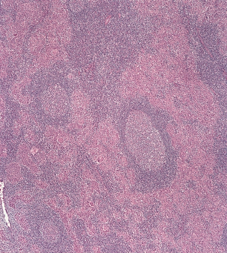 Undifferentiated nasopharyngeal carcinoma – low power
 -
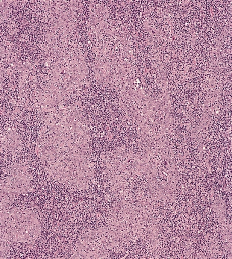 Undifferentiated nasopharyngeal carcinoma – med. power
 -
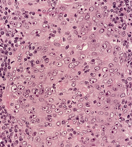 Undifferentiated nasopharyngeal carcinoma – high power


Immunohistochemistry
Immunohistochemistry stains for nasopharyngeal carcinoma include:
- EBER positive
- p16 negative[4]
References
- ↑ Abdel Khalek Abdel Razek, A.; King, A. (2012). “MRI and CT of nasopharyngeal carcinoma”. AJR Am J Roentgenol. 198 (1): 11–8. doi:10.2214/AJR.11.6954. PMID 22194474. Unknown parameter
|month=ignored (help) - ↑ Nasopharyngeal carcinoma http://librepathology.org/wiki/index.php/Nasopharyngeal_carcinoma
- ↑ Richard Cote, Saul Suster, Lawrence Weiss, Noel Weidner (Editor). Modern Surgical Pathology (2 Volume Set). London: W B Saunders. ISBN 0-7216-7253-1.
- ↑ Gulley ML, Nicholls JM, Schneider BG, Amin MB, Ro JY, Geradts J (1998). “Nasopharyngeal carcinomas frequently lack the p16/MTS1 tumor suppressor protein but consistently express the retinoblastoma gene product”. Am. J. Pathol. 152 (4): 865–9. PMC 1858242. PMID 9546345. Unknown parameter
|month=ignored (help)
Editor-In-Chief: C. Michael Gibson, M.S., M.D. [1] Associate Editor(s)-in-Chief: Homa Najafi, M.D.[2]Faizan Sheraz, M.D. [3]
Causes
Overview
Common causes of nasopharyngeal carcinoma include Epstein Barr virus (EBV infection), Human Papillomavirus (HPV infection), and consumption of salted fish as a source of N-nitrosamine.
Causes
Common Causes
Common causes of nasopharyngeal carcinoma may include:[1][2][3][4][5][6][7][8][9][10]
- EBV infection: Role of EBV infection in the pathogenesis of nasopharyngeal carcinoma is established. Detection of EBV DNA and EBV gene expression even in the first phases of tumor transformation in all tumor cells confirmed this role.
- HPV infection: HPV infection is associated with the non-endemic form of nasopharyngeal carcinoma. The patients with HPV positive nasopharyngeal carcinoma has poor prognosis rather than the EBV positive ones.
- Consumption of salted fish: N-nitrosamine in salted fish acts as a carcinogenic factor on nasopharyngeal cells. Individuals who start consumption of salted fish from their childhood have a greater risk to develop NPC rather than the ones who start in their adulthood.
References
- ↑ Jia, Wei-Hua; Qin, Hai-De (2012). “Non-viral environmental risk factors for nasopharyngeal carcinoma: A systematic review”. Seminars in Cancer Biology. 22 (2): 117–126. doi:10.1016/j.semcancer.2012.01.009. ISSN 1044-579X.
- ↑ Jia, Wei-Hua; Luo, Xiang-Yu; Feng, Bing-Jian; Ruan, Hong-Lian; Bei, Jin-Xin; Liu, Wen-Sheng; Qin, Hai-De; Feng, Qi-Sheng; Chen, Li-Zhen; Yao, Shugart Yin; Zeng, Yi-Xin (2010). “Traditional Cantonese diet and nasopharyngeal carcinoma risk: a large-scale case-control study in Guangdong, China”. BMC Cancer. 10 (1). doi:10.1186/1471-2407-10-446. ISSN 1471-2407.
- ↑ Guo, Xiuchan; Johnson, Randall C.; Deng, Hong; Liao, Jian; Guan, Li; Nelson, George W.; Tang, Mingzhong; Zheng, Yuming; de The, Guy; O’Brien, Stephen J.; Winkler, Cheryl A.; Zeng, Yi (2009). “Evaluation of nonviral risk factors for nasopharyngeal carcinoma in a high-risk population of Southern China”. International Journal of Cancer. 124 (12): 2942–2947. doi:10.1002/ijc.24293. ISSN 0020-7136.
- ↑ Maxwell, Jessica H.; Kumar, Bhavna; Feng, Felix Y.; McHugh, Jonathan B.; Cordell, Kitrina G.; Eisbruch, Avraham; Worden, Francis P.; Wolf, Gregory T.; Prince, Mark E.; Moyer, Jeffrey S.; Teknos, Theodoros N.; Chepeha, Douglas B.; Stoerker, Jay; Walline, Heather; Carey, Thomas E.; Bradford, Carol R. (2009). “HPV-positive/p16-positive/EBV-negative nasopharyngeal carcinoma in white North Americans”. Head & Neck: NA–NA. doi:10.1002/hed.21216. ISSN 1043-3074.
- ↑ Chan, Yap-Hang; Lo, Ching-Man; Lau, Hiu-Ying; Lam, Tai-Hing (2014). “Vertically transmitted nasopharyngeal infection of the human papillomavirus: Does it play an aetiological role in nasopharyngeal cancer?”. Oral Oncology. 50 (5): 326–329. doi:10.1016/j.oraloncology.2013.12.025. ISSN 1368-8375.
- ↑ Dogan, Snjezana; Hedberg, Matthew L.; Ferris, Robert L.; Rath, Tanya J.; Assaad, Adel M.; Chiosea, Simion I. (2014). “Human papillomavirus and Epstein-Barr virus in nasopharyngeal carcinoma in a low-incidence population”. Head & Neck. 36 (4): 511–516. doi:10.1002/hed.23318. ISSN 1043-3074.
- ↑ Stenmark, Matthew H.; McHugh, Jonathan B.; Schipper, Matthew; Walline, Heather M.; Komarck, Christine; Feng, Felix Y.; Worden, Francis P.; Wolf, Gregory T.; Chepeha, Douglas B.; Prince, Mark E.; Bradford, Carol R.; Mukherji, Suresh K.; Eisbruch, Avraham; Carey, Thomas E. (2014). “Nonendemic HPV-Positive Nasopharyngeal Carcinoma: Association With Poor Prognosis”. International Journal of Radiation Oncology*Biology*Physics. 88 (3): 580–588. doi:10.1016/j.ijrobp.2013.11.246. ISSN 0360-3016.
- ↑ Radha Raghupathy, Edwin Pun Hui & Anthony Tak Cheung Chan (2014). “Epstein-Barr virus as a paradigm in nasopharyngeal cancer: from lab to clinic”. American Society of Clinical Oncology educational book. American Society of Clinical Oncology. Annual Meeting: 149–153. doi:10.14694/EdBook_AM.2014.34.149. PMID 24857071.
- ↑ Pathmanathan, Rajadurai; Prasad, Umapati; Sadler, Robert; Flynn, Kathryn; Raab-Traub, Nancy (1995). “Clonal Proliferations of Cells Infected with Epstein–Barr Virus in Preinvasive Lesions Related to Nasopharyngeal Carcinoma”. New England Journal of Medicine. 333 (11): 693–698. doi:10.1056/NEJM199509143331103. ISSN 0028-4793.
- ↑ A. S. Chan, K. F. To, K. W. Lo, K. F. Mak, W. Pak, B. Chiu, G. M. Tse, M. Ding, X. Li, J. C. Lee & D. P. Huang (2000). “High frequency of chromosome 3p deletion in histologically normal nasopharyngeal epithelia from southern Chinese”. Cancer research. 60 (19): 5365–5370. PMID 11034072. Unknown parameter
|month=ignored (help)
Editor-In-Chief: C. Michael Gibson, M.S., M.D. [1] Associate Editor(s)-in-Chief: Homa Najafi, M.D.[2]Faizan Sheraz, M.D. [3]
Differentiating Nasopharyngeal Carcinoma from other Diseases

Overview
Nasopharyngeal carcinoma must be differentiated from other congenital abnormalities, inflammatory, and malignant lesions of neck.
Differentiating nasopharyngeal carcinoma from other neck masses
Nasopharyngeal carcinoma must be differentiated from other congenital abnormalities, inflammatory, and malignant lesions of neck.
| Category | Diseases | Benign/
Malignant |
Clinical manifestation | Paraclinical findings | Gold standard diagnosis | Associated findings | ||||||||
|---|---|---|---|---|---|---|---|---|---|---|---|---|---|---|
| Demography | History | Symptoms | Signs | Lab findings | Histopathology | Imaging | ||||||||
| Pain | Dysphagia | Mass exam | Others | |||||||||||
| Congenital | Branchial cleft cyst[1] |
|
|
− | ± |
|
|
− |
|
|
− | |||
| Thyroglossal duct cyst[2][3] |
|
|
− | − |
|
− | − |
|
|
− | − | |||
| Hemangioma[4] |
|
− | − |
|
|
|
|
|
||||||
| Vascular malformation[5][6] |
|
|
± | − |
|
|
|
|
|
− | ||||
| Category | Diseases | Benign | Demography | History | Pain | Dysphagia | Mass exam | Others | Lab findings | Histopathology | Imaging | Gold standard diagnosis | Associated findings | |
| Congenital | Lymphatic malformation[7][8] |
|
− | + |
|
|
− |
|
|
− | ||||
| Laryngocele[9][10][11] |
|
− | + |
|
|
− | − | |||||||
| Ranula[12][13] | − | − |
|
− | − |
|
|
− | − | |||||
| Category | Diseases | Benign | Demography | History | Pain | Dysphagia | Mass exam | Others | Lab findings | Histopathology | Imaging | Gold standard diagnosis | Associated findings | |
| Congenital | Teratoma[14][15] |
|
|
− | − |
|
− |
|
|
|
− | − | ||
| Dermoid cyst[16][17] | − | − |
|
|
− |
|
|
− | − | |||||
| Thymic cyst[18] | − | − |
|
− | − |
|
|
− | − | |||||
| Category | Diseases | Benign | Demography | History | Pain | Dysphagia | Mass exam | Others | Lab findings | Histopathology | Imaging | Gold standard diagnosis | Associated findings | |
| Inflammatory | Acute sialadenitis[19] |
|
|
+ | – |
|
|
|
|
− | ||||
| Chronic sialadenitis[20] |
|
|
+ | − |
|
|
|
|
|
− | ||||
| Reactive viral lymphadenopathy | CMV[21] |
|
|
− | − |
|
|
|
|
|
− | |||
| EBV[22][23] |
|
− | − |
|
|
|
|
|
− | |||||
| HIV[24] |
|
− | − |
|
|
|
|
|
|
− | ||||
| Viral URI[25] | − | − |
|
|
|
|
|
− | − | |||||
| Category | Diseases | Benign | Demography | History | Pain | Dysphagia | Mass exam | Others | Lab findings | Histopathology | Imaging | Gold standard diagnosis | Associated findings | |
| Inflammatory | Bacterial lymphadenopathy | Tularemia[26][27] |
|
+ | − |
|
|
|
|
− | ||||
| Brucellosis[28] |
|
+ | − |
|
|
|
|
− | ||||||
| Cat-scratch disease[29][30] |
|
+ | − |
|
|
− | − |
| ||||||
| Actinomycosis[31][32] | − | − |
|
|
|
− | ||||||||
| Mycobacterial infections[22][33][34] | − | − |
|
|
|
− | ||||||||
| Streptococcal infection[21][35] |
|
|
+ | + |
|
− | ||||||||
| Category | Diseases | Benign | Demography | History | Pain | Dysphagia | Mass exam | Others | Lab findings | Histopathology | Imaging | Gold standard diagnosis | Associated findings | |
| Inflammatory | Parasitic lymphadenopathy | Toxoplasma gondii[36][37] |
|
|
+ | − |
|
|
|
− | ||||
| Sarcoidosis_National_Heart,_Lung,_and_Blood_Institute_(NHLBI)-38|[38][19] |
|
− | − |
|
|
|
| |||||||
| Sjögren syndrome[39] |
|
− | + |
|
|
|
|
− | ||||||
| Castleman disease (angiofollicular lymphoproliferative disease)[40][41] |
|
|
− | − |
|
|
|
|||||||
| Category | Diseases | Benign | Demography | History | Pain | Dysphagia | Mass exam | Others | Lab findings | Histopathology | Imaging | Gold standard diagnosis | Associated findings | |
| Inflammatory | Kikuchi disease (histiocytic necrotizing lymphadenitis)[42] |
|
+ | − |
|
|
|
− | ||||||
| Kimura disease[43] |
|
|
− | − |
|
|
|
− | ||||||
| Rosai-Dorfman disease_Genetic_and_Rare_Diseases_Information_Center_(GARD)_–_an_NCATS_Program-44|[44][45] |
|
− | − |
|
|
− | − | − | − | |||||
| Kawasaki disease_Kawasaki_Disease_|_CDC-46|[46]_National_Heart,_Lung,_and_Blood_Institute_(NHLBI)-47|[47] |
|
|
− | − |
|
− |
|
− | ||||||
| Category | Diseases | Benign or Malignant | Demography | History | Pain | Dysphagia | Mass exam | Others | Lab findings | Histopathology | Imaging | Gold standard diagnosis | Associated findings | |
| Neoplasm | Salivary gland neoplasm | Pleomorphic adenoma[48][49] |
|
|
− | + |
|
− | − |
|
− | |||
| Warthin’s tumor[50]_Genetic_and_Rare_Diseases_Information_Center_(GARD)_–_an_NCATS_Program-51|[51] |
|
|
− | + |
|
− | − |
|
− | |||||
| Oncocytoma |
|
|
± | ± |
|
|
|
|
|
– | ||||
| Monomorphic adenoma [53][54][55] |
|
|
± | ± |
|
|
|
|
– | |||||
| Mucoepidermoid carcinoma |
|
|
± | ± |
|
|
− |
|
|
| ||||
| Category | Diseases | Benign | Demography | History | Pain | Dysphagia | Mass exam | Others | Lab findings | Histopathology | Imaging | Gold standard diagnosis | Associated findings | |
| Neoplasm | Salivary gland neoplasm | Adenoid cystic carcinoma [57] |
|
|
± | ± |
|
|
− |
|
|
− | ||
| Adenocarcinoma |
|
|
− | − |
|
|
|
|
|
− | ||||
| Salivary duct cancer[59][60][61] |
(Highly aggressive) |
|
|
± | ± |
|
|
|
|
|
− | |||
| Squamous cell carcinoma[62][63] |
|
+ | − |
|
|
|
|
|
− | |||||
| Category | Diseases | Benign | Demography | History | Pain | Dysphagia | Mass exam | Others | Lab findings | Histopathology | Imaging | Gold standard diagnosis | Associated findings | |
| Neoplasm | Hypopharyngeal cancer[64][65][66] |
|
− | + |
|
− |
|
|
− | |||||
| Parathyroid cancer[67][68][69] |
|
|
+ | + |
|
|
|
|
|
| ||||
| Carotid body tumors[70][71][72][73] |
|
|
+ | − |
|
|
|
|
− | |||||
| Paraganglioma[74][75][76] |
|
|
− | − |
|
− |
|
|
|
− | ||||
| Category | Diseases | Benign | Demography | History | Pain | Dysphagia | Mass exam | Others | Lab findings | Histopathology | Imaging | Gold standard diagnosis | Associated findings | |
| Neoplasm | Schwannoma[77][78][79] |
|
|
+ | ± |
|
|
|
|
|
||||
| Lymphoma [80][81][82][83][84][85] |
|
|
− | ± |
|
|
|
|
|
| ||||
| Liposarcoma [86][87][88][89] |
|
± | − |
|
|
|
|
|
− | |||||
| Category | Diseases | Benign | Demography | History | Pain | Dysphagia | Mass exam | Others | Lab findings | Histopathology | Imaging | Gold standard diagnosis | Associated findings | |
| Neoplasm | Lipoma [90][91][92] |
|
± | − |
|
|
|
|
| |||||
| Glomus vagale, glomus jugulare tumors[93][94][95][96][97][98] |
|
− | ± |
|
|
|
|
|
− | |||||
| Metastatic head and neck cancer[99][100] |
|
|
− | ± |
|
|
|
|
|
− | ||||
| Category | Diseases | Benign | Demography | History | Pain | Dysphagia | Mass exam | Others | Lab findings | Histopathology | Imaging | Gold standard diagnosis | Associated findings | |
| Other | Laryngeal cancer[101][102] | Benign/Malignant |
|
|
± | ± |
|
human papillomavirus (HPV) infection |
|
|
|
− | ||
| Arteriovenous fistula |
|
|
− | − |
|
|
− | |||||||
| Thyroid nodule/ Goiter |
|
|
± | ± |
|
|
|
|
|
|
− | |||
| Category | Diseases | Benign | Demography | History | Pain | Dysphagia | Mass exam | Others | Lab findings | Histopathology | Imaging | Gold standard diagnosis | Associated findings | |
References
- ↑ Nahata, Vaishali (2016). “Branchial cleft cyst”. Indian Journal of Dermatology. 61 (6): 701. doi:10.4103/0019-5154.193718. ISSN 0019-5154.
- ↑ Amos J, Shermetaro C. PMID 30085599. Missing or empty
|title=(help) - ↑ Deaver MJ, Silman EF, Lotfipour S (August 2009). “Infected thyroglossal duct cyst”. West J Emerg Med. 10 (3): 205. PMC 2729228. PMID 19718389.
- ↑ Léauté-Labrèze, C.; Prey, S.; Ezzedine, K. (2011). “Infantile haemangioma: Part I. Pathophysiology, epidemiology, clinical features, life cycle and associated structural abnormalities”. Journal of the European Academy of Dermatology and Venereology. 25 (11): 1245–1253. doi:10.1111/j.1468-3083.2011.04102.x. ISSN 0926-9959.
- ↑ Cox JA, Bartlett E, Lee EI (May 2014). “Vascular malformations: a review”. Semin Plast Surg. 28 (2): 58–63. doi:10.1055/s-0034-1376263. PMC 4078214. PMID 25045330.
- ↑ Behravesh S, Yakes W, Gupta N, Naidu S, Chong BW, Khademhosseini A, Oklu R (December 2016). “Venous malformations: clinical diagnosis and treatment”. Cardiovasc Diagn Ther. 6 (6): 557–569. doi:10.21037/cdt.2016.11.10. PMC 5220204. PMID 28123976.
- ↑ Cox JA, Bartlett E, Lee EI (May 2014). “Vascular malformations: a review”. Semin Plast Surg. 28 (2): 58–63. doi:10.1055/s-0034-1376263. PMC 4078214. PMID 25045330.
- ↑ Guruprasad Y, Chauhan DS (September 2012). “Cervical cystic hygroma”. J Maxillofac Oral Surg. 11 (3): 333–6. doi:10.1007/s12663-010-0149-x. PMC 3428451. PMID 23997487.
- ↑ Werner RL, Schroeder JW, Castle JT (March 2014). “Bilateral laryngoceles”. Head Neck Pathol. 8 (1): 110–3. doi:10.1007/s12105-013-0478-4. PMC 3950389. PMID 23881550.
- ↑ Prasad KC, Vijayalakshmi S, Prasad SC (December 2008). “Laryngoceles – presentations and management”. Indian J Otolaryngol Head Neck Surg. 60 (4): 303–8. doi:10.1007/s12070-008-0108-8. PMC 3476818. PMID 23120570.
- ↑ Mahdoufi R, Barhmi I, Tazi N, Abada R, Roubal M, Mahtar M (July 2017). “Mixed Pyolaryngocele: A Rare Case of Deep Neck Infection”. Iran J Otorhinolaryngol. 29 (93): 225–228. PMC 5554815. PMID 28819622.
- ↑ Packiri S, Gurunathan D, Selvarasu K (September 2017). “Management of Paediatric Oral Ranula: A Systematic Review”. J Clin Diagn Res. 11 (9): ZE06–ZE09. doi:10.7860/JCDR/2017/28498.10622. PMC 5713871. PMID 29207849.
- ↑ Kokong D, Iduh A, Chukwu I, Mugu J, Nuhu S, Augustine S (June 2017). “Ranula: Current Concept of Pathophysiologic Basis and Surgical Management Options”. World J Surg. 41 (6): 1476–1481. doi:10.1007/s00268-017-3901-2. PMC 5422487. PMID 28194490.
- ↑ Chauhan DS, Guruprasad Y, Inderchand S (September 2011). “Congenital nasopharyngeal teratoma with a cleft palate: case report and a 7 year follow up”. J Maxillofac Oral Surg. 10 (3): 253–6. doi:10.1007/s12663-010-0140-6. PMC 3238564. PMID 22942597.
- ↑ Bahgat M, Bahgat Y, Bahgat A (July 2012). “Oropharyngeal teratoma, a rare cause of airway obstruction in neonates”. BMJ Case Rep. 2012. doi:10.1136/bcr-2012-006580. PMC 4543570. PMID 22814615.
- ↑ Paradis, Josée; Koltai, Peter J. (2015). “Pediatric Teratoma and Dermoid Cysts”. Otolaryngologic Clinics of North America. 48 (1): 121–136. doi:10.1016/j.otc.2014.09.009. ISSN 0030-6665.
- ↑ Gaddikeri S, Vattoth S, Gaddikeri RS, Stuart R, Harrison K, Young D, Bhargava P (2014). “Congenital cystic neck masses: embryology and imaging appearances, with clinicopathological correlation”. Curr Probl Diagn Radiol. 43 (2): 55–67. doi:10.1067/j.cpradiol.2013.12.001. PMID 24629659.
- ↑ Gaddikeri, Santhosh; Vattoth, Surjith; Gaddikeri, Ramya S.; Stuart, Royal; Harrison, Keith; Young, Daniel; Bhargava, Puneet (2014). “Congenital Cystic Neck Masses: Embryology and Imaging Appearances, With Clinicopathological Correlation”. Current Problems in Diagnostic Radiology. 43 (2): 55–67. doi:10.1067/j.cpradiol.2013.12.001. ISSN 0363-0188.
- ↑ 19.0 19.1 Abdel Razek A, Mukherji S (June 2017). “Imaging of sialadenitis”. Neuroradiol J. 30 (3): 205–215. doi:10.1177/1971400916682752. PMC 5480791. PMID 28059621. Vancouver style error: initials (help)
- ↑ Orlandi MA, Pistorio V, Guerra PA (2013). “Ultrasound in sialadenitis”. J Ultrasound. 16 (1): 3–9. doi:10.1007/s40477-013-0002-4. PMC 3774898. PMID 24046793.
- ↑ 21.0 21.1 Mohseni S, Shojaiefard A, Khorgami Z, Alinejad S, Ghorbani A, Ghafouri A (March 2014). “Peripheral lymphadenopathy: approach and diagnostic tools”. Iran J Med Sci. 39 (2 Suppl): 158–70. PMC 3993046. PMID 24753638.
- ↑ 22.0 22.1 Mohseni S, Shojaiefard A, Khorgami Z, Alinejad S, Ghorbani A, Ghafouri A (March 2014). “Peripheral lymphadenopathy: approach and diagnostic tools”. Iran J Med Sci. 39 (2 Suppl): 158–70. PMC 3993046. PMID 24753638.
- ↑ Stuhlmann-Laeisz C, Oschlies I, Klapper W (December 2014). “Detection of EBV in reactive and neoplastic lymphoproliferations in adults-when and how?”. J Hematop. 7 (4): 165–170. doi:10.1007/s12308-014-0209-0. PMC 4243011. PMID 25478033.
- ↑ Moonim MT, Alarcon L, Freeman J, Mahadeva U, van der Walt JD, Lucas SB (March 2010). “Identifying HIV infection in diagnostic histopathology tissue samples–the role of HIV-1 p24 immunohistochemistry in identifying clinically unsuspected HIV infection: a 3-year analysis”. Histopathology. 56 (4): 530–41. doi:10.1111/j.1365-2559.2010.03513.x. PMID 20459560.
- ↑ Thomas M, Bomar PA. PMID 30422556. Missing or empty
|title=(help) - ↑ Grunow R, Splettstoesser W, McDonald S, Otterbein C, O’Brien T, Morgan C, Aldrich J, Hofer E, Finke EJ, Meyer H (January 2000). “Detection of Francisella tularensis in biological specimens using a capture enzyme-linked immunosorbent assay, an immunochromatographic handheld assay, and a PCR”. Clin. Diagn. Lab. Immunol. 7 (1): 86–90. PMC 95828. PMID 10618283.
- ↑ Koç, Sema (2012). “Clinical and laboratory findings of tularemia: a retrospective analysis”. The Turkish Journal of Ear Nose and Throat: 26–31. doi:10.5606/kbbihtisas.2012.005. ISSN 1300-7475.
- ↑ Golshani M, Buozari S (November 2017). “A review of Brucellosis in Iran: Epidemiology, Risk Factors, Diagnosis, Control, and Prevention”. Iran. Biomed. J. 21 (6): 349–59. PMC 5572431. PMID 28766326.
- ↑ “Cat-Scratch Disease in the United States, 2005–2013 – Volume 22, Number 10—October 2016 – Emerging Infectious Diseases journal – CDC”.
- ↑ Hansmann, Y.; DeMartino, S.; Piemont, Y.; Meyer, N.; Mariet, P.; Heller, R.; Christmann, D.; Jaulhac, B. (2005). “Diagnosis of Cat Scratch Disease with Detection of Bartonella henselae by PCR: a Study of Patients with Lymph Node Enlargement”. Journal of Clinical Microbiology. 43 (8): 3800–3806. doi:10.1128/JCM.43.8.3800-3806.2005. ISSN 0095-1137.
- ↑ Valour F, Sénéchal A, Dupieux C, Karsenty J, Lustig S, Breton P, Gleizal A, Boussel L, Laurent F, Braun E, Chidiac C, Ader F, Ferry T (2014). “Actinomycosis: etiology, clinical features, diagnosis, treatment, and management”. Infect Drug Resist. 7: 183–97. doi:10.2147/IDR.S39601. PMC 4094581. PMID 25045274.
- ↑ Bonnefond S, Catroux M, Melenotte C, Karkowski L, Rolland L, Trouillier S, Raffray L (June 2016). “Clinical features of actinomycosis: A retrospective, multicenter study of 28 cases of miscellaneous presentations”. Medicine (Baltimore). 95 (24): e3923. doi:10.1097/MD.0000000000003923. PMC 4998488. PMID 27311002.
- ↑ Suskind DL, Handler SD, Tom LW, Potsic WP, Wetmore RF (July 1997). “Nontuberculous mycobacterial cervical adenitis”. Clin Pediatr (Phila). 36 (7): 403–9. doi:10.1177/000992289703600705. PMID 9241478.
- ↑ Drobniewski FA, Caws M, Gibson A, Young D (March 2003). “Modern laboratory diagnosis of tuberculosis”. Lancet Infect Dis. 3 (3): 141–7. PMID 12614730.
- ↑ Kenealy T (November 2007). “Sore throat”. BMJ Clin Evid. 2007. PMC 2943825. PMID 19450346.
- ↑ Kumar GG, Mahadevan A, Guruprasad AS, Kovoor JM, Satishchandra P, Nath A, Ranga U, Shankar SK (June 2010). “Eccentric target sign in cerebral toxoplasmosis: neuropathological correlate to the imaging feature”. J Magn Reson Imaging. 31 (6): 1469–72. doi:10.1002/jmri.22192. PMC 2908244. PMID 20512900.
- ↑ [+https://www.cdc.gov/parasites/toxoplasmosis/diagnosis.html “CDC – Toxoplasmosis – Diagnosis”] Check
|url=value (help). - _National_Heart,_Lung,_and_Blood_Institute_(NHLBI)_38-0|↑ “Sarcoidosis | National Heart, Lung, and Blood Institute (NHLBI)”.
- ↑ Mavragani CP, Moutsopoulos HM (October 2014). “Sjögren syndrome”. CMAJ. 186 (15): E579–86. doi:10.1503/cmaj.122037. PMC 4203623. PMID 24566651.
- ↑ Dispenzieri A, Armitage JO, Loe MJ, Geyer SM, Allred J, Camoriano JK, Menke DM, Weisenburger DD, Ristow K, Dogan A, Habermann TM (November 2012). “The clinical spectrum of Castleman’s disease”. Am. J. Hematol. 87 (11): 997–1002. doi:10.1002/ajh.23291. PMC 3900496. PMID 22791417.
- ↑ Saeed-Abdul-Rahman I, Al-Amri AM (September 2012). “Castleman disease”. Korean J Hematol. 47 (3): 163–77. doi:10.5045/kjh.2012.47.3.163. PMC 3464333. PMID 23071471.
- ↑ Bosch X, Guilabert A (May 2006). “Kikuchi-Fujimoto disease”. Orphanet J Rare Dis. 1: 18. doi:10.1186/1750-1172-1-18. PMC 1481509. PMID 16722618.
- ↑ AlGhamdi FE, Al-Khatib TA, Marzouki HZ, AlGarni MA (March 2016). “Kimura disease: No age or ethnicity limit”. Saudi Med J. 37 (3): 315–9. doi:10.15537/smj.2016.3.14448. PMC 4800898. PMID 26905356.
- _Genetic_and_Rare_Diseases_Information_Center_(GARD)_–_an_NCATS_Program_44-0|↑ “Rosai-Dorfman disease | Genetic and Rare Diseases Information Center (GARD) – an NCATS Program”.
- ↑ Foucar E, Rosai J, Dorfman R (February 1990). “Sinus histiocytosis with massive lymphadenopathy (Rosai-Dorfman disease): review of the entity”. Semin Diagn Pathol. 7 (1): 19–73. PMID 2180012.
- _Kawasaki_Disease_|_CDC_46-0|↑ “About Kawasaki Disease | Kawasaki Disease | CDC”.
- _National_Heart,_Lung,_and_Blood_Institute_(NHLBI)_47-0|↑ “Kawasaki Disease | National Heart, Lung, and Blood Institute (NHLBI)”.
- ↑ Debnath SC, Adhyapok AK (June 2010). “Pleomorphic adenoma (benign mixed tumour) of the minor salivary glands of the upper lip”. J Maxillofac Oral Surg. 9 (2): 205–8. doi:10.1007/s12663-010-0052-5. PMC 3244097. PMID 22190789.
- ↑ Kato H, Kawaguchi M, Ando T, Mizuta K, Aoki M, Matsuo M (August 2018). “Pleomorphic adenoma of salivary glands: common and uncommon CT and MR imaging features”. Jpn J Radiol. 36 (8): 463–471. doi:10.1007/s11604-018-0747-y. PMID 29845358.
- ↑ Chulam TC, Noronha Francisco AL, Goncalves Filho J, Pinto Alves CA, Kowalski LP (December 2013). “Warthin’s tumour of the parotid gland: our experience”. Acta Otorhinolaryngol Ital. 33 (6): 393–7. PMID 24376295.
- _Genetic_and_Rare_Diseases_Information_Center_(GARD)_–_an_NCATS_Program_51-0|↑ “Warthin tumor | Genetic and Rare Diseases Information Center (GARD) – an NCATS Program”.
- ↑ Chen B, Hentzelman JI, Walker RJ, Lai JP (2016). “Oncocytoma of the Submandibular Gland: Diagnosis and Treatment Based on Clinicopathology”. Case Rep Otolaryngol. 2016: 8719030. doi:10.1155/2016/8719030. PMC 5045990. PMID 27722003.
- ↑ Kim KH, Sung MW, Kim JW, Koo JW (July 2000). “Pleomorphic adenoma of the trachea”. Otolaryngol Head Neck Surg. 123 (1 Pt 1): 147–8. doi:10.1067/mhn.2000.102809. PMID 10889498.
- ↑ Pramod Krishna B (June 2013). “Pleomorphic Adenoma of Minor Salivary Gland in a 14 year Old Child”. J Maxillofac Oral Surg. 12 (2): 228–31. doi:10.1007/s12663-010-0125-5. PMC 3681990. PMID 24431845.
- ↑ Kessler AT, Bhatt AA (2018). “Review of the Major and Minor Salivary Glands, Part 2: Neoplasms and Tumor-like Lesions”. J Clin Imaging Sci. 8: 48. doi:10.4103/jcis.JCIS_46_18. PMC 6251244. PMID 30546932.
- ↑ Chenevert J, Barnes LE, Chiosea SI (February 2011). “Mucoepidermoid carcinoma: a five-decade journey”. Virchows Arch. 458 (2): 133–40. doi:10.1007/s00428-011-1040-y. PMID 21243374.
- ↑ Jones AV, Craig GT, Speight PM, Franklin CD (April 2008). “The range and demographics of salivary gland tumours diagnosed in a UK population”. Oral Oncol. 44 (4): 407–17. doi:10.1016/j.oraloncology.2007.05.010. PMID 17825603.
- ↑ Beltran D, Faquin WC, Gallagher G, August M (March 2006). “Selective immunohistochemical comparison of polymorphous low-grade adenocarcinoma and adenoid cystic carcinoma”. J. Oral Maxillofac. Surg. 64 (3): 415–23. doi:10.1016/j.joms.2005.11.027. PMID 16487803.
- ↑ Mlika M, Kourda N, Zidi Y, Aloui R, Zneidi N, Rammeh S, Zermani R, Jilani SB (January 2012). “Salivary duct carcinoma of the parotid gland”. J Oral Maxillofac Pathol. 16 (1): 134–6. doi:10.4103/0973-029X.92992. PMC 3303509. PMID 22434951.
- ↑ Schmitt NC, Kang H, Sharma A (November 2017). “Salivary duct carcinoma: An aggressive salivary gland malignancy with opportunities for targeted therapy”. Oral Oncol. 74: 40–48. doi:10.1016/j.oraloncology.2017.09.008. PMC 5685667. PMID 29103750.
- ↑ Simpson RH (July 2013). “Salivary duct carcinoma: new developments–morphological variants including pure in situ high grade lesions; proposed molecular classification”. Head Neck Pathol. 7 Suppl 1: S48–58. doi:10.1007/s12105-013-0456-x. PMC 3712088. PMID 23821208.
- ↑ Manvikar V, Ramulu S, Ravishanker ST, Chakravarthy C (May 2014). “Squamous cell carcinoma of submandibular salivary gland: A rare case report”. J Oral Maxillofac Pathol. 18 (2): 299–302. doi:10.4103/0973-029X.140909. PMC 4196305. PMID 25328317.
- ↑ Ying YL, Johnson JT, Myers EN (July 2006). “Squamous cell carcinoma of the parotid gland”. Head Neck. 28 (7): 626–32. doi:10.1002/hed.20360. PMID 16475198.
- ↑ Helliwell TR (February 2003). “acp Best Practice No 169. Evidence based pathology: squamous carcinoma of the hypopharynx”. J. Clin. Pathol. 56 (2): 81–5. PMC 1769882. PMID 12560383.
- ↑ International Journal of Recent Scientific Research. doi:10.24327/IJRSR. ISSN 0976-3031. Missing or empty
|title=(help) - ↑ Maasland, Denise HE; van den Brandt, Piet A; Kremer, Bernd; Goldbohm, R Alexandra; Schouten, Leo J (2014). “Alcohol consumption, cigarette smoking and the risk of subtypes of head-neck cancer: results from the Netherlands Cohort Study”. BMC Cancer. 14 (1). doi:10.1186/1471-2407-14-187. ISSN 1471-2407.
- ↑ Wei CH, Harari A (March 2012). “Parathyroid carcinoma: update and guidelines for management”. Curr Treat Options Oncol. 13 (1): 11–23. doi:10.1007/s11864-011-0171-3. PMID 22327883.
- ↑ Sahasranam P, Tran MT, Mohamed H, Friedman TC (August 2007). “Multiglandular parathyroid carcinoma: a case report and brief review”. South. Med. J. 100 (8): 841–4. doi:10.1097/SMJ.0b013e318073ca37. PMID 17713315.
- ↑ Holmes EC, Morton DL, Ketcham AS (April 1969). “Parathyroid carcinoma: a collective review”. Ann. Surg. 169 (4): 631–40. PMC 1387475. PMID 4886854.
- ↑ Sajid MS, Hamilton G, Baker DM (August 2007). “A multicenter review of carotid body tumour management”. Eur J Vasc Endovasc Surg. 34 (2): 127–30. doi:10.1016/j.ejvs.2007.01.015. PMID 17400487.
- ↑ Boedeker CC, Ridder GJ, Schipper J (2005). “Paragangliomas of the head and neck: diagnosis and treatment”. Fam. Cancer. 4 (1): 55–9. doi:10.1007/s10689-004-2154-z. PMID 15883711.
- ↑ Pellitteri PK, Rinaldo A, Myssiorek D, Gary Jackson C, Bradley PJ, Devaney KO, Shaha AR, Netterville JL, Manni JJ, Ferlito A (July 2004). “Paragangliomas of the head and neck”. Oral Oncol. 40 (6): 563–75. doi:10.1016/j.oraloncology.2003.09.004. PMID 15063383.
- ↑ Darouassi Y, Alaoui M, Mliha Touati M, Al Maghraoui O, En-Nouali A, Bouaity B, Ammar H (August 2017). “Carotid Body Tumors: A Case Series and Review of the Literature”. Ann Vasc Surg. 43: 265–271. doi:10.1016/j.avsg.2017.03.167. PMID 28478173.
- ↑ Neumann HP, Pawlu C, Peczkowska M, Bausch B, McWhinney SR, Muresan M, Buchta M, Franke G, Klisch J, Bley TA, Hoegerle S, Boedeker CC, Opocher G, Schipper J, Januszewicz A, Eng C (August 2004). “Distinct clinical features of paraganglioma syndromes associated with SDHB and SDHD gene mutations”. JAMA. 292 (8): 943–51. doi:10.1001/jama.292.8.943. PMID 15328326.
- ↑ Erickson D, Kudva YC, Ebersold MJ, Thompson GB, Grant CS, van Heerden JA, Young WF (November 2001). “Benign paragangliomas: clinical presentation and treatment outcomes in 236 patients”. J. Clin. Endocrinol. Metab. 86 (11): 5210–6. doi:10.1210/jcem.86.11.8034. PMID 11701678.
- ↑ O’Riordain DS, Young WF, Grant CS, Carney JA, van Heerden JA (September 1996). “Clinical spectrum and outcome of functional extraadrenal paraganglioma”. World J Surg. 20 (7): 916–21, discussion 922. PMID 8678971.
- ↑ Hilton DA, Hanemann CO (April 2014). “Schwannomas and their pathogenesis”. Brain Pathol. 24 (3): 205–20. doi:10.1111/bpa.12125. PMID 24450866.
- ↑ Albert P, Patel J, Badawy K, Weissinger W, Brenner M, Bourhill I, Parnell J (2017). “Peripheral Nerve Schwannoma: A Review of Varying Clinical Presentations and Imaging Findings”. J Foot Ankle Surg. 56 (3): 632–637. doi:10.1053/j.jfas.2016.12.003. PMID 28237565.
- ↑ Wong B, Bathala S, Grant D (January 2017). “Laryngeal schwannoma: a systematic review”. Eur Arch Otorhinolaryngol. 274 (1): 25–34. doi:10.1007/s00405-016-4013-6. PMID 27020268. Vancouver style error: initials (help)
- ↑ Anderson T, Chabner BA, Young RC, Berard CW, Garvin AJ, Simon RM, DeVita VT (December 1982). “Malignant lymphoma. 1. The histology and staging of 473 patients at the National Cancer Institute”. Cancer. 50 (12): 2699–707. PMID 7139563.
- ↑ Anderson T, Chabner BA, Young RC, Berard CW, Garvin AJ, Simon RM, DeVita VT (December 1982). “Malignant lymphoma. 1. The histology and staging of 473 patients at the National Cancer Institute”. Cancer. 50 (12): 2699–707. PMID 7139563.
- ↑ Negri E, Little D, Boiocchi M, La Vecchia C, Franceschi S (August 2004). “B-cell non-Hodgkin’s lymphoma and hepatitis C virus infection: a systematic review”. Int. J. Cancer. 111 (1): 1–8. doi:10.1002/ijc.20205. PMID 15185336.
- ↑ Moormeier JA, Williams SF, Golomb HM (February 1990). “The staging of non-Hodgkin’s lymphomas”. Semin. Oncol. 17 (1): 43–50. PMID 2406917.
- ↑ Negri E, Little D, Boiocchi M, La Vecchia C, Franceschi S (August 2004). “B-cell non-Hodgkin’s lymphoma and hepatitis C virus infection: a systematic review”. Int. J. Cancer. 111 (1): 1–8. doi:10.1002/ijc.20205. PMID 15185336.
- ↑ Anderson T, Chabner BA, Young RC, Berard CW, Garvin AJ, Simon RM, DeVita VT (December 1982). “Malignant lymphoma. 1. The histology and staging of 473 patients at the National Cancer Institute”. Cancer. 50 (12): 2699–707. PMID 7139563.
- ↑ Evans HL (January 2007). “Atypical lipomatous tumor, its variants, and its combined forms: a study of 61 cases, with a minimum follow-up of 10 years”. Am. J. Surg. Pathol. 31 (1): 1–14. doi:10.1097/01.pas.0000213406.95440.7a. PMID 17197914.
- ↑ Conyers R, Young S, Thomas DM (2011). “Liposarcoma: molecular genetics and therapeutics”. Sarcoma. 2011: 483154. doi:10.1155/2011/483154. PMC 3021868. PMID 21253554.
- ↑ Alaggio R, Coffin CM, Weiss SW, Bridge JA, Issakov J, Oliveira AM, Folpe AL (May 2009). “Liposarcomas in young patients: a study of 82 cases occurring in patients younger than 22 years of age”. Am. J. Surg. Pathol. 33 (5): 645–58. doi:10.1097/PAS.0b013e3181963c9c. PMID 19194281.
- ↑ Serpell JW, Chen RY (July 2007). “Review of large deep lipomatous tumours”. ANZ J Surg. 77 (7): 524–9. doi:10.1111/j.1445-2197.2007.04042.x. PMID 17610686.
- ↑ de Bree E, Karatzanis A, Hunt JL, Strojan P, Rinaldo A, Takes RP, Ferlito A, de Bree R (May 2015). “Lipomatous tumours of the head and neck: a spectrum of biological behaviour”. Eur Arch Otorhinolaryngol. 272 (5): 1061–77. doi:10.1007/s00405-014-3065-8. PMID 24800932.
- ↑ Rydholm A, Berg NO (December 1983). “Size, site and clinical incidence of lipoma. Factors in the differential diagnosis of lipoma and sarcoma”. Acta Orthop Scand. 54 (6): 929–34. PMID 6670522.
- ↑ Myhre-Jensen O (June 1981). “A consecutive 7-year series of 1331 benign soft tissue tumours. Clinicopathologic data. Comparison with sarcomas”. Acta Orthop Scand. 52 (3): 287–93. PMID 7282321.
- ↑ Urquhart AC, Johnson JT, Myers EN, Schechter GL (April 1994). “Glomus vagale: paraganglioma of the vagus nerve”. Laryngoscope. 104 (4): 440–5. doi:10.1288/00005537-199404000-00008. PMID 8164483.
- ↑ Valavanis A, Schubiger O, Oguz M (1983). “High-resolution CT investigation of nonchromaffin paragangliomas of the temporal bone”. AJNR Am J Neuroradiol. 4 (3): 516–9. PMID 6308990.
- ↑ Urquhart AC, Johnson JT, Myers EN, Schechter GL (April 1994). “Glomus vagale: paraganglioma of the vagus nerve”. Laryngoscope. 104 (4): 440–5. doi:10.1288/00005537-199404000-00008. PMID 8164483.
- ↑ Stein PP, Black HR (January 1991). “A simplified diagnostic approach to pheochromocytoma. A review of the literature and report of one institution’s experience”. Medicine (Baltimore). 70 (1): 46–66. PMID 1988766.
- ↑ Sajid MS, Hamilton G, Baker DM (August 2007). “A multicenter review of carotid body tumour management”. Eur J Vasc Endovasc Surg. 34 (2): 127–30. doi:10.1016/j.ejvs.2007.01.015. PMID 17400487.
- ↑ Boedeker CC, Ridder GJ, Schipper J (2005). “Paragangliomas of the head and neck: diagnosis and treatment”. Fam. Cancer. 4 (1): 55–9. doi:10.1007/s10689-004-2154-z. PMID 15883711.
- ↑ Gluckman JL, Robbins KT, Fried MP (1990). “Cervical metastatic squamous carcinoma of unknown or occult primary source”. Head Neck. 12 (5): 440–3. PMID 2211107.
- ↑ Waltonen JD, Ozer E, Hall NC, Schuller DE, Agrawal A (October 2009). “Metastatic carcinoma of the neck of unknown primary origin: evolution and efficacy of the modern workup”. Arch. Otolaryngol. Head Neck Surg. 135 (10): 1024–9. doi:10.1001/archoto.2009.145. PMID 19841343.
- ↑ Feldman PS, Kaplan MJ, Johns ME, Cantrell RW (November 1983). “Fine-needle aspiration in squamous cell carcinoma of the head and neck”. Arch Otolaryngol. 109 (11): 735–42. PMID 6639441.
- ↑ Grénman R, Koivunen P, Minn H (2015). “[Laryngeal cancer in Finland]”. Duodecim (in Finnish). 131 (4): 331–7. PMID 26237923.
- ↑ Guneyli S, Cinar C, Bozkaya H, Korkmaz M, Oran I (September 2016). “Endovascular management of congenital arteriovenous fistulae in the neck”. Diagn Interv Imaging. 97 (9): 871–5. doi:10.1016/j.diii.2015.08.006. PMID 26972281.
- ↑ Gobin YP, Garcia de la Fuente JA, Herbreteau D, Houdart E, Merland JJ (November 1993). “Endovascular treatment of external carotid-jugular fistulae in the parotid region”. Neurosurgery. 33 (5): 812–6. PMID 8264877.
- ↑ Madjar S, Weissberg D (July 1995). “Retrosternal goiter”. Chest. 108 (1): 78–82. PMID 7606997.
- ↑ Hedayati N, McHenry CR (March 2002). “The clinical presentation and operative management of nodular and diffuse substernal thyroid disease”. Am Surg. 68 (3): 245–51, discussion 251–2. PMID 11893102.
- ↑ Hughes K, Eastman C (August 2012). “Goitre – causes, investigation and management”. Aust Fam Physician. 41 (8): 572–6. PMID 23145396.
- ↑ Hermus AR, Huysmans DA (August 2000). “[Diagnosis and therapy of patients with euthyroid goiter]”. Ned Tijdschr Geneeskd (in Dutch; Flemish). 144 (34): 1623–7. PMID 10972051.
Editor-In-Chief: C. Michael Gibson, M.S., M.D. [1] Associate Editor(s)-in-Chief: Maneesha Nandimandalam, M.B.B.S.[2], Gertrude Djouka, M.D.[3], Qurrat-ul-ain Abid, M.D.[4], Homa Najafi, M.D.[5], Faizan Sheraz, M.D. [6]
Epidemiology and Demographics
Overview
The incidence of nasopharyngeal carcinoma is different based on the geographic areas. The incidence in the USA is low, in Alaska and Greenland is moderate and the highest incidence is seen in southeast Asia, North Africa, southern China and middle east. The incidence of this disease increases with increasing age in low- risk population. However, in high-risk populations, the maximum incidence is seen in individuals around ages 50 to 59 and then the incidence decreases by increasing age. Nasopharyngeal carcinoma usually affects individuals of the Asian race, and is more commonly affected by male than female. The majority of nasopharyngeal carcinoma cases are reported in southern China, Southeast Asia, North Africa and also is more seen in Inuit Indians.
Epidemiology and Demographics
Incidence
- The incidence of nasopharyngeal carcinoma is different based on the geographic areas. The incidence in the USA is approximately 0.2 to 0.5 per 100,000 individuals, in Alaska and Greenland is 15 to 20 per 100,000 individuals and the highest incidence is seen in southeast Asia, North Africa, southern China and middle east with the incidence of 80 per 100,000 individuals.[1][2]
Case-fatality rate/Mortality rate
- In 2008 , the incidence of nasopharyngeal carcinoma is approximately 0.84 per 100,000 individuals with mortality rate of 61.13%.[3]
Age
- The incidence of nasopharyngeal carcinoma increases with increasing age in low- risk population. However, in high-risk populations, the maximum incidence is seen in individuals around ages 50 to 59 and then the incidence decreases by increasing age.[4][5]
Race
Gender
- Nasopharyngeal carcinoma is more commonly affected by male than female. The male to female ratio is approximately 2-3 to 1.[7]
Region
- The majority of nasopharyngeal carcinoma cases are reported in southern China, Southeast Asia, North Africa and also is more seen in Inuit Indians.[8][9]
References
- ↑ Caponigro, Francesco; Longo, Francesco; Ionna, Franco; Perri, Francesco (2010). “Treatment approaches to nasopharyngeal carcinoma: a review”. Anti-Cancer Drugs. 21 (5): 471–477. doi:10.1097/CAD.0b013e328337160e. ISSN 0959-4973.
- ↑ Lee, Anne W.M.; Ng, W.T.; Chan, Y.H.; Sze, Henry; Chan, Connie; Lam, T.H. (2012). “The battle against nasopharyngeal cancer”. Radiotherapy and Oncology. 104 (3): 272–278. doi:10.1016/j.radonc.2012.08.001. ISSN 0167-8140.
- ↑ Jemal, Ahmedin; Bray, Freddie; Center, Melissa M.; Ferlay, Jacques; Ward, Elizabeth; Forman, David (2011). “Global cancer statistics”. CA: A Cancer Journal for Clinicians. 61 (2): 69–90. doi:10.3322/caac.20107. ISSN 0007-9235.
- ↑ Lee, Jivianne T.; Ko, Clifford Y. (2016). “Has survival improved for nasopharyngeal carcinoma in the United States?”. Otolaryngology-Head and Neck Surgery. 132 (2): 303–308. doi:10.1016/j.otohns.2004.09.018. ISSN 0194-5998.
- ↑ Lee, Anne W.M.; Foo, William; Mang, Oscar; Sze, W.M.; Chappell, Rick; Lau, W.H.; Ko, W.M. (2003). “Changing epidemiology of nasopharyngeal carcinoma in Hong Kong over a 20-year period (1980-99): An encouraging reduction in both incidence and mortality”. International Journal of Cancer. 103 (5): 680–685. doi:10.1002/ijc.10894. ISSN 0020-7136.
- ↑ Wang, Yu; Zhang, Yawei; Ma, Shuangge (2013). “Racial differences in nasopharyngeal carcinoma in the United States”. Cancer Epidemiology. 37 (6): 793–802. doi:10.1016/j.canep.2013.08.008. ISSN 1877-7821.
- ↑ Jia, Wei-Hua; Qin, Hai-De (2012). “Non-viral environmental risk factors for nasopharyngeal carcinoma: A systematic review”. Seminars in Cancer Biology. 22 (2): 117–126. doi:10.1016/j.semcancer.2012.01.009. ISSN 1044-579X.
- ↑ Chang, E. T.; Adami, H.-O. (2006). “The Enigmatic Epidemiology of Nasopharyngeal Carcinoma”. Cancer Epidemiology Biomarkers & Prevention. 15 (10): 1765–1777. doi:10.1158/1055-9965.EPI-06-0353. ISSN 1055-9965.
- ↑ Muhyi Al-Sarraf & Maryada S. Reddy (2002). “Nasopharyngeal carcinoma”. Current treatment options in oncology. 3 (1): 21–32. PMID 12057084. Unknown parameter
|month=ignored (help)
Editor-In-Chief: C. Michael Gibson, M.S., M.D. [1] Associate Editor(s)-in-Chief: Homa Najafi, M.D.[2]Faizan Sheraz, M.D. [3]
Risk Factors
Overview
Common risk factors in the development of nasopharyngeal carcinoma include the family history of cancer, smoking, some disease in ear, nose, and throat, inadequate consumption of fresh fruits and vegetables, heavily alcohol consuming, herbal products and exposure to wood dust.
Risk Factors
Common Risk Factors
- Common risk factors in the development of nasopharyngeal carcinoma include:[1][2][3][4][5][6][7][8][9][10][11][12][13][14]
- History of infection with Epstein-Barr virus (EBV)
- History of infection with Human papillomavirus (HPV)
- Diet
- Consumption of salt-cured food
- High consumption of preserved or fermented foods, including meats, eggs, fruits, and vegetables, which contain high levels of nitrosamines as well as bacterial mutagens, direct genotoxins, and EBV-reactivating substances.
- Herbal products such as herbal medicine and traditional Chinese herbal tea.
- Consumption among the Maghrebian population from Tunisia, Algeria, and Morocco of rancid butter and sheep’s fat, which contain of butyric acid.
- Heavily alcohol consuming
- Family history of cancer (especially nasopharyngeal carcinoma)
- Tobacco smoking
- Some diseases in the ear, nose or throat include:
- Exposure to wood dust
References
- ↑ Chang, E. T.; Adami, H.-O. (2006). “The Enigmatic Epidemiology of Nasopharyngeal Carcinoma”. Cancer Epidemiology Biomarkers & Prevention. 15 (10): 1765–1777. doi:10.1158/1055-9965.EPI-06-0353. ISSN 1055-9965.
- ↑ Friborg, Jeppe; Wohlfahrt, Jan; Koch, Anders; Storm, Hans; Olsen, Ove R.; Melbye, Mads (2005). “Cancer Susceptibility in Nasopharyngeal Carcinoma Families—A Population-Based Cohort Study”. Cancer Research. 65 (18): 8567–8572. doi:10.1158/0008-5472.CAN-04-4208. ISSN 0008-5472.
- ↑ Guo, Xiuchan; Johnson, Randall C.; Deng, Hong; Liao, Jian; Guan, Li; Nelson, George W.; Tang, Mingzhong; Zheng, Yuming; de The, Guy; O’Brien, Stephen J.; Winkler, Cheryl A.; Zeng, Yi (2009). “Evaluation of nonviral risk factors for nasopharyngeal carcinoma in a high-risk population of Southern China”. International Journal of Cancer. 124 (12): 2942–2947. doi:10.1002/ijc.24293. ISSN 0020-7136.
- ↑ Ji, Xuemei; Zhang, Weidong; Xie, Conghua; Wang, Bicheng; Zhang, Gong; Zhou, Fuxiang (2011). “Nasopharyngeal carcinoma risk by histologic type in central China: Impact of smoking, alcohol and family history”. International Journal of Cancer. 129 (3): 724–732. doi:10.1002/ijc.25696. ISSN 0020-7136.
- ↑ J. M. Yuan, X. L. Wang, Y. B. Xiang, Y. T. Gao, R. K. Ross & M. C. Yu (2000). “Non-dietary risk factors for nasopharyngeal carcinoma in Shanghai, China”. International journal of cancer. 85 (3): 364–369. PMID 10652428. Unknown parameter
|month=ignored (help) - ↑ Wiwat Ekburanawat, Chatchai Ekpanyaskul, Paul Brennan, Chaiyaporn Kanka, Kitisak Tepsuwan, Somnuk Temiyastith, Tawin Klinvimol, Surathat Pongnikorn & Suleeporn Sangrajrang (2010). “Evaluation of non-viral risk factors for nasopharyngeal carcinoma in Thailand: results from a case-control study”. Asian Pacific journal of cancer prevention : APJCP. 11 (4): 929–932. PMID 21133603.
- ↑ Marron, M.; Boffetta, P.; Zhang, Z.-F.; Zaridze, D.; Wunsch-Filho, V.; Winn, D. M; Wei, Q.; Talamini, R.; Szeszenia-Dabrowska, N.; Sturgis, E. M; Smith, E.; Schwartz, S. M; Rudnai, P.; Purdue, M. P; Olshan, A. F; Eluf-Neto, J.; Muscat, J.; Morgenstern, H.; Menezes, A.; McClean, M.; Matos, E.; Mates, I. N.; Lissowska, J.; Levi, F.; Lazarus, P.; Vecchia, C. L.; Koifman, S.; Kelsey, K.; Herrero, R.; Hayes, R. B; Franceschi, S.; Fernandez, L.; Fabianova, E.; Daudt, A. W; Maso, L. D.; Curado, M. P.; Cadoni, G.; Chen, C.; Castellsague, X.; Boccia, S.; Benhamou, S.; Ferro, G.; Berthiller, J.; Brennan, P.; Moller, H.; Hashibe, M. (2009). “Cessation of alcohol drinking, tobacco smoking and the reversal of head and neck cancer risk”. International Journal of Epidemiology. 39 (1): 182–196. doi:10.1093/ije/dyp291. ISSN 0300-5771.
- ↑ A. Hildesheim, S. West, E. DeVeyra, M. F. De Guzman, A. Jurado, C. Jones, J. Imai & Y. Hinuma (1992). “Herbal medicine use, Epstein-Barr virus, and risk of nasopharyngeal carcinoma”. Cancer research. 52 (11): 3048–3051. PMID 1317256. Unknown parameter
|month=ignored (help) - ↑ S. West, A. Hildesheim & M. Dosemeci (1993). “Non-viral risk factors for nasopharyngeal carcinoma in the Philippines: results from a case-control study”. International journal of cancer. 55 (5): 722–727. PMID 7503957. Unknown parameter
|month=ignored (help) - ↑ Hsu, W.-L.; Chen, J.-Y.; Chien, Y.-C.; Liu, M.-Y.; You, S.-L.; Hsu, M.-M.; Yang, C.-S.; Chen, C.-J. (2009). “Independent Effect of EBV and Cigarette Smoking on Nasopharyngeal Carcinoma: A 20-Year Follow-Up Study on 9,622 Males without Family History in Taiwan”. Cancer Epidemiology Biomarkers & Prevention. 18 (4): 1218–1226. doi:10.1158/1055-9965.EPI-08-1175. ISSN 1055-9965.
- ↑ M. C. Yu, D. H. Garabrant, T. B. Huang & B. E. Henderson (1990). “Occupational and other non-dietary risk factors for nasopharyngeal carcinoma in Guangzhou, China”. International journal of cancer. 45 (6): 1033–1039. PMID 2351484. Unknown parameter
|month=ignored (help) - ↑ R. W. Armstrong, P. B. Imrey, M. S. Lye, M. J. Armstrong, M. C. Yu & S. Sani (2000). “Nasopharyngeal carcinoma in Malaysian Chinese: occupational exposures to particles, formaldehyde and heat”. International journal of epidemiology. 29 (6): 991–998. PMID 11101539. Unknown parameter
|month=ignored (help) - ↑ Yang, X.(R. (2005). “Evaluation of Risk Factors for Nasopharyngeal Carcinoma in High-Risk Nasopharyngeal Carcinoma Families in Taiwan”. Cancer Epidemiology Biomarkers & Prevention. 14 (4): 900–905. doi:10.1158/1055-9965.EPI-04-0680. ISSN 1055-9965.
- ↑ A. Hildesheim, M. Dosemeci, C. C. Chan, C. J. Chen, Y. J. Cheng, M. M. Hsu, I. H. Chen, B. F. Mittl, B. Sun, P. H. Levine, J. Y. Chen, L. A. Brinton & C. S. Yang (2001). “Occupational exposure to wood, formaldehyde, and solvents and risk of nasopharyngeal carcinoma”. Cancer epidemiology, biomarkers & prevention : a publication of the American Association for Cancer Research, cosponsored by the American Society of Preventive Oncology. 10 (11): 1145–1153. PMID 11700262. Unknown parameter
|month=ignored (help)
Editor-In-Chief: C. Michael Gibson, M.S., M.D. [1] Associate Editor(s)-in-Chief: Homa Najafi, M.D.[2]Faizan Sheraz, M.D. [3]
Screening
Overview
Screening for nasopharyngeal carcinoma only is done in endemic areas. Different methods, such as measurement the titre of different types of antibodies against EBV and plasma EBV DNA, and endoscopic examination of nasopharynx can be used for screening.
Screening
Screening for nasopharyngeal carcinoma only is done in endemic areas, the screening methods which can be used are:[1][2][3][4][5][6][7][8]
- Measuring the titre levels of:
- Endoscopic examination of nasopharynx
References
- ↑ Altun, M.; Fandi, A.; Dupuis, O.; Cvitkovic, E.; Krajina, Z.; Eschwege, F. (1995). “Undifferentiated nasopharyngeal cancer (UCNT): Current diagnostic and therapeutic aspects”. International Journal of Radiation Oncology*Biology*Physics. 32 (3): 859–877. doi:10.1016/0360-3016(95)00516-2. ISSN 0360-3016.
- ↑ Wang, W.-Y.; Twu, C.-W.; Chen, H.-H.; Jan, J.-S.; Jiang, R.-S.; Chao, J. Y.C.; Liang, K.-L.; Chen, K.-W.; Wu, C.-T.; Lin, J.-C. (2010). “Plasma EBV DNA Clearance Rate as a Novel Prognostic Marker for Metastatic/Recurrent Nasopharyngeal Carcinoma”. Clinical Cancer Research. 16 (3): 1016–1024. doi:10.1158/1078-0432.CCR-09-2796. ISSN 1078-0432.
- ↑ Y. Zeng, L. G. Zhang, Y. C. Wu, Y. S. Huang, N. Q. Huang, J. Y. Li, Y. B. Wang, M. K. Jiang, Z. Fang & N. N. Meng (1985). “Prospective studies on nasopharyngeal carcinoma in Epstein-Barr virus IgA/VCA antibody-positive persons in Wuzhou City, China”. International journal of cancer. 36 (5): 545–547. PMID 4055129. Unknown parameter
|month=ignored (help) - ↑ Y. S. Zong, J. S. Sham, M. H. Ng, X. T. Ou, Y. Q. Guo, S. A. Zheng, J. S. Liang & H. Qiu (1992). “Immunoglobulin A against viral capsid antigen of Epstein-Barr virus and indirect mirror examination of the nasopharynx in the detection of asymptomatic nasopharyngeal carcinoma”. Cancer. 69 (1): 3–7. PMID 1309307. Unknown parameter
|month=ignored (help) - ↑ Wei-min Cheng, Kwok Hung Chan, Hong-lin Chen, Rui-xian Luo, S. Park Ng, Winsie Luk, Bo-jian Zheng, Ming-fang Ji, Jin-sheng Liang, Jonathan S. T. Sham, De-Kun Wang, Yong-sheng Zong & Mun Hon Ng (2002). “Assessing the risk of nasopharyngeal carcinoma on the basis of EBV antibody spectrum”. International journal of cancer. 97 (4): 489–492. PMID 11802211. Unknown parameter
|month=ignored (help) - ↑ Chien, Yin-Chu; Chen, Jen-Yang; Liu, Mei-Ying; Yang, Hwai-I; Hsu, Mow-Ming; Chen, Chien-Jen; Yang, Czau-Siung (2001). “Serologic Markers of Epstein–Barr Virus Infection and Nasopharyngeal Carcinoma in Taiwanese Men”. New England Journal of Medicine. 345 (26): 1877–1882. doi:10.1056/NEJMoa011610. ISSN 0028-4793.
- ↑ Yu, K. J.; Hsu, W.-L.; Pfeiffer, R. M.; Chiang, C.-J.; Wang, C.-P.; Lou, P.-J.; Cheng, Y.-J.; Gravitt, P.; Diehl, S. R.; Goldstein, A. M.; Chen, C.-J.; Hildesheim, A. (2011). “Prognostic Utility of Anti-EBV Antibody Testing for Defining NPC Risk among Individuals from High-Risk NPC Families”. Clinical Cancer Research. 17 (7): 1906–1914. doi:10.1158/1078-0432.CCR-10-1681. ISSN 1078-0432.
- ↑ Leung, S.-f. (2004). “Improved Accuracy of Detection of Nasopharyngeal Carcinoma by Combined Application of Circulating Epstein-Barr Virus DNA and Anti-Epstein-Barr Viral Capsid Antigen IgA Antibody”. Clinical Chemistry. 50 (2): 339–345. doi:10.1373/clinchem.2003.022426. ISSN 0009-9147.
Editor-In-Chief: C. Michael Gibson, M.S., M.D. [1] Associate Editor(s)-in-Chief: Homa Najafi, M.D.[2]Faizan Sheraz, M.D. [3]
Natural History, Complications and Prognosis
Overview
If left untreated nasopharyngeal carcinoma produces few symptoms early in the course of the disease. Non-keratinizing nasopharyngeal carcinoma is associated with a 5 year survival rate of 65%. The common complications of nasopharyngeal carcinoma’s treatment include Radiation necrosis of the temporal lobes, Cranial nerve palsies, encephalomyelopathy, Atrophy and fibrosis of the muscles of mastication, atrophy of salivary glands, hearing loss, and osteoradionecrosis.
Natural History
- If left untreated nasopharyngeal carcinoma produces few symptoms early in its course.
- Once the tumor has expanded from its site of origin in the lateral wall of the nasopharynx, it may obstruct the nasal passages and cause nasal discharge or epistaxis.[1]
Complications
Complications of radiotherapy include:[2][3][4][5][6][7]
- Neurologic complications:
- Radiation necrosis of the temporal lobes
- Cranial nerve palsies
- Encephalomyelopathy
- Non- neurologic complications:
- Atrophy and fibrosis of the muscles of mastication
- Atrophy of salivary glands
- Hearing loss
- Osteoradionecrosis
Complications of chemotherapy include:[8]
- Suppression of bone marrow
Prognosis
Prognosis of nasopharyngeal carcinoma is influenced by tumor type:[9][10]
- Type I: Keratinizing squamous cell carcinoma – 42% 5 year survival
- Type II: Non-keratinizing squamous cell carcinoma – 65% 5 year survival
- Type III: Undifferentiated carcinoma – 14% 5 year survival
References
- ↑ Petersson, Fredrik (2015). “Nasopharyngeal carcinoma: A review”. Seminars in Diagnostic Pathology. 32 (1): 54–73. doi:10.1053/j.semdp.2015.02.021. ISSN 0740-2570.
- ↑ A. W. Lee, S. C. Law, S. H. Ng, D. K. Chan, Y. F. Poon, W. Foo, S. Y. Tung, F. K. Cheung & J. H. Ho (1992). “Retrospective analysis of nasopharyngeal carcinoma treated during 1976-1985: late complications following megavoltage irradiation”. The British journal of radiology. 65 (778): 918–928. doi:10.1259/0007-1285-65-778-918. PMID 1422667. Unknown parameter
|month=ignored (help) - ↑ A. W. Lee, S. H. Ng, J. H. Ho, V. K. Tse, Y. F. Poon, C. C. Tse, G. K. Au, S. K. O, W. H. Lau & W. W. Foo (1988). “Clinical diagnosis of late temporal lobe necrosis following radiation therapy for nasopharyngeal carcinoma”. Cancer. 61 (8): 1535–1542. PMID 3349419. Unknown parameter
|month=ignored (help) - ↑ A. W. Lee, W. Foo, R. Chappell, J. F. Fowler, W. M. Sze, Y. F. Poon, S. C. Law, S. H. Ng, S. K. O, S. Y. Tung, W. H. Lau & J. H. Ho (1998). “Effect of time, dose, and fractionation on temporal lobe necrosis following radiotherapy for nasopharyngeal carcinoma”. International journal of radiation oncology, biology, physics. 40 (1): 35–42. PMID 9422555. Unknown parameter
|month=ignored (help) - ↑ Anne W. M. Lee, Dora L. W. Kwong, Sing Fai Leung, Stewart Y. Tung, Wai Man Sze, Jonathan S. T. Sham, Peter M. L. Teo, To Wai Leung, Po Man Wu, Rick Chappell, Lester J. Peters & John F. Fowler (2002). “Factors affecting risk of symptomatic temporal lobe necrosis: significance of fractional dose and treatment time”. International journal of radiation oncology, biology, physics. 53 (1): 75–85. PMID 12007944. Unknown parameter
|month=ignored (help) - ↑ J. Chong, L. K. Hinckley & L. E. Ginsberg (2000). “Masticator space abnormalities associated with mandibular osteoradionecrosis: MR and CT findings in five patients”. AJNR. American journal of neuroradiology. 21 (1): 175–178. PMID 10669246. Unknown parameter
|month=ignored (help) - ↑ Chong, V.F.H.; Ong, C.K. (2008). “Nasopharyngeal carcinoma”. European Journal of Radiology. 66 (3): 437–447. doi:10.1016/j.ejrad.2008.03.029. ISSN 0720-048X.
- ↑ Mould, R F; Tai, T H P (2002). “Nasopharyngeal carcinoma: treatments and outcomes in the 20th century”. The British Journal of Radiology. 75 (892): 307–339. doi:10.1259/bjr.75.892.750307. ISSN 0007-1285.
- ↑ Richard Cote, Saul Suster, Lawrence Weiss, Noel Weidner (Editor). Modern Surgical Pathology (2 Volume Set). London: W B Saunders. ISBN 0-7216-7253-1.
- ↑ Treatment and prognosis of nasopharyngeal carcinoma. Dr Bruno Di Muzio and Dr Frank Gaillard et al. Radiopaedia 2015. http://radiopaedia.org/articles/nasopharyngeal-carcinoma
Editor-In-Chief: C. Michael Gibson, M.S., M.D. [1] Associate Editor(s)-in-Chief: Homa Najafi, M.D.[2]Faizan Sheraz, M.D. [3]
Diagnosis
Diagnosis
Staging | History and Symptoms | Physical Examination | Staging | Laboratory Findings | Electrocardiogram | Chest X Ray | CT | MRI | Echocardiography or Ultrasound | Other Imaging Findings | Other Diagnostic Studies
Treatment
Treatment
Medical therapy | Interventions | Surgery | Primary Prevention | Secondary Prevention | Cost-Effectiveness of Therapy | Future or Investigational Therapies
Editor-In-Chief: C. Michael Gibson, M.S., M.D. [1] Associate Editor(s)-in-Chief: Homa Najafi, M.D.[2]Michael Maddaleni, B.S., Faizan Sheraz, M.D. [3]