
Pericarditis
For patient information, click here.
Editor-In-Chief: C. Michael Gibson, M.S., M.D. [1]; Associate Editor(s)-in-Chief: Homa Najafi, M.D.[2]Varun Kumar, M.B.B.S.; Cafer Zorkun, M.D., Ph.D. [3]
Synonyms and keywords:acute pericarditis, chronic pericarditis, idiopathic pericarditis, recurrent pericarditis, chronic effusive pericarditis, chronic constrictive pericarditis, inflammation of the pericardium, pericardial inflammation, inflammation of the pericardial sac, serous pericarditis, purulent pericarditis, hemorrhagic pericarditis, fibrinous pericarditis, caseous pericarditis, bacterial pericarditis, viral pericarditis, fungal pericarditis, parasitic pericarditis, autoimmune pericarditis, neoplastic pericarditis, metabolic pericarditis, traumatic pericarditis, iatrogenic pericarditis, drug-related pericarditis, postoperative pericarditis, post-operative pericarditis, post-surgery pericarditis, postsurgery pericarditis, acute recurrent pericarditis, radiation induced pericarditis, radiation-induced pericarditis, uremic pericarditis, radiation induced constrictive pericarditis, children pericarditis, pericarditis in children
Overview
Editor-In-Chief: C. Michael Gibson, M.S., M.D. [1]Associate Editor(s)-in-Chief: Homa Najafi, M.D.[2]
Overview
Pericarditis is a condition in which the sac-like covering surrounding the heart (the pericardium) becomes inflamed. Symptoms of pericarditis include chest pain which increases with deep breathing and lying flat.
Historical Perspective
The pericardium is a double-walled sac that contains the heart and the roots of the great vessels. Morphologically, it is a conical-shaped, double-walled fibro-serous membrane. It rests posteriorly to the sternum at the level of second to sixth costal cartilages and T5-T8 vertebrae.
Classification
Pericarditis may be classified according to duration of the disease and recurrence into acute, Incessant, recurrent and chronic. Moreover, pericarditis can be classified based on the etiology in two groups of infectious and non-infectious causes.
Pathophysiology
Pericarditis is inflammation of the pericardium, which is the double-walled sac that contains the heart and the roots of the great vessels. There can be an accompanying accumulation of fluid that can be either serous (free flowing fluid) or fibrinous (an exudate, which is a thick fluid composed of proteins, fibrin strands, inflammatory cells, cell breakdown products, and sometimes bacteria). Vascular congestion of the pericardium is also present. The underlying myocardium may or may not be inflamed as well. If the myocardium is involved in the inflammatory process, it is called myopericarditis, and CK and troponin levels may be elevated. Cardiotropic viruses usually spread to the myocardium and pericardium hematogenously and cause acute inflammation with infiltration of polymorphonuclear (PMN) leukocytes and pericardial vascularization. Most patients with viral pericarditis recover completely with few developing recurrences. Some patients develop constrictive pericarditis which could be disabling. Bacterial pericarditis results from contiguous spread of infection within the chest, either de novo or after surgery or trauma, spread from infective endocarditis, hematogenous, or direct inoculation as a result of penetrating injury or cardiothoracic surgery.
Causes
The causes of pericarditis can be divided into infectious and non-infectious ones. Infectious causes include bacterial, viral, fungal and, parasitic. While, non-infectious causes include autoimmune, neoplastic, metabolic, traumatic and iatrogenic, and drug-related. Acute myocardial infarction, Addisonian crisis, aortic dissection and rupture, blunt or penetrating chest trauma, esophageal perforation, gastric perforation, and myocardial rupture are life threatening causes of pericarditis. Common causes of pericarditis include viral, bacterial organisms, neoplasms, autoimmune and renal failure.
Differentiating Pericarditis from other Diseases
Pericarditis must be differentiated from diseases presenting with chest pain, shortness of breath and tachypnea which include myocardial infarction, pulmonary embolism, congestive heart failure, pneumonia, vasculitis, and chronic obstructive pulmonary disease (COPD). Manifestation of the pericarditis can help in differentiation from myocardial infarction. Moreover, other differential diagnosis include aortic stenosis, coronary artery vasospasm, esophageal rupture, esophageal spasm, esophagitis,acute gastritis, gastroesophageal reflux disease, and peptic ulcer disease should be considered.
Epidemiology and Demographics
The incidence of acute pericarditis is approximately 27.7 per 100,000 individuals annually. The recurrence of disease is seen in almost 30% of patients after first episode. The mortality rate of acute pericarditis is approximately 1.1% in developed countries. Patients of all age groups may develop acute pericarditis. Although it commonly affects men in 20 to 50 years of age. Pericarditis in developed countries is most commonly due to malignancy or viral infection. It usually follows respiratory infections, most commonly echovirus or coxsackie virus. In children, it is most commonly caused by adenovirus or coxsackie virus. In developing countries pericarditis is usually secondary to tuberculosis or HIV infection. Tuberculous pericarditis, caused by Mycobacterium tuberculosis, is found in approximately 1% of all autopsied cases of TB and in 1% to 2% of instances of pulmonary TB.
Risk Factors
Screening
There is insufficient evidence to recommend routine screening for pericarditis.
Natural History, Complications and Prognosis
Pericarditis is often self-limited and most people recover in 2 weeks to 3 months. However, the condition can be complicated by significant fluid buildup around the heart (a pericardial effusion or cardiac tamponade) and may require urgent intervention including pericardiocentesis. If scarring of the sac around the heart (the pericardium) occurs, then this is called constrictive pericarditis which may require surgical stripping of the scar.
Diagnosis
Diagnostic Study of Choice
History and Symptoms
Patients with pericarditis commonly present with chest pain that changes with position, cough, fever, breathlessness, and fatigue are the other common symptoms. Less common symptoms include palpitations, hiccup, odynophagia, faint, dizziness, and abdominal pain which is seen mostly in children.
Physical Examination
A careful physical examination must be performed to exclude the presence of cardiac tamponade, a dangerous complication of pericarditis. If cardiac tamponade is present, then pulsus paradoxus, hypotension, an elevated jugular venous pressure and peripheral edema may be present.
Laboratory Findings
Non-specific markers of inflammation are generally elevated in pericarditis. These include the leukocyte count, C-reactive protein, and ESR. The cardiac troponin is elevated if there is an injury to the underlying myocardium, a condition termed as myopericarditis. Diagnostic pericardiocentesis and biopsy help in identifying an underlying infectious or malignant process.Non-specific markers of inflammation are generally elevated in pericarditis. These include the leukocyte count, C-reactive protein, and ESR. The cardiac troponin is elevated if there is an injury to the underlying myocardium, a condition termed as myopericarditis. Diagnostic pericardiocentesis and biopsy help in identifying an underlying infectious or malignant process.
Electrocardiogram
In the presence of a large effusion or tamponade, there may be diminished voltage and electrical alternans (alternation of QRS complex amplitude or axis between beats).
X-ray
A flask-shaped, enlarged cardiac silhouette will be observed on chest x-ray in pericarditis complicated with pericardial effusion or tamponade. A mass may also be seen when malignancy is the cause. Calcification of pericardium may be noted in constrictive pericarditis.
Echocardiography and Ultrasound
Echocardiography is generally performed to assess for the presence of a pericardial effusion and to assess and monitor its size. Echocardiography is critical in confirming the clinical suspicion cardiac tamponade.
CT scan
On CT, pericardial fluid adds to the thickness of pericardium as both have the similar signal intensities. In pericarditis, pericardium can generate an intermediate signal intensity and may enhance after gadolinium administration. In pericardial effusion, hemorrhagic effusions can be differentiated from a transudate or an exudate based on signal characteristics (high signal on T1-weighted images) or density (high-density clot on CT). CT is superior to MRI in the visualization of pericardial calcification which is often seen in the patient with pericardial constriction. CT imaging also helps in detecting the presence of tumors and the extent of metastasis of the neoplasm.
MRI
On MRI, normal pericardium appears as a thin dark band that is bordered by a bright band on both sides on T1 weighted spin imaging. These surrounding bright bands are associated with the surrounding epicardial and pericardial fat. Following the administration of gadolinium, pericardium may appear thick and inflamed in the setting of pericarditis. Lower intensity signal is observed in constrictive pericarditis than in acute pericarditis. Comprehensive visualization of the LV endocardium and the physiologic consequences of abnormal pericardial thickening can also be obtained without exposure to ionizing radiation.
Other Imaging Findings
Other diagnostic studies
Echocardiography guided pericardiocentesis may be helpful in the diagnosis of the pericarditis etiology. Pericardial fluid aspiration for cytology and immunohistochemistry analysis should be done in pericarditis with effusion. Pericardiocentesis should be done in patients with high susceptibility of neoplastic pericarditis which cytology analysis for malignancy was negative.
Treatment
Medical Therapy
The management of pericarditis depends on whether the patient has an uncomplicated vs. complicated disease course. Uncomplicated pericarditis is generally treated with non-steroidal anti-inflammatory drugs, such as Ibuprofen in cases of either viral or idiopathic pericarditis, and Aspirin in cases of post-MI pericarditis. Pericarditis complicated with either effusion or cardiac tamponade is generally treated with urgent pericardiocentesis in the case of cardiac tamponade, antibiotics in the case of purulent pericardial effusion, and either steroids or colchicine among patients with recurrent or refractory disease.
Interventions
Surgery
Pericardiocentesis
Percutaneous pericardiocentesis is a procedure where fluid is aspirated from the pericardium (the sac enveloping the heart) using a needle via a percutaneous approach. Pericardiocentesis can provide a diagnostic sampling of pericardial fluid and can be used as a therapeutic maneuver to evacuate pericardial fluid and lower the pericardial pressure.
Pericardial Window
Creation of a pericardial window is a cardiac surgical procedure in which an opening is made in the pericardium to drain fluid that has accumulated around the heart by creating a fistula or “window” from the pericardial space to the peritoneal cavity. Flow of fluid into the peritoneal cavity prevents the accumulation of fluid around the heart (a pericardial effusion), which might cause compression and impaired filling of the heart (cardiac tamponade), a dangerous complication. The procedure is performed for both diagnostic and therapeutic purposes. The creation of a pericardial window is usually performed by a cardiac surgeon or thoracic surgeon who makes an incision, commonly sub-xiphoid, and cuts a small hole in the pericardium. This surgery is performed with local anesthesia. An incision is made either below the sternum, or alternately between the ribs of the left chest. The resection can be with scissors, cautery, a stapling device, or a harmonic scalpel, with no one technique demonstrably better than another. It is best to have a combination of techniques available to resect the pericardium adequately. The surgeon may place a catheter in the pericardial window so that fluid can continue to drain for a short period of time after the surgery. Chest tubes are removed in 2-3 days once the drainage is less than 200cc/24hrs.
Pericardial Stripping
Pericardiectomy is the surgical removal of part or most of the pericardium. This operation is performed to relieve constrictive pericarditis or to remove a pericardium that is calcified and fibrous. Constrictive pericarditis is a progressive disease without spontaneous reversal of pericardial thickening. Some patients can be medically managed for several years. Edema can be controlled with diuretics and slowing the heart rate can maximize the diastolic filling time. Many patients eventually develop significant debility from impaired cardiac output and elevated right and left sided filling pressures. The definitive treatment for constrictive pericarditis is pericardiectomy which is also known as pericardial stripping. This is a surgical procedure where the entire pericardium is peeled away from the heart. Due to the significant risks involved with pericardial stripping, many patients are treated medically, with judicious use of diuretics.
Primary Prevention
Secondary Prevention
References
Pericardial Anatomy
Editor-In-Chief: C. Michael Gibson, M.S., M.D. [1]; Assistant Editor(s)-in-Chief: Rim Halaby
Overview
The pericardium is a double-walled sac that contains the heart and the roots of the great vessels. Morphologically, it is a conical-shaped, double-walled fibro-serous membrane. It rests posteriorly to the sternum at the level of second to sixth costal cartilages and T5-T8 vertebrae.
Layers
- The pericardium is made up of two layers:
- Fibrous pericardium
- Serous pericardium
- Smooth internal layer made up of 2 components:
- Parietal: reflects onto fibrous pericardium
- Visceral: reflects onto heart and great vessels and forms the epicardium, the external layer of the heart wall
- Smooth internal layer made up of 2 components:
- Pericardial cavity: Potential space between parietal and visceral layers. It contains a serous fluid film that occupies the cavity and functions as lubricant against friction by all chest movements.[1][2][3]
Pericardial Sinuses
- There are two small chambers or sinuses located where the visceral and parietal pericardia are continuous with one another within the pericardial cavity.
- Transverse sinus:
- Located posterior to the pulmonary trunk and ascending aorta at the level between the superior vena cava and aortic arch
- Formed after dorsal mesocardium rupture embryonically
- Functional role is to allow the unhindered expansion of great arteries posteriorly during cardiac systole
- Utilized surgically to pass surgical clamps or place ligatures around great arteries.
- Oblique sinus:
- A blind recess (cul-de-sac) posterior to the left atrium between superior vena cava, right and left pulmonary veins inferior to the transverse sinus
- Formed embryonically by the incorporation of the pulmonary vein tributaries into the left atrium
- Functional role believed to be the expansion of the left atrium upon normal collapse of the thorax[4][5][6]


Diseases of the Pericardium
- Pericarditis is an inflammatory condition of the pericardium.
- Pericardial effusion is fluid accumulation in the pericardial sac.
- Constrictive pericarditis occurs when there is a scar encasing, the heart that chronically constricts the filling of the heart.
- Cardiac tamponade is a medical emergency in which fluid in the pericardial sac acutely restricts the filling of the heart. This requires surgical drainage or pericardiocentesis.
Additional Images
-
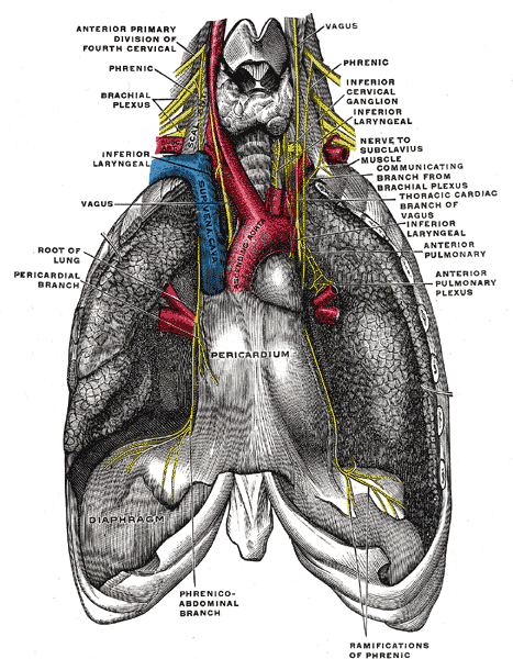 The phrenic nerve and its relations with the vagus nerve.
 -
 Thoracic portion of the sympathetic trunk.
 -
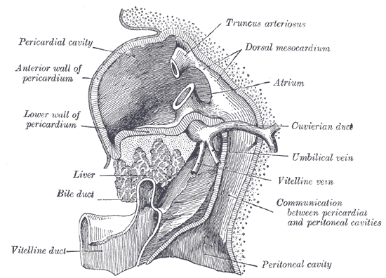 Liver with the septum transversum. Human embryo 3 mm long.
 -
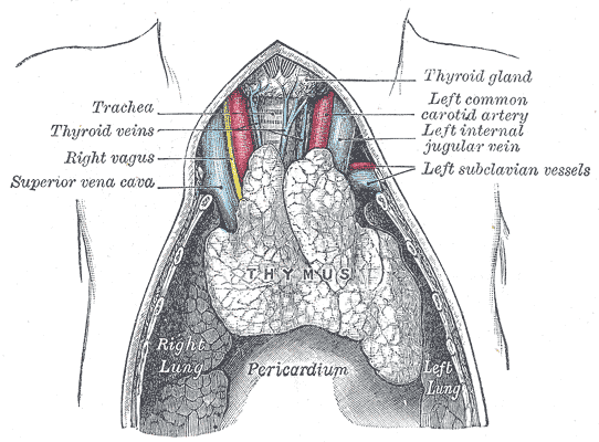 The thymus of a full-time fetus, exposed in situ.


References
- ↑ Kishore, K. (2003). The Heart of Structural Development: The Functional Basis of the Location and Morphology of the Human Vascular Pump. J Postgrad Med, 49:282-4.
- ↑ Moore, K. L., Agur, A. M., & Dalley, A. F. (2011). Essential Clinical Anatomy – Fourth Edition. Lippincott Williams & Wilkins.
- ↑ Tank, P. W. (2009). Grant’s Dissector – Fourteenth Edition. Lippincott Williams & Wilkins.
de:Herzbeutel it:Pericardio la:Pericardium ms:Perikardium nl:Pericard nn:Hjartepose fi:Perikardium
Classification
Editor-In-Chief: C. Michael Gibson, M.S., M.D. [1]; Associate Editor(s)-in-Chief: Homa Najafi, M.D.[2]
Overview
Pericarditis may be classified according to duration of the disease and recurrence into acute, Incessant, recurrent and chronic. Moreover, pericarditis can be classified based on the etiology in two groups of infectious and non-infectious causes.
Classification
- Pericarditis may be classified according to duration of the disease and recurrence into four groups:[1][2][3][4]
| Pericarditis classification based on duration | |||||||||||||||||||||||||||||||||||||||||||||||||
| Acute: New-onset disease which lasts < 4-6 weeks | Incessant: Pericarditis lasting for >4–6 weeks but <3 months without remission | Recurrent: Recurrence after the first episode of acute pericarditis 4–6 weeks or longer interval | Chronic: Pericarditis lasts for >3 months | ||||||||||||||||||||||||||||||||||||||||||||||
- Moreover, pericarditis can be classified based on the etiology in two groups:[1][2][3][5]
- Infectious:
- Non-infectious:
- Autoimmune
- Neoplastic
- Metabolic
- Traumatic and iatrogenic
- Drug-related
| Pericarditis classification based on etiology | |||||||||||||||||||||||||||||||||||||||||||||||||||||||||||||||||||||||||||||||||||||
| Infectious causes | Non-infectious causes | ||||||||||||||||||||||||||||||||||||||||||||||||||||||||||||||||||||||||||||||||||||
| Viral:
Enteroviruses(coxsackieviruses, echoviruses) Herpes viruses(EBV, CMV, HHV-6) Adenoviruses Parvovirus B19 | Bacterial:
Mycobacterium tuberculosis Coxiella burnetii Borrelia burgdorferi | Fungal:
Histoplasma species Aspergillus species Blastomyces species Candida species | Parasitic:
Echinococcus species Toxoplasma species | Autoimmune:
Systemic autoimmune and auto-inflammatory diseases Systemic vasculitides Sarcoidosis Familial Mediterranean fever IBD Still disease | Neoplastic:
Primary tumours (pericardial mesothelioma) secondary metastatic tumors( lung and breast cancer, lymphoma) | Metabolic:
Uraemia Myxoedema Anorexia nervosa | Traumatic and Iatrogenic | Drug-related | Others:
Amyloidosis Aortic dissection Pulmonary arterial Hypertension Chronic heart failure Congenital absence of the pericardium | ||||||||||||||||||||||||||||||||||||||||||||||||||||||||||||||||||||||||||||
References
- ↑ 1.0 1.1 Imazio, Massimo (2012). “Contemporary management of pericardial diseases”. Current Opinion in Cardiology. 27 (3): 308–317. doi:10.1097/HCO.0b013e3283524fbe. ISSN 0268-4705.
- ↑ 2.0 2.1 Imazio, Massimo; Spodick, David H.; Brucato, Antonio; Trinchero, Rita; Adler, Yehuda (2010). “Controversial Issues in the Management of Pericardial Diseases”. Circulation. 121 (7): 916–928. doi:10.1161/CIRCULATIONAHA.108.844753. ISSN 0009-7322.
- ↑ 3.0 3.1 Imazio, Massimo; Brucato, Antonio; DeRosa, Francesco Giuseppe; Lestuzzi, Chiara; Bombana, Enrico; Scipione, Federica; Leuzzi, Stefano; Cecchi, Enrico; Trinchero, Rita; Adler, Yehuda (2009). “Aetiological diagnosis in acute and recurrent pericarditis: when and how”. Journal of Cardiovascular Medicine. 10 (3): 217–230. doi:10.2459/JCM.0b013e328322f9b1. ISSN 1558-2027.
- ↑ Imazio, Massimo; Belli, Riccardo; Brucato, Antonio; Cemin, Roberto; Ferrua, Stefania; Beqaraj, Federico; Demarie, Daniela; Ferro, Silvia; Forno, Davide; Maestroni, Silvia; Cumetti, Davide; Varbella, Ferdinando; Trinchero, Rita; Spodick, David H; Adler, Yehuda (2014). “Efficacy and safety of colchicine for treatment of multiple recurrences of pericarditis (CORP-2): a multicentre, double-blind, placebo-controlled, randomised trial”. The Lancet. 383 (9936): 2232–2237. doi:10.1016/S0140-6736(13)62709-9. ISSN 0140-6736.
- ↑ Sliwa, Karen; Mocumbi, Ana Olga (2009). “Forgotten cardiovascular diseases in Africa”. Clinical Research in Cardiology. 99 (2): 65–74. doi:10.1007/s00392-009-0094-1. ISSN 1861-0684.
Pathophysiology
Editor-In-Chief: C. Michael Gibson, M.S., M.D. [1]; Associate Editor(s)-In-Chief: Varun Kumar, M.B.B.S., Cafer Zorkun, M.D., Ph.D. [2], Rim Halaby Homa Najafi, M.D.[3]
Overview
Pericarditis is inflammation of the pericardium, which is the double-walled sac that contains the heart and the roots of the great vessels. There can be an accompanying accumulation of fluid that can be either serous (free flowing fluid) or fibrinous (an exudate, which is a thick fluid composed of proteins, fibrin strands, inflammatory cells, cell breakdown products, and sometimes bacteria). Vascular congestion of the pericardium is also present. The underlying myocardium may or may not be inflamed as well. If the myocardium is involved in the inflammatory process, it is called myopericarditis, and CK and troponin levels may be elevated. Cardiotropic viruses usually spread to the myocardium and pericardium hematogenously and cause acute inflammation with infiltration of polymorphonuclear (PMN) leukocytes and pericardial vascularization. Most patients with viral pericarditis recover completely with few developing recurrences. Some patients develop constrictive pericarditis which could be disabling. Bacterial pericarditis results from contiguous spread of infection within the chest, either de novo or after surgery or trauma, spread from infective endocarditis, hematogenous, or direct inoculation as a result of penetrating injury or cardiothoracic surgery.
Pathophysiology
Anatomy and Physiology of Pericardium
Layers of the Pericardium
- The pericardium is made up of two layers:[1][2][3]
- Fibrous pericardium:
- Hard protective external layer
- Attached to sternum anteriorly by sterno–pericardial ligaments and fused with the central tendon of the diaphragm and great vessels to allow mobility of the pericardial sac against sudden cardiac overfilling
- Serous pericardium:
- Smooth internal layer made up of 2 components:
- Parietal: reflects onto fibrous pericardium
- Visceral: reflects onto heart and great vessels and forms the epicardium, the external layer of the heart wall
- Smooth internal layer made up of 2 components:
- Fibrous pericardium:
- Pericardial cavity: Potential space between parietal and visceral layers. It contains a serous fluid film that occupies the cavity and functions as lubricant against friction by all chest movements.
Pericardial Sinuses
- There are two small chambers or sinuses located where the visceral and parietal pericardium are continuous with one another within the pericardial cavity.[4][5][6]
- Transverse sinus:
- Located posterior to the pulmonary trunk and ascending aorta at the level between the superior vena cava and aortic arch
- Formed after dorsal myocardium ruptured embryonically
- Functional role is to allow the unhindered expansion of great arteries posteriorly during cardiac systole
- Utilized surgically to pass surgical clamps or place ligatures around great arteries.
- Oblique sinus:
- A blind recess (cul-de-sac) posterior to the left atrium between superior vena cava, right and left pulmonary veins inferior to the transverse sinus
- Formed embryonically by the incorporation of the pulmonary vein tributaries into the left atrium
- Functional role believed to be the expansion of the left atrium upon normal collapse of the thorax
Diseases of the Pericardium
- Pericarditis is an inflammatory condition of the pericardium.
- Pericardial effusion is fluid accumulation in the pericardial sac.
- Constrictive pericarditis occurs when there is a scar encasing, the heart that chronically constricts the filling of the heart.
- Cardiac tamponade is a medical emergency in which fluid in the pericardial sac acutely restricts the filling of the heart. This requires surgical drainage or pericardiocentesis.
Additional Images
-
The phrenic nerve and its relations with the vagus nerve.
-
Thoracic portion of the sympathetic trunk.
-
Liver with the septum transversum. Human embryo 3 mm long.
-
The thymus of a full-time fetus, exposed in situ.
Pathogenesis
- Cardiotropic viruses usually spread to the myocardium and pericardium hematogenously and cause acute inflammation with infiltration of polymorphonuclear (PMN) leukocytes and pericardial vascularization. This may cause pericardial effusion and fibrinous change of the pericardium. The pericardium may also develop a serous or hemorrhagic effusion. Most patients with viral pericarditis recover completely with few developing recurrences. Some patients develop constrictive pericarditis which could be disabling.[7][8][9]
- Contiguous spread of infection within the chest, either de novo or after surgery or trauma.
- Spread from infective endocarditis
- Hematogenous spread of infection
- Direct inoculation as a result of penetrating injury or cardiothoracic surgery
- Tuberculous pericarditis develops from lymphatic spread of peritracheal, peribronchial or mediastinal lymph nodes or by contiguous spread from a focus of infection in the lung or pleura. There are four pathologic stages observed:[15][16][17]
- Stage 1: Presence of diffuse fibrin deposition, granulomas and abundant mycobacterium.
- Stage 2: Development of serous or serosanguineous pericardial effusion with a predominantly lymphocytic exudate with monocytes and foam cells.
- Stage 3: Absorption of effusion with the organization of granulomatous caseation and thickening of pericardium secondary to deposition of fibrin and collagen.
- Stage 4: Development of constrictive pericarditis. The pericardial space is obliterated by dense adhesions with marked thickening of the parietal layer and replacement of granulomas by fibrous tissue.
- These types of granulomatous pericarditis also occur with fungal infections, rheumatoid arthritis (RA), and sarcoidosis.
- Pericarditis in renal failure is thought to result from inflammation of the visceral and parietal layers of the pericardium by metabolic toxins such as urea, creatinine, uric acid, methylguanidine, guanidinoacetate, parathyroid hormone, beta2-microglobulin, and others. It may be associated with hemorrhagic or serous effusion.
Gross Pathology Images
-
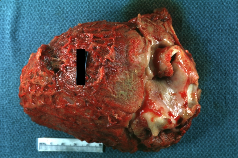
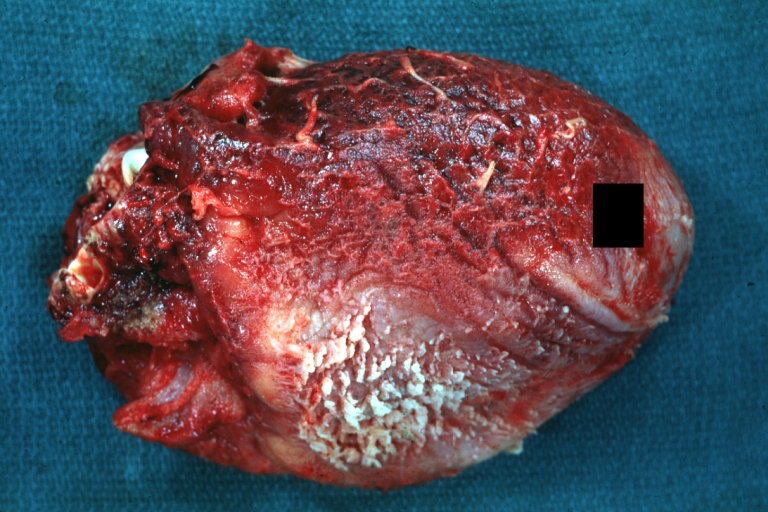
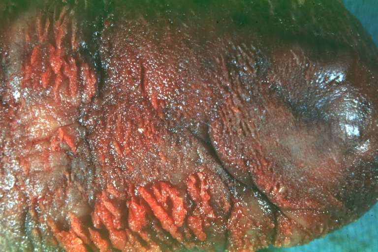
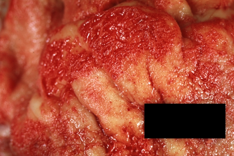
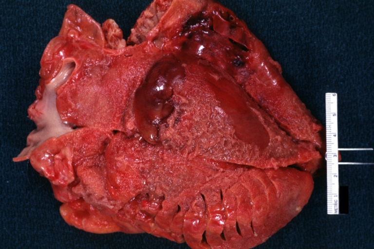
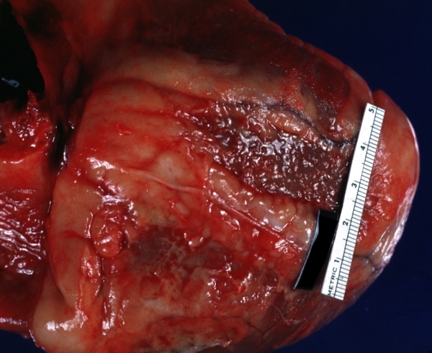
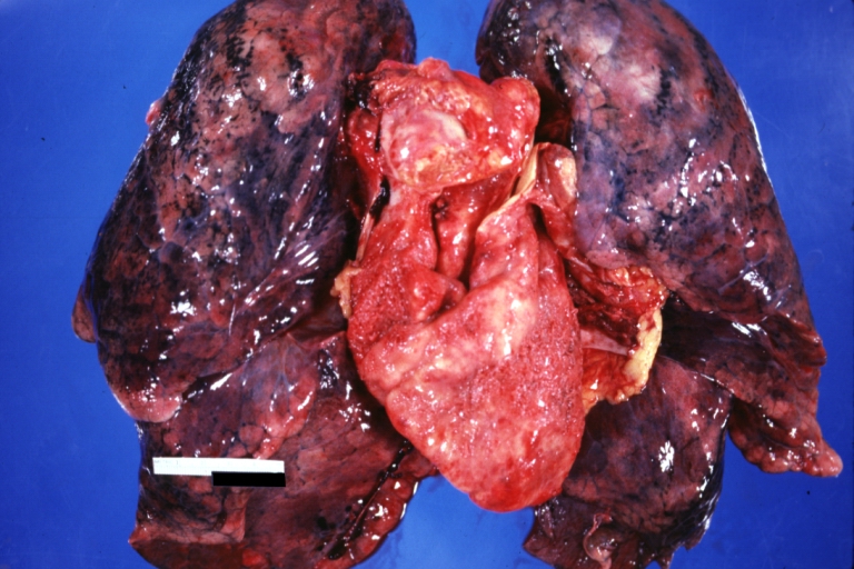
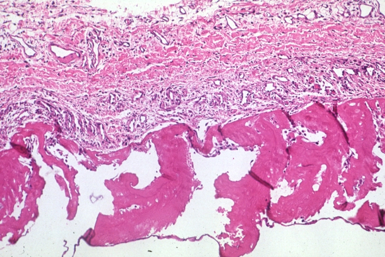
 Fibrinous pericarditis: Gross, natural color, an excellent example of bread and butter appearance. Uremia, chronic glomerulonephritis and sepsis.
 -
 Fibrinous pericarditis: Gross, a good example (bread and butter appearance).
 -
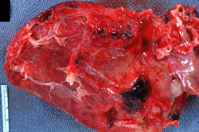 Fibrinous pericarditis: Gross, an excellent example.


-
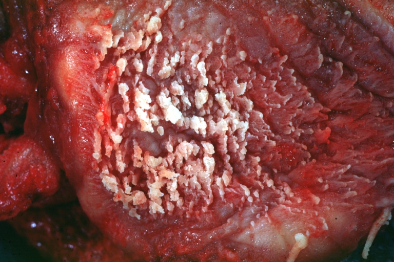 Fibrinous pericarditis: Gross, an excellent example, close-up view of fibrin.
 -
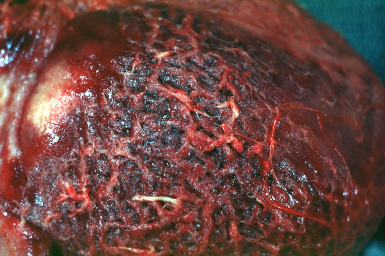 Fibrinous pericarditis: Gross, an excellent example, close-up view.
 -
 Fibrinous pericarditis: Gross, an excellent example.


-
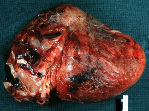
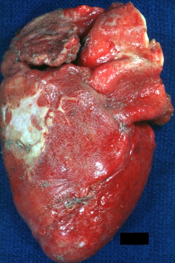
 Fibrinous pericarditis: Gross, external view of localized pericarditis over an acute infarction.
 -
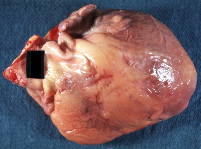
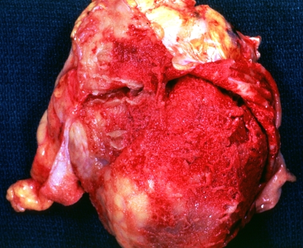
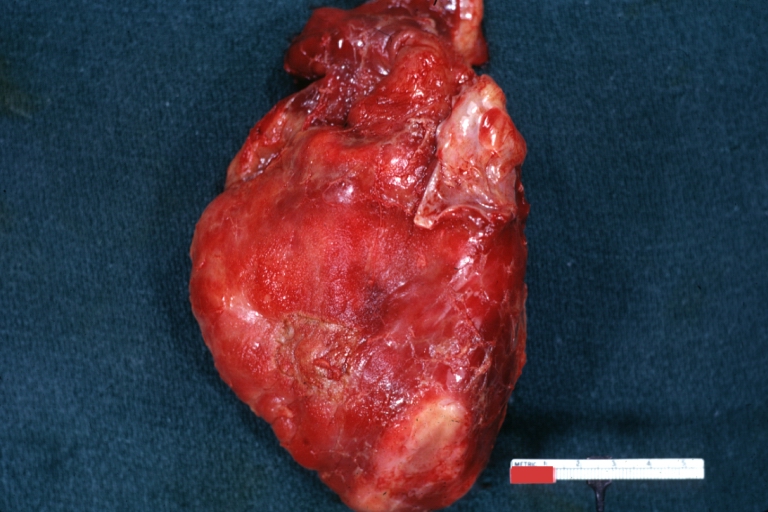
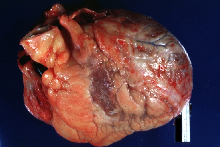
 Fibrinous pericarditis: Gross, intact heart, good example.
 -
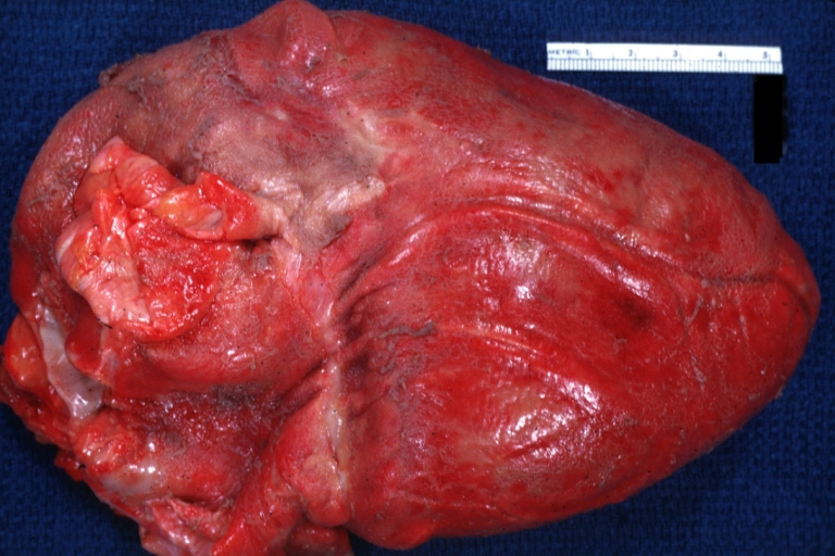
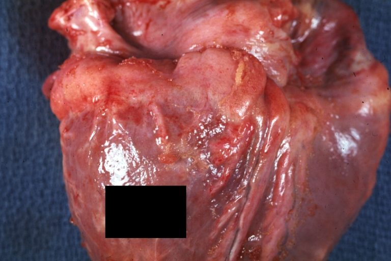 Fibrinous pericarditis: Gross, good example, mild, with small amount of fibrin.


-
 Fibrinous pericarditis: Gross, close-up, an excellent example of color and detail.
 -
 Fibrinous pericarditis: Gross, a good example.
 -
 Fibrinous pericarditis: Gross, a good example, very mild case.


-
 Fibrinous pericarditis: Gross, an excellent example.
 -
 Fibrinous pericarditis: Gross, a close-up view, an excellent illustration of fibrinous exudate.
 -
 Pericarditis in uremia.


-
 Fibrinous pericarditis: Gross, fixed tissue (note to color changes), a close-up view of fibrin on epicardial surface of heart, a typical example.
 -
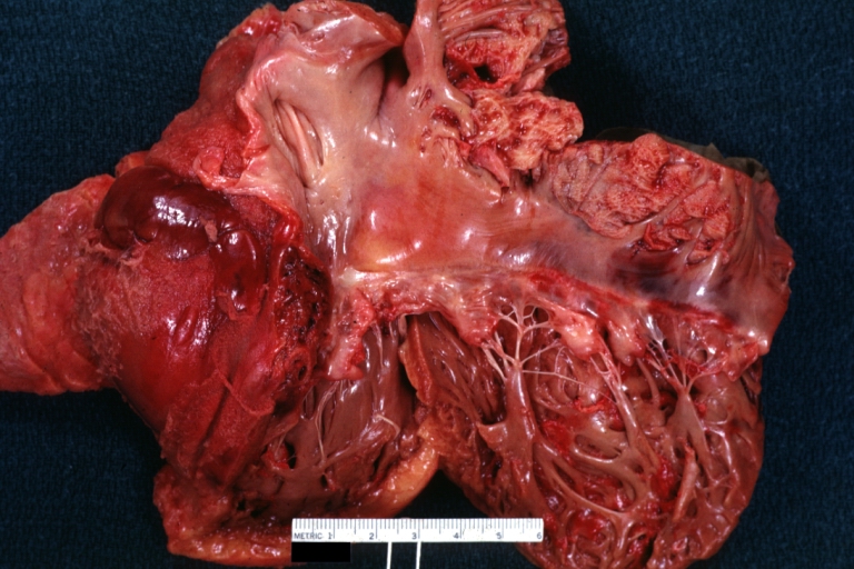 Fibrinous pericarditis: Gross, natural color, large right atrial thrombus and fibrinous pericarditis. Normal tricuspid valve with some aging changes (good example).
 -
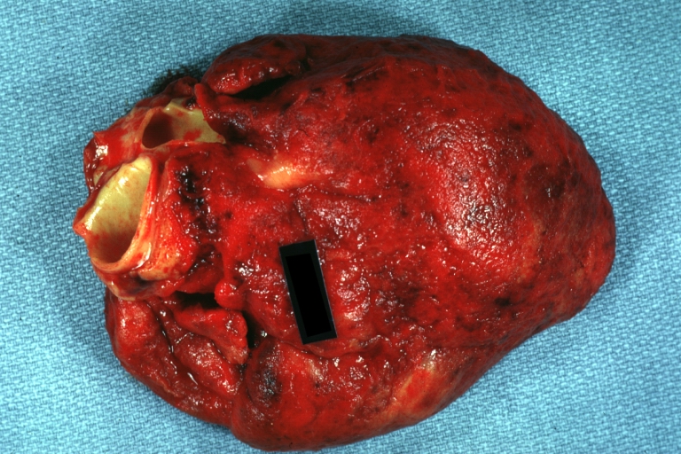
 Fibrinous pericarditis: Gross, natural color.


-
 Fibrinous pericarditis: Gross, natural color, an excellent example.
 -
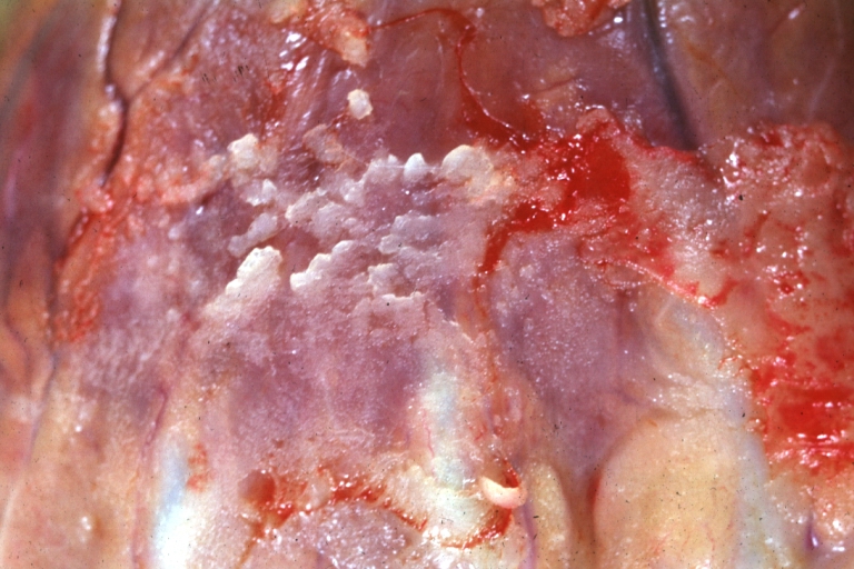 Fibrinous pericarditis: Gross, natural color, very close-up photo showing fibrinous exudate simulating frost (an excellent example).
 -
 Rheumatoid fibrinous pericarditis: Gross, natural color, a typical lesion in 22 years old white female due to juvenile rheumatoid arthritis.


-
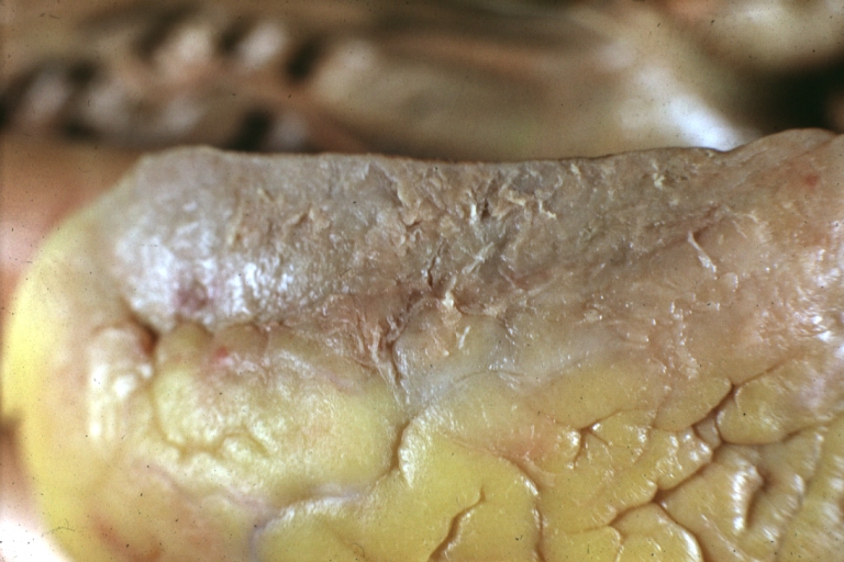
 Fibrinous pericarditis: Gross, natural color, close-up view of minimal fibrinous exudate on epicardial surface due to terminal renal failure.
 -
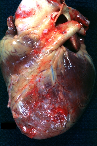
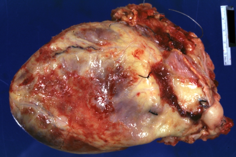
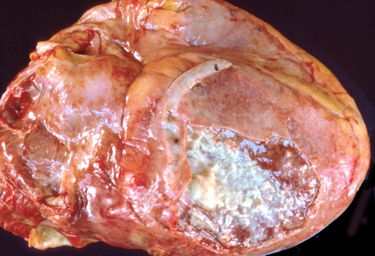
 Fibrinous pericarditis: Gross, natural color, anterior view of heart with mild fibrinous exudate over epicardium due to terminal renal failure.
 -
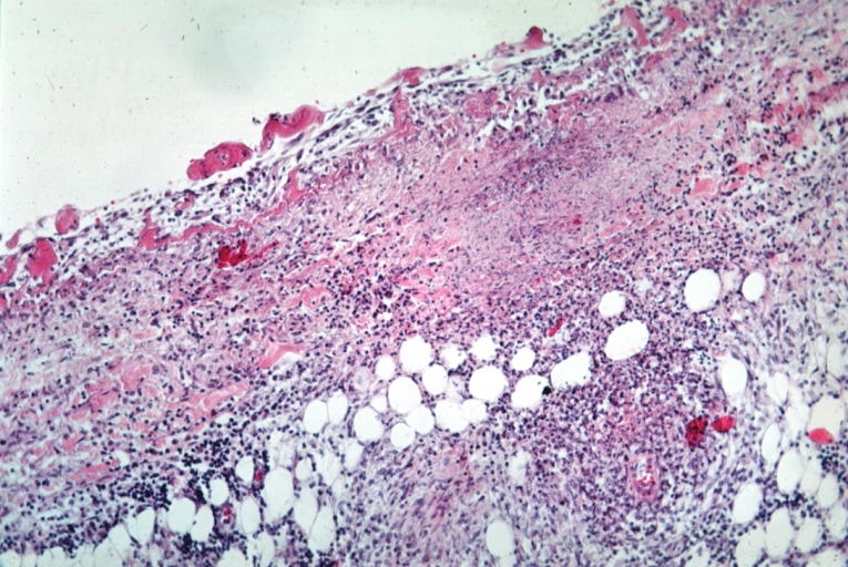
 Tuberculous pericarditis: Gross, natural color, shaggy hemorrhagic exudate. This case is much more hemorrhagic than the typical tuberculous pericarditis.


-
 Heart transplant: Gross, natural color, external view of heart. Two months after transplantation with fibrinous pericarditis.
 -
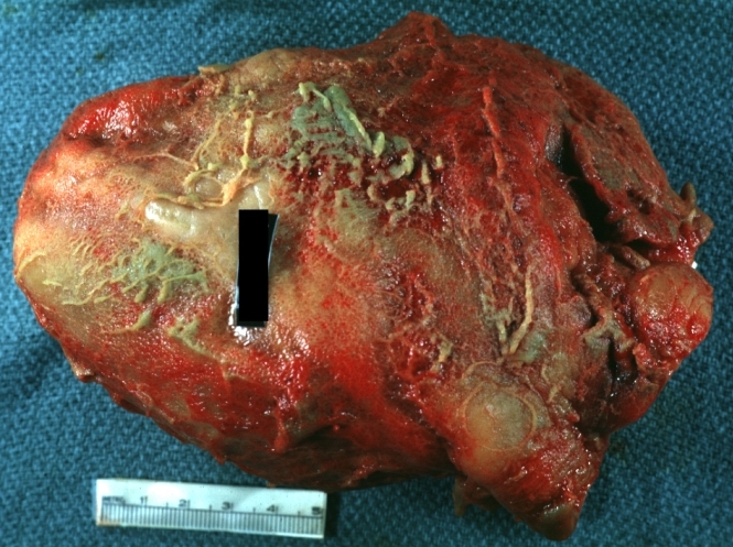
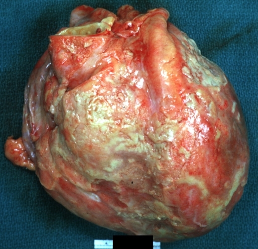
 Neoplastic pericarditis: Gross, natural color, shaggy pericarditis. Primer is adenocarcinoma of the lung.
 -
 Heart: Septic pericarditis.


Microscopic Pathology Images
-
 Tuberculous pericarditis.
 -
 Tuberculous pericarditis.
 -
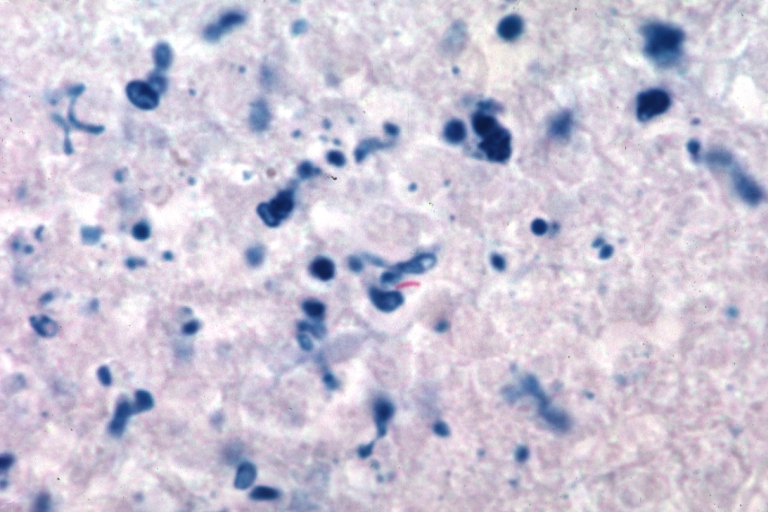 Tuberculous pericarditis: Micro oil acid fast stain. The organism easily seen.


-
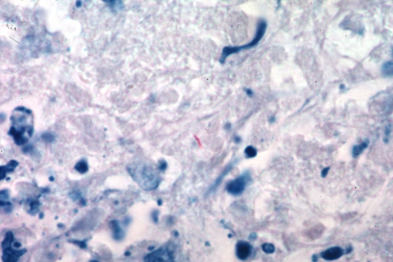 Tuberculous pericarditis: Micro oil acid fast stain. The organism easily seen.
 -
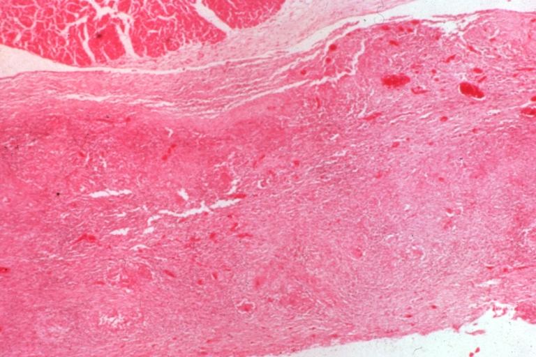
 Uremic pericarditis: Micro med mag, H&E. A good example
 -
 Tuberculous pericarditis: Micro med mag, H&E, a typical lesion


-
 Fibrinous pericarditis.
 -
 Pericarditis fibrinosa (Fibrinous pericarditis).
 -
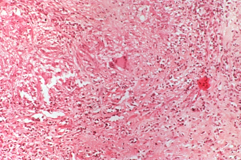
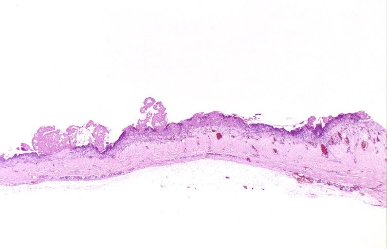
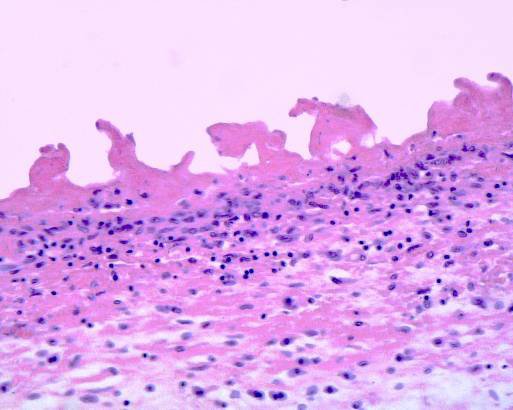
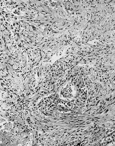 Malignant Mesothelioma, Biphasic Type: Pericardium: This tumor has epithelioid cells (lower half) surrounded by spindled cells. The patient was a 46-year-old woman with constrictive pericarditis; the pericardium was studded with coalescing tumor nodules.


Videos
| AKS7kSl4x5k}} | Acute Fibrinous Pericarditis
{{#ev:youtube|5fz_W1YxbC8}} |
|---|
References
- ↑ Kishore, K. (2003). The Heart of Structural Development: The Functional Basis of the Location and Morphology of the Human Vascular Pump. J Postgrad Med, 49:282-4.
- ↑ Moore, K. L., Agur, A. M., & Dalley, A. F. (2011). Essential Clinical Anatomy – Fourth Edition. Lippincott Williams & Wilkins.
- ↑ Tank, P. W. (2009). Grant’s Dissector – Fourteenth Edition. Lippincott Williams & Wilkins.
- ↑ Matthews JD, Cameron SJ, George M (1970). “Constrictive pericarditis following Coxsackie virus infection”. Thorax. 25 (5): 624–6. PMC 472200. PMID 5489188.
- ↑ Ilan Y, Oren R, Ben-Chetrit E (1991). “Acute pericarditis: etiology, treatment and prognosis. A study of 115 patients”. Jpn Heart J. 32 (3): 315–21. PMID 1920818.
- ↑ Shabetai R (1990). “Acute pericarditis”. Cardiol Clin. 8 (4): 639–44. PMID 2249218.
- ↑ Klacsmann PG, Bulkley BH, Hutchins GM (1977). “The changed spectrum of purulent pericarditis: an 86 year autopsy experience in 200 patients”. Am J Med. 63 (5): 666–73. PMID 930941.
- ↑ Kauffman CA, Watanakunakorn C, Phair JP (1973). “Purulent pneumococcal pericarditis. A continuing problem in the antibiotic era”. Am J Med. 54 (6): 743–50. PMID 4200204.
- ↑ Rubin RH, Moellering RC (1975). “Clinical, microbiologic and therapeutic aspects of purulent pericarditis”. Am J Med. 59 (1): 68–78. PMID 1138554.
- ↑ Ribeiro P, Shapiro L, Nihoyannopoulos P, Gonzalez A, Oakley CM (1985). “Pericarditis in infective endocarditis”. Eur Heart J. 6 (11): 975–8. PMID 4076207.
- ↑ Roberts WC, Buchbinder NA (1972). “Right-sided valvular infective endocarditis. A clinicopathologic study of twelve necropsy patients”. Am J Med. 53 (1): 7–19. PMID 4402567.
- ↑ Peel AA (1948). “TUBERCULOUS PERICARDITIS”. Br Heart J. 10 (3): 195–207. PMC 481044. PMID 18610109.
- ↑ Permanyer-Miralda G, Sagristá-Sauleda J, Soler-Soler J (1985). “Primary acute pericardial disease: a prospective series of 231 consecutive patients”. Am J Cardiol. 56 (10): 623–30. PMID 4050698.
- ↑ Mayosi BM, Burgess LJ, Doubell AF (2005). “Tuberculous pericarditis”. Circulation. 112 (23): 3608–16. doi:10.1161/CIRCULATIONAHA.105.543066. PMID 16330703.
Causes
HIV | Post-MI | Post-pericardiotomy | Radiation | Tuberculosis | Uremia | Malignancy
Editor-In-Chief: C. Michael Gibson, M.S., M.D. [1]; Associate Editor(s)-in-Chief: Mugilan Poongkunran M.B.B.S [2] Ahmed Zaghw, M.D. [3] Homa Najafi, M.D.[4]
Overview
The causes of pericarditis can be divided into infectious and non-infectious ones. Infectious causes include bacterial, viral, fungal and, parasitic. While, non-infectious causes include autoimmune, neoplastic, metabolic, traumatic and iatrogenic, and drug-related. Acute myocardial infarction, Addisonian crisis, aortic dissection and rupture, blunt or penetrating chest trauma, esophageal perforation, gastric perforation, and myocardial rupture are life threatening causes of pericarditis. Common causes of pericarditis include viral, bacterial organisms, neoplasms, autoimmune and renal failure.
Causes
Life Threatening Causes
Life-threatening causes include conditions which may result in death or permanent disability within 24 hours if left untreated and include:[1][2][3][4]
- Acute myocardial infarction
- Addisonian crisis
- Aortic dissection
- Aortic rupture
- Blunt or penetrating chest trauma
- Esophogeal perforation
- Gastric perforation
- Myocardial rupture
Common Causes
- Acute myocardial infarction
- Cardiac catheterization
- Staphylococcus aureus (MSSA, MRSA)
- Streptococcus pneumoniae
- Neisseria meningitidis
- Enterobacteriaceae
- Tuberculosis
- Histoplasmosis
- Coxsackie B virus
- Echovirus
- HIV
- Adenovirus
- Influenza
- Neoplasm
- Renal Failure
- Sulfa drugs
- Aminosalicylic acid
- Amiodarone
- Systemic lupus erythematosus
- Rheumatoid arthritis
- Idiopathic
Causes by Organ System
Causes in Alphabetical Order
- 5-Fluorouracil
- Actinomycosis
- Acute myocardial infarction
- Acute rheumatic fever
- Addisonian crisis
- Adenovirus
- Amebiasis
- Aminosalicylic acid
- Amiodarone
- Amyloidosis
- Angioma
- Ankylosing spondylitis
- Anticoagulants
- Aortic dissection
- Aortic rupture
- Asbestosis
- Behcet’s disease
- Benign obstruction of thoracic duct
- Blunt or penetrating chest trauma
- Borrelia
- Breast cancer
- Bromocriptine
- Carcinoid
- Cardiac catheterization
- Cardiopulmonary resuscitation
- Cathether ablation for arrhythmias
- Certolizumab pegol
- Chlamydia psittaci
- Cholesterol pericarditis
- Chylopericardium
- Coccidioidomycosis
- Collagen vascular disease
- Coronary artery bypass grafting
- Coxsackie B virus
- Cromolyn sodium
- Cyclophosphamide
- Cyclosporine
- Cytarabine
- Cytomegalovirus
- Dantrolene
- Daunorubicin
- Dermatomyositis
- Dialysis
- Dissecting aortic aneurysm
- Doxorubicin
- Dressler’s syndrome
- EBV
- Echinococcosis
- ECHO virus
- Endocarditis
- Esophageal rupture
- Esophogeal perforation
- Familial mediterranean fever
- Fibroma
- Francisella
- Gastric perforation
- Gaucher disease
- Heart surgery
- Herpes viruses
- Histoplasmosis
- HIV
- Hydantoin
- Hydralazine
- Hypothyroidism
- Idiopathic
- Infectious mononucleosis
- Inflammatory bowel disease
- Influenza virus
- Isoniazid
- Jacobs arthropathy-camptodactyly syndrome
- Kaposi’s sarcoma
- Kawasaki disease
- Leiomyoma
- Legionella
- Leukemia
- Lipoma
- Lung cancer
- Lymphoma
- Melanoma
- Meningococci
- Mesalazine
- Mesothelioma
- Methyldopa
- Methysergide
- Minoxidil
- Mixed connective tissue disease
- Mulibrey nanism syndrome
- Mumps virus
- Mycoplasma
- Myocardial rupture
- Myocarditis
- Myxedema
- Neisseria gonorrhoeae
- Neoplasia that has spread to the pericardium,
- Ovarian cancer
- Pacemaker syndrome
- Pancreatic-pericardial fistula
- Penicillin
- Percutaneous coronary intervention
- Pergolide
- Phenylbutazone
- Pneumococci
- Pneumonia
- Polyarteritis nodosa
- Polymyositis
- Polytetrafluoroethylene inhalation
- Postpericardiotomy syndrome
- Practolol
- Procainamide
- Radiation therapy
- Recurrent hereditary polyserositis
- Rhabdomyosarcoma
- Reiter’s syndrome
- Renal failure
- Reserpine
- Rheumatoid arthritis
- Rickettsia
- Sarcoidosis
- Sarcoma
- Scleroderma
- Serum sickness
- Silicosis
- Sipple syndrome
- Staphylococci
- Streptococci
- Streptokinase
- Streptomycin
- Sulfa drugs
- Systemic lupus erythematosus
- TAVI
- Temporal arteritis
- Teratoma
- Thiazides
- Thiouracil
- Thoracic surgery
- Thorax trauma
- Thrombolytics
- Tocainide
- Toxoplasmosis
- Treponema pallidum
- Tuberculosis
- Ureaplasma
- Uremia
- Vaccines (smallpox, yellow fever)
- Valvuloplasty
- Varicella virus
- Wegener’s granulomatosis
- Whipple’s Disease
Summery of causes
| Pericarditis classification based on etiology | |||||||||||||||||||||||||||||||||||||||||||||||||||||||||||||||||||||||||||||||||||||
| Infectious causes | Non-infectious causes | ||||||||||||||||||||||||||||||||||||||||||||||||||||||||||||||||||||||||||||||||||||
| Viral:
Enteroviruses(coxsackieviruses, echoviruses) Herpes viruses(EBV, CMV, HHV-6) Adenoviruses Parvovirus B19 | Bacterial:
Mycobacterium tuberculosis Coxiella burnetii Borrelia burgdorferi | Fungal:
Histoplasma species Aspergillus species Blastomyces species Candida species | Parasitic:
Echinococcus species Toxoplasma species | Autoimmune:
Systemic autoimmune and auto-inflammatory diseases Systemic vasculitides Sarcoidosis Familial Mediterranean fever IBD Still disease | Neoplastic:
Primary tumours (pericardial mesothelioma) secondary metastatic tumors( lung and breast cancer, lymphoma) | Metabolic:
Uraemia Myxoedema Anorexia nervosa | Traumatic and Iatrogenic | Drug-related | Others:
Amyloidosis Aortic dissection Pulmonary arterial Hypertension Chronic heart failure Congenital absence of the pericardium | ||||||||||||||||||||||||||||||||||||||||||||||||||||||||||||||||||||||||||||
References
- ↑ Imazio, Massimo (2012). “Contemporary management of pericardial diseases”. Current Opinion in Cardiology. 27 (3): 308–317. doi:10.1097/HCO.0b013e3283524fbe. ISSN 0268-4705.
- ↑ Imazio, Massimo; Spodick, David H.; Brucato, Antonio; Trinchero, Rita; Adler, Yehuda (2010). “Controversial Issues in the Management of Pericardial Diseases”. Circulation. 121 (7): 916–928. doi:10.1161/CIRCULATIONAHA.108.844753. ISSN 0009-7322.
- ↑ Imazio, Massimo; Brucato, Antonio; DeRosa, Francesco Giuseppe; Lestuzzi, Chiara; Bombana, Enrico; Scipione, Federica; Leuzzi, Stefano; Cecchi, Enrico; Trinchero, Rita; Adler, Yehuda (2009). “Aetiological diagnosis in acute and recurrent pericarditis: when and how”. Journal of Cardiovascular Medicine. 10 (3): 217–230. doi:10.2459/JCM.0b013e328322f9b1. ISSN 1558-2027.
- ↑ Sliwa, Karen; Mocumbi, Ana Olga (2009). “Forgotten cardiovascular diseases in Africa”. Clinical Research in Cardiology. 99 (2): 65–74. doi:10.1007/s00392-009-0094-1. ISSN 1861-0684.
Differentiating Pericarditis from other Diseases
Editor-In-Chief: C. Michael Gibson, M.S., M.D. [2]; Associate Editor(s)-in-Chief: Syed Hassan A. Kazmi BSc, MD [3] Homa Najafi, M.D.[4]
Overview
Pericarditis must be differentiated from diseases presenting with chest pain, shortness of breath and tachypnea which include myocardial infarction, pulmonary embolism, congestive heart failure, pneumonia, vasculitis, and chronic obstructive pulmonary disease (COPD). Manifestation of the pericarditis can help in differentiation from myocardial infarction. Moreover, other differential diagnosis include aortic stenosis, coronary artery vasospasm, esophageal rupture, esophageal spasm, esophagitis,acute gastritis, gastroesophageal reflux disease, and peptic ulcer disease should be considered.
Differentiating Pericarditis from other Diseases
- Pericarditis must be differentiated from diseases presenting with chest pain, shortness of breath and tachypnea.
- For a full discussion of the differential diagnosis of chest pain click here
- For an expert algorithm that aids in the diagnosis of the cause of chest pain click here
- Pericarditis must be differentiated from myocardial infarction as an important cause of chest pain.The differentiating features include:[1]
| Characteristic/Parameter | Pericarditis | Myocardial infarction |
|---|---|---|
| Pain description | Sharp, pleuritic, retro-sternal (under the sternum) or left precordial (left chest) pain. | Crushing, pressure-like, heavy pain. Described as “elephant on the chest“. |
| Radiation | Pain radiates to the trapezius ridge (to the lowest portion of the scapula on the back) or no radiation. | Pain radiates to the jaw, or the left or arm, or does not radiate. |
| Exertion | Does not change the pain | Can increase the pain |
| Position | Pain is worse supine or upon inspiration (breathing in) | Not positional |
| Onset/duration | Sudden pain, that lasts for hours or sometimes days before a patient comes to the ER | Sudden or chronically worsening pain that can come and go in paroxysms or it can last for hours before the patient decides to come to the ER |
Differentiating pericarditis from other diseases on the basis of chest pain, shortness of breath, and tachypnea
The differentials include the following:[2][3][4][5][6][7][8][9][10][11][12][13][14][15][16][17][18][19][20][21]
| Diseases | Diagnostic tests | Physical Examination | Symptoms | Past medical history | Other Findings | ||||||||||
|---|---|---|---|---|---|---|---|---|---|---|---|---|---|---|---|
| CT scan and MRI | EKG | Chest X-ray | Tachypnea | Tachycardia | Fever | Chest Pain | Hemoptysis | Dyspnea on Exertion | Wheezing | Chest Tenderness | Nasalopharyngeal Ulceration | Carotid Bruit | |||
| Pulmonary embolism |
|
|
|
✔ | ✔ | ✔ (Low grade) | ✔ | ✔ (In case of massive PE) | ✔ | – | – | – | – |
|
|
| Congestive heart failure |
|
✔ | ✔ | ✔ | – | – | ✔ | – | – | – | – |
|
| ||
| Percarditis |
|
|
|
✔ | ✔ | ✔ (Low grade) | ✔ (Relieved by sitting up and leaning forward) | – | ✔ | – | – | – | – |
|
|
| Pneumonia |
|
|
|
✔ | ✔ | ✔ | ✔ | – | ✔ | ✔ | – | – | – |
|
|
| Vasculitis |
|
|
✔ | ✔ | ✔ | ✔ | ✔ | ✔ | – | ✔ | ✔ | ✔ |
|
||
| Chronic obstructive pulmonary disease (COPD) |
|
|
✔ | ✔ | – | – | – | ✔ | ✔ | – | – | – |
|
| |
Other differentials
Pericarditis also resembles the following disorders and needs to be differentiated from them:
- Angina pectoris
- Aortic stenosis
- Coronary artery vasospasm
- Esophageal rupture
- Esophageal spasm
- Esophagitis
- Gastritis, acute
- Gastroesophageal reflux disease
- Peptic ulcer disease
References
- ↑ American College of Physicians (ACP). Medical Knowledge Self-Assessment Program (MKSAP-15): Cardiovascular Medicine. “Pericardial disease.” p. 64. ISBN 978-934465-28-8 [1]
- ↑ Brenes-Salazar JA (2014). “Westermark’s and Palla’s signs in acute and chronic pulmonary embolism: Still valid in the current computed tomography era”. J Emerg Trauma Shock. 7 (1): 57–8. doi:10.4103/0974-2700.125645. PMC 3912657. PMID 24550636.
- ↑ “CT Angiography of Pulmonary Embolism: Diagnostic Criteria and Causes of Misdiagnosis | RadioGraphics”.
- ↑ Bĕlohlávek J, Dytrych V, Linhart A (2013). “Pulmonary embolism, part I: Epidemiology, risk factors and risk stratification, pathophysiology, clinical presentation, diagnosis and nonthrombotic pulmonary embolism”. Exp Clin Cardiol. 18 (2): 129–38. PMC 3718593. PMID 23940438.
- ↑ “Pulmonary Embolism: Symptoms – National Library of Medicine – PubMed Health”.
- ↑ Ramani GV, Uber PA, Mehra MR (2010). “Chronic heart failure: contemporary diagnosis and management”. Mayo Clin. Proc. 85 (2): 180–95. doi:10.4065/mcp.2009.0494. PMC 2813829. PMID 20118395.
- ↑ Blinderman CD, Homel P, Billings JA, Portenoy RK, Tennstedt SL (2008). “Symptom distress and quality of life in patients with advanced congestive heart failure”. J Pain Symptom Manage. 35 (6): 594–603. doi:10.1016/j.jpainsymman.2007.06.007. PMC 2662445. PMID 18215495.
- ↑ Hawkins NM, Petrie MC, Jhund PS, Chalmers GW, Dunn FG, McMurray JJ (2009). “Heart failure and chronic obstructive pulmonary disease: diagnostic pitfalls and epidemiology”. Eur. J. Heart Fail. 11 (2): 130–9. doi:10.1093/eurjhf/hfn013. PMC 2639415. PMID 19168510.
- ↑ Takasugi JE, Godwin JD (1998). “Radiology of chronic obstructive pulmonary disease”. Radiol. Clin. North Am. 36 (1): 29–55. PMID 9465867.
- ↑ Wedzicha JA, Donaldson GC (2003). “Exacerbations of chronic obstructive pulmonary disease”. Respir Care. 48 (12): 1204–13, discussion 1213–5. PMID 14651761.
- ↑ Nakawah MO, Hawkins C, Barbandi F (2013). “Asthma, chronic obstructive pulmonary disease (COPD), and the overlap syndrome”. J Am Board Fam Med. 26 (4): 470–7. doi:10.3122/jabfm.2013.04.120256. PMID 23833163.
- ↑ Khandaker MH, Espinosa RE, Nishimura RA, Sinak LJ, Hayes SN, Melduni RM, Oh JK (2010). “Pericardial disease: diagnosis and management”. Mayo Clin. Proc. 85 (6): 572–93. doi:10.4065/mcp.2010.0046. PMC 2878263. PMID 20511488.
- ↑ Bogaert J, Francone M (2013). “Pericardial disease: value of CT and MR imaging”. Radiology. 267 (2): 340–56. doi:10.1148/radiol.13121059. PMID 23610095.
- ↑ Gharib AM, Stern EJ (2001). “Radiology of pneumonia”. Med. Clin. North Am. 85 (6): 1461–91, x. PMID 11680112.
- ↑ Schmidt WA (2013). “Imaging in vasculitis”. Best Pract Res Clin Rheumatol. 27 (1): 107–18. doi:10.1016/j.berh.2013.01.001. PMID 23507061.
- ↑ Suresh E (2006). “Diagnostic approach to patients with suspected vasculitis”. Postgrad Med J. 82 (970): 483–8. doi:10.1136/pgmj.2005.042648. PMC 2585712. PMID 16891436.
- ↑ Stein PD, Dalen JE, McIntyre KM, Sasahara AA, Wenger NK, Willis PW (1975). “The electrocardiogram in acute pulmonary embolism”. Prog Cardiovasc Dis. 17 (4): 247–57. PMID 123074.
- ↑ Warnier MJ, Rutten FH, Numans ME, Kors JA, Tan HL, de Boer A, Hoes AW, De Bruin ML (2013). “Electrocardiographic characteristics of patients with chronic obstructive pulmonary disease”. COPD. 10 (1): 62–71. doi:10.3109/15412555.2012.727918. PMID 23413894.
- ↑ Stein PD, Matta F, Ekkah M, Saleh T, Janjua M, Patel YR, Khadra H (2012). “Electrocardiogram in pneumonia”. Am. J. Cardiol. 110 (12): 1836–40. doi:10.1016/j.amjcard.2012.08.019. PMID 23000104.
- ↑ Hazebroek MR, Kemna MJ, Schalla S, Sanders-van Wijk S, Gerretsen SC, Dennert R, Merken J, Kuznetsova T, Staessen JA, Brunner-La Rocca HP, van Paassen P, Cohen Tervaert JW, Heymans S (2015). “Prevalence and prognostic relevance of cardiac involvement in ANCA-associated vasculitis: eosinophilic granulomatosis with polyangiitis and granulomatosis with polyangiitis”. Int. J. Cardiol. 199: 170–9. doi:10.1016/j.ijcard.2015.06.087. PMID 26209947.
- ↑ Dennert RM, van Paassen P, Schalla S, Kuznetsova T, Alzand BS, Staessen JA, Velthuis S, Crijns HJ, Tervaert JW, Heymans S (2010). “Cardiac involvement in Churg-Strauss syndrome”. Arthritis Rheum. 62 (2): 627–34. doi:10.1002/art.27263. PMID 20112390.
Epidemiology and Demographics
Editor-In-Chief: C. Michael Gibson, M.S., M.D. [1]; Associate Editor(s)-in-Chief: Varun Kumar, M.B.B.S. Homa Najafi, M.D.[2]
Overview
The incidence of acute pericarditis is approximately 27.7 per 100,000 individuals annually. The recurrence of disease is seen in almost 30% of patients after first episode. The mortality rate of acute pericarditis is approximately 1.1% in developed countries. Patients of all age groups may develop acute pericarditis. Although it commonly affects men in 20 to 50 years of age. Pericarditis in developed countries is most commonly due to malignancy or viral infection. It usually follows respiratory infections, most commonly echovirus or coxsackie virus. In children, it is most commonly caused by adenovirus or coxsackie virus. In developing countries pericarditis is usually secondary to tuberculosis or HIV infection. Tuberculous pericarditis, caused by Mycobacterium tuberculosis, is found in approximately 1% of all autopsied cases of TB and in 1% to 2% of instances of pulmonary TB.
Epidemiology and Demographics
Incidence
- The incidence of acute pericarditis is approximately 27.7 per 100,000 individuals annually.[1][2][3][4]
- The incidence of hospitalization for acute pericarditis was estimated to be 3.32 cases per 100,000 individuals annually.
- The recurrence of pericarditis is seen in almost 30% of patients after first episode of disease.
Case-fatality rate/Mortality rate
- The mortality rate of acute pericarditis is approximately 1.1% in developed countries.[2]
Age
- Patients of all age groups may develop acute pericarditis. Although it commonly affects people in 20 to 50 years of age.[5]
Race
Gender
Developed Countries
- Pericarditis in developed countries is most commonly due to malignancy or viral infection.[6][7][8]
- It usually follows respiratory infections, most commonly echovirus or coxsackie virus.
- In children, it is most commonly caused by adenovirus or coxsackie virus.
- The incidence and prevalence of viral pericarditis vary with season and region.
Developing Countries
- In developing countries pericarditis is usually secondary to tuberculosis or HIV infection.[9][10][11][12]
- Tuberculous pericarditis, caused by Mycobacterium tuberculosis, is found in approximately 1% of all autopsied cases of TB and in 1% to 2% of instances of pulmonary TB. It accounted for 69.5% (162 of 233) of cases referred for diagnostic pericardiocentesis in a study in Western Cape Province of South Africa, while the same accounts for 4% of cases in developed countries.
References
- ↑ Imazio, M; Cecchi, E; Demichelis, B; Chinaglia, A; Ierna, S; Demarie, D; Ghisio, A; Pomari, F; Belli, R; Trinchero, R (2007). “Myopericarditis versus viral or idiopathic acute pericarditis”. Heart. 94 (4): 498–501. doi:10.1136/hrt.2006.104067. ISSN 1355-6037.
- ↑ 2.0 2.1 Kytö, Ville; Sipilä, Jussi; Rautava, Päivi (2014). “Clinical Profile and Influences on Outcomes in Patients Hospitalized for Acute Pericarditis”. Circulation. 130 (18): 1601–1606. doi:10.1161/CIRCULATIONAHA.114.010376. ISSN 0009-7322.
- ↑ Imazio, Massimo; Bobbio, Marco; Cecchi, Enrico; Demarie, Daniela; Demichelis, Brunella; Pomari, Franco; Moratti, Mauro; Gaschino, Gianni; Giammaria, Massimo; Ghisio, Aldo; Belli, Riccardo; Trinchero, Rita (2005). “Colchicine in Addition to Conventional Therapy for Acute Pericarditis”. Circulation. 112 (13): 2012–2016. doi:10.1161/CIRCULATIONAHA.105.542738. ISSN 0009-7322.
- ↑ Imazio, Massimo; Brucato, Antonio; Cemin, Roberto; Ferrua, Stefania; Maggiolini, Stefano; Beqaraj, Federico; Demarie, Daniela; Forno, Davide; Ferro, Silvia; Maestroni, Silvia; Belli, Riccardo; Trinchero, Rita; Spodick, David H.; Adler, Yehuda (2013). “A Randomized Trial of Colchicine for Acute Pericarditis”. New England Journal of Medicine. 369 (16): 1522–1528. doi:10.1056/NEJMoa1208536. ISSN 0028-4793.
- ↑ Ariyarajah, Vignendra; Spodick, David H. (2007). “Acute Pericarditis”. Cardiology in Review. 15 (1): 24–30. doi:10.1097/01.crd.0000210645.89717.34. ISSN 1061-5377.
- ↑ Troughton RW, Asher CR, Klein AL (2004). “Pericarditis”. Lancet. 363 (9410): 717–27. doi:10.1016/S0140-6736(04)15648-1. PMID 15001332.
- ↑ Little WC, Freeman GL (2006). “Pericardial disease”. Circulation. 113 (12): 1622–32. doi:10.1161/CIRCULATIONAHA.105.561514. PMID 16567581.
- ↑ Imazio M, Brucato A, Adler Y, Brambilla G, Artom G, Cecchi E; et al. (2007). “Prognosis of idiopathic recurrent pericarditis as determined from previously published reports”. Am J Cardiol. 100 (6): 1026–8. doi:10.1016/j.amjcard.2007.04.047. PMID 17826391.
- ↑ Sagristà-Sauleda J, Permanyer-Miralda G, Soler-Soler J (1988). “Tuberculous pericarditis: ten year experience with a prospective protocol for diagnosis and treatment”. J Am Coll Cardiol. 11 (4): 724–8. PMID 3351140.
- ↑ Chen Y, Brennessel D, Walters J, Johnson M, Rosner F, Raza M (1999). “Human immunodeficiency virus-associated pericardial effusion: report of 40 cases and review of the literature”. Am Heart J. 137 (3): 516–21. PMID 10047635.
- ↑ Fowler NO (1991). “Tuberculous pericarditis”. JAMA. 266 (1): 99–103. PMID 2046135.
- ↑ Reuter H, Burgess LJ, Doubell AF (2005). “Epidemiology of pericardial effusions at a large academic hospital in South Africa”. Epidemiol Infect. 133 (3): 393–9. PMC 2870262. PMID 15962545.
Natural History, Complications and Prognosis
Pericardial Effusion | Cardiac Tamponade | Constrictive Pericarditis
Editor-In-Chief: C. Michael Gibson, M.S., M.D. [1]; Associate Editor(s)-in-Chief: Varun Kumar, M.B.B.S. Homa Najafi, M.D.[2]
Overview
Pericarditis can lead to the development of pericardial effusion, cardiac tamponade and constrictive pericarditis. Cardiac tamponade, or compression of the heart by fluid in the pericardial sac, reduces the ability of the heart to pump blood. It is a medical emergency that requires urgent pericardiocentesis or a pericardial window. The prognosis depends on the complications of pericarditis, the underlying etiology, and the associated co-morbidities. Pericarditis secondary to malignancy, MI, autoimmune disease and renal failure carries a poor prognosis.
Natural History, Complications, and Prognosis
Natural History
Pericarditis is inflammation of the pericardium, the double-walled sac that contains the heart and the roots of the great vessels. There can be an accompanying accumulation of fluid that can be either serous (free flowing fluid) or fibrinous (an exudate, which is a thick fluid composed of proteins, fibrin strands, inflammatory cells, cell breakdown products, and sometimes bacteria), which leads to development of pericardial effusion and cardiac tamponade. Vascular congestion of the pericardium is also present. The underlying myocardium may or may not be inflamed as well. If the myocardium is involved in the inflammatory process, it is called myopericarditis, and the CK and troponin levels may be elevated. Subsequent scarring of the pericardium may lead to constrictive pericarditis.
Complications
Common complications of pericarditis include:[1]
- Pericardial Effusion
- Many forms of pericarditis can be complicated by significant fluid buildup around the heart, which is known as a pericardial effusion.
- Pericardial Tamponade
- If the fluid accumulates too rapidly or is too large, then cardiac tamponade, a condition in which the heart is compressed by the fluid and cannot pump enough blood forward, may occur. Cardiac tamponade may require urgent intervention including pericardiocentesis or a pericardial window. This complication is more common in patients with specific underlying etiologies such as malignancy, tuberculosis, or purulent pericarditis. It rarely occurs in idiopathic pericarditis.
- Constrictive Pericarditis
- If scarring of the sac around the heart (pericardium) occurs, then this is called constrictive pericarditis which may require surgical stripping of the scar (pericardiectomy).
Prognosis
The prognosis associated with pericarditis depends on the underlying cause and associated condition(s).[2][3][4][5]
- Idiopathic Pericarditis:
- Idiopathic pericarditis is often self-limited and most patients recover in 2 weeks to 3 months. Idiopathic or viral pericarditis is associated with a favorable long-term prognosis with few developing recurrences. Approximately 15-30% of patients with idiopathic acute pericarditis who are not treated with colchicine will develop recurrent pericarditis.
- Post MI Pericarditis or Dressler’s Syndrome:
- Post MI pericarditis is usually associated with larger infarcts, and therefore these patients have a poorer long term prognosis.
- Tuberculous Pericarditis:
- The mortality rate associated with tuberculous pericarditis in the preantibiotic era was 80-90%. The mortality is 17-34% if the tuberculous pericarditis is associated with HIV.
- Traumatic Pericardial Injury:
- In penetrating injuries, pericardial effusion and tamponade may develop rapidly. Early detection and early treatment of cardiac tamponade is associated with a good prognosis. Minor perforations, isolated right ventricular wounds, and a systolic blood pressure more than 50 mm Hg are all associated with better outcomes.
- Malignant Pericarditis:
- Pericarditis associated with malignancy is associated with poorer outcomes and a more complicated course.
- Autoimmune Disease:
- Pericarditis associated with scleroderma and rheumatic fever is associated with worse outcomes.
- Renal Failure:
- Pericarditis associated with renal failure is associated with significant morbidity and may result in hemorrhagic pericarditis.
References
- ↑ Mayosi BM, Burgess LJ, Doubell AF (2005). “Tuberculous pericarditis”. Circulation. 112 (23): 3608–16. doi:10.1161/CIRCULATIONAHA.105.543066. PMID 16330703.
- ↑ Ilan Y, Oren R, Ben-Chetrit E (1991). “Acute pericarditis: etiology, treatment and prognosis. A study of 115 patients”. Jpn Heart J. 32 (3): 315–21. PMID 1920818.
- ↑ Shabetai R (1990). “Acute pericarditis”. Cardiol Clin. 8 (4): 639–44. PMID 2249218.
- ↑ Harvey AM, Whitehill MR. Tuberculous pericarditis. Medicine. 1937; 16: 45–94<ref name=”pmid10908256″>Hakim JG, Ternouth I, Mushangi E, Siziya S, Robertson V, Malin A (2000). “Double blind randomised placebo controlled trial of adjunctive prednisolone in the treatment of effusive tuberculous pericarditis in HIV seropositive patients”. Heart. 84 (2): 183–8. PMC 1760932. PMID 10908256.
- ↑ Nicholls, AJ. Heart and Circulation. In: Handbook of Dialysis, Daugirdas, JT, Ing, TS (Eds), Little, Brown and Co., New York 1994. p.149.
Diagnosis
Diagnosis
History and Symptoms | Physical Examination | Laboratory Findings | Electrocardiogram | EKG Examples | Chest X Ray | MRI | CT | Echocardiography | Other Imaging Findings
Treatment
Treatment
Medical Therapy | Pericardiocentesis | Pericardial Window | Pericardial Stripping | Treatment Related Videos
Looking for the patient version?
© 2026 MyEClinic – IFTM Institut für Telematik in der Medizin GmbH